Review
. 2019 Jun 13;24(12):2211.
doi: 10.3390/molecules24122211.

Current Aspects of siRNA Bioconjugate for In Vitro and In Vivo Delivery

Affiliations
Review

Current Aspects of siRNA Bioconjugate for In Vitro and In Vivo Delivery

Wanyi Tai. Molecules. .

Abstract

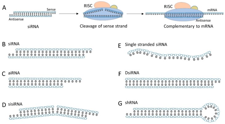
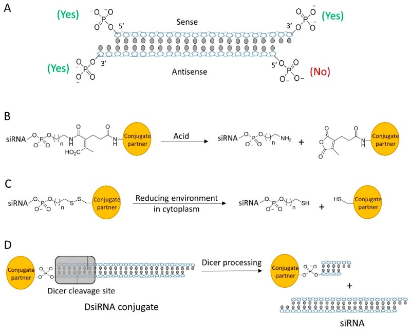
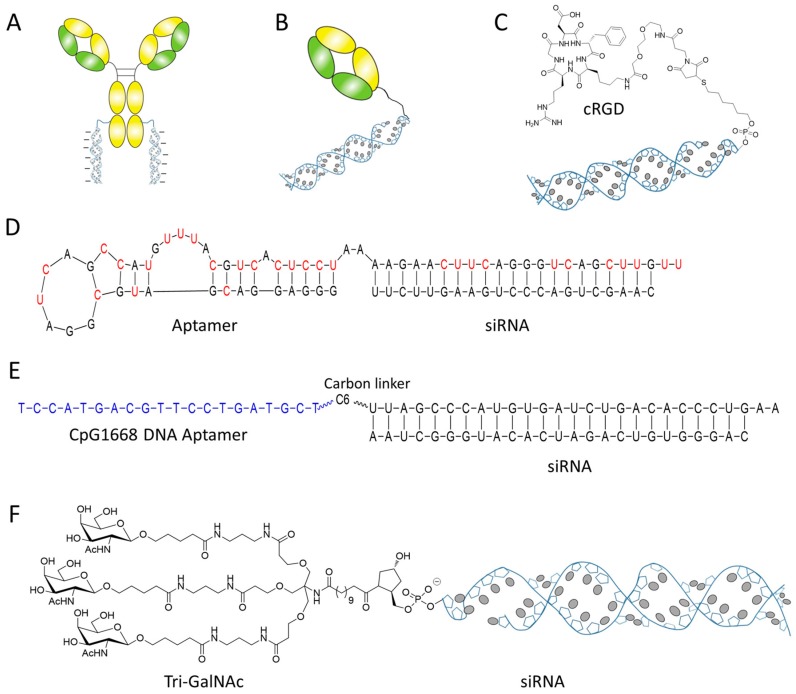
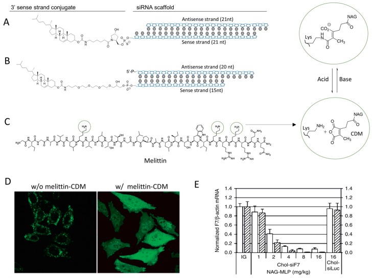
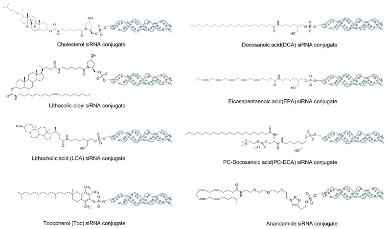
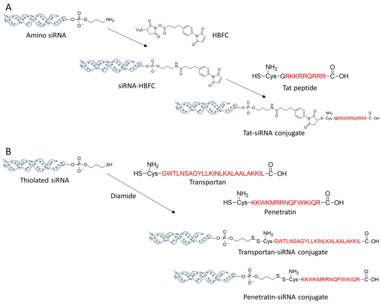
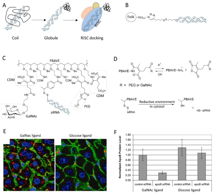
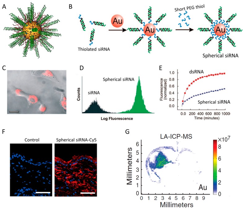
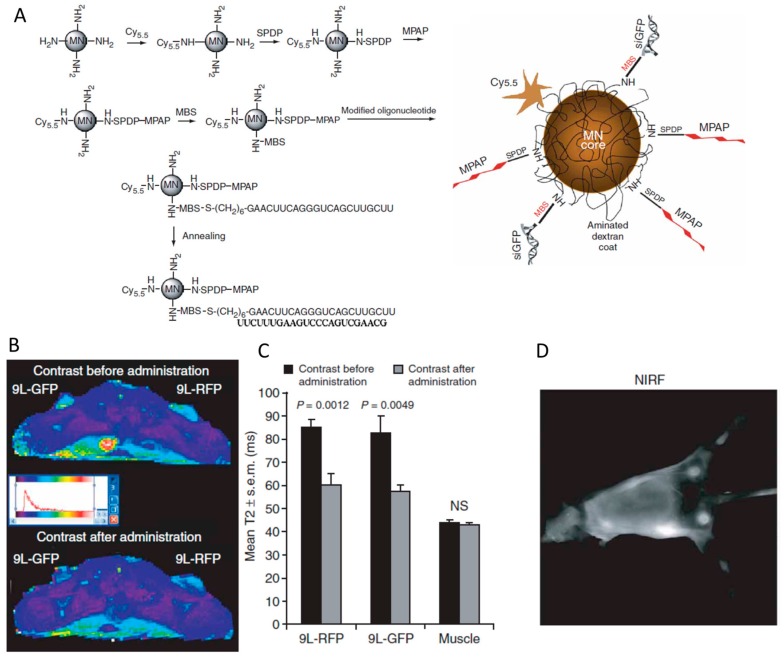
Studies on siRNA delivery have seen intense growth in the past decades since siRNA has emerged as a new class of gene therapeutics for the treatment of various diseases. siRNA bioconjugate, as one of the major delivery strategies, offers the potential to enhance and broaden pharmacological properties of siRNA, while minimizing the heterogeneity and stability-correlated toxicology. This review summarizes the recent developments of siRNA bioconjugate, including the conjugation with antibody, peptide, aptamer, small chemical, lipidoid, cell-penetrating peptide polymer, and nanoparticle. These siRNA bioconjugate, either administrated alone or formulated with other agents, could significantly improve pharmacokinetic behavior, enhance the biological half-life, and increase the targetability while maintaining sufficient gene silencing activity, with a concomitant improvement of the therapeutic outcomes and diminishment of adverse effects. This review emphasizes the delivery application of these siRNA bioconjugates, especially the conjugation strategy that control the integrity, stability and release of siRNA bioconjugates. The limitations conferred by these conjugation strategies have also been covered.

Keywords: asymmetric siRNA; lipid siRNA conjugate; siRNA bioconjugate; siRNA scaffold; spherical siRNA.


Conflict of interest statement

The author declares no conflict of interest.

Figures

Figure 1
Figure 1
(A) A simplified schematic for the RNAi pathway. (B) The canonical siRNA that has 19 bp sequence plus a unilateral 2 nt overhang at 3′ end. (C) Asymmetric siRNA (aiRNA) with a 20 nt antisense strand and 15 nt sense strand. (D) Small internally segmented RNA (sisiRNA) containing the segmented sense strand. (E) The single-stranded siRNA that can engaging RNAi machinery. (F) Dicer substrate siRNA (DsiRNA) can be processed into mature canonical siRNA by Dicer cleavage. (G) Short hairpin RNA (shRNA) has a siRNA duplex plus a hairpin loop.
Figure 2
Figure 2
(A) siRNA contains 4 terminal phosphate groups for modification, but only 3 sites are tolerable for bioconjugation. (B) Acid-sensitive linker cleaved by acid. (C) Disulfide linker can be cleaved in cytosol to release siRNA. (D) DsiRNA conjugate is processed by Dicer to release mature siRNA.
Figure 3
Figure 3
siRNA conjugated with ligands. (A) siRNA is directly conjugated with antibody via cleavable or noncleavable linker. (B) siRNA conjugated with immunoprotein Fab. (C) A targeting peptide ligand cRGD was conjugated with siRNA by noncleavable thioester bond. (D) The sequence and structure of a PSMA targeting aptamer siRNA chimera. (E) siRNA is conjugated with a DNA aptamer by carbon linker. (F) The structure of tri-GalNAc siRNA conjugate.
Figure 4
Figure 4
(A) The chemical structure of cholesterol siRNA conjugate. (B) The structure of cholesterol conjugated with aiRNA. (C) The chemical structure of pore forming peptide melittin and the mechanism of pH-sensitive CDM chemistry. The three potential modification sites of melittin are cyclized. (D) Melittin–CDM derivative enhances the escape efficacy of cargo from endosome/lysosome. FITC labeled cargo shows a punctuated distribution in cells (left), but localizes to the whole cell in the assistance of melittin–CDM derivative. (E) NAG-MLP (melittin–CDM derivative, dosing from 1 to 16 mg/kg) dramatically improves the in vivo gene silencing effect of chol-siRNA (chol-siF7). Adapted with permission from [86,87].
Figure 5
Figure 5
The chemical structures of the lipids utilized for siRNA bioconjugate.
Figure 6
Figure 6
(A) The conjugation strategy of Tat-siRNA. (B) Transportan and penetratin were conjugated with siRNA by the disulfide linkers.
Figure 7
Figure 7
(A) Controlling gene silencing activity of siRNA by conjugation with thermoresponsive polymer that has coil–globule transition behavior. (B) The structure of folic acid PEG siRNA conjugate. (C) The chemical structure of dynamic polyconjugate (DPC) using PBAVE as backbone polymer. (D) Depicted is the decomposition of DPC inside cells, mainly by reactions of CDM under acid condition and disulfide under reductive environment in cytosol. (E) Targeted delivery of DPC to liver was mediated by the ligand GalNac. Glucose ligand was used as a control. (F) Knockdown of target gene expression in livers of mice after i.v. injection of DPC. Adapted with permission from [111].
Figure 8
Figure 8
(A) The schematic structure of spherical siRNA nanoparticle. (B) Preparation of spherical siRNA by immobilizing siRNA and short-PEG to Au nanoparticle surface in two steps. (C,D) Spherical siRNA (Cy3-labeled) showed strong cell uptake on HeLa cells. (E) The structure of spherical siRNA (RNA–Au NPs) can protect siRNA (dsRNA) from degradation in serum. (F) Spherical siRNA penetrates through mouse skin and delivers siRNA into in the cytoplasm of epidermal cells. (G) The localization of Au content in coronal brain sections of mice injected intracranially with spherical siRNA. Adapted with permission from [117,119,121,126].
Figure 9
Figure 9
(A) The synthesis of the magnetic nanoparticle (MN) by decorating Cy5.5, MPAP, peptide and siRNA step by step. (B) Magnetic resonance imaging of mice bearing bilateral 9L-GFP and 9L-RFP tumors before and 24 h after the administration of MN-siRNA conjugate. (C) The tumors exhibit a significant drop in T2 relaxivity, whereas the muscle tissue remained unchanged. (D) In vivo NIR imaging of the tumors in mice. Adapted with permission from [132].

References

    1. Lares M.R., Rossi J.J., Ouellet D.L. RNAi and small interfering RNAs in human disease therapeutic applications. Trends Biotechnol. 2010;28:570–579. doi: 10.1016/j.tibtech.2010.07.009. - DOI - PMC - PubMed
    1. Whitehead K.A., Langer R., Anderson D.G. Knocking down barriers: Advances in siRNA delivery. Nat. Rev. Drug Discov. 2009;8:129–138. doi: 10.1038/nrd2742. - DOI - PMC - PubMed
    1. Kedmi R., Veiga N., Ramishetti S., Goldsmith M., Rosenblum D., Dammes N., Hazan-Halevy I., Nahary L., Leviatan-Ben-Arye S., Harlev M., et al. A modular platform for targeted RNAi therapeutics. Nat. Nanotechnol. 2018;13:214–219. doi: 10.1038/s41565-017-0043-5. - DOI - PubMed
    1. Kristen A.V., Ajroud-Driss S., Conceicao I., Gorevic P., Kyriakides T., Obici L. Patisiran, an RNAi therapeutic for the treatment of hereditary transthyretin-mediated amyloidosis. Neurodegener. Dis. Manag. 2019;9:5–23. doi: 10.2217/nmt-2018-0033. - DOI - PubMed
    1. Jenny K.W., Lam A.J.W. What is the Future of SiRNA Therapeutics. J. Drug Des. Res. 2014;1:1005.

LinkOut - more resources