Review
. 2019 Jun 13;24(12):2216.
doi: 10.3390/molecules24122216.

Porous Silicon-Based Aptasensors: The Next Generation of Label-Free Devices for Health Monitoring

Affiliations
Review

Porous Silicon-Based Aptasensors: The Next Generation of Label-Free Devices for Health Monitoring

Monica Terracciano et al. Molecules. .

Abstract

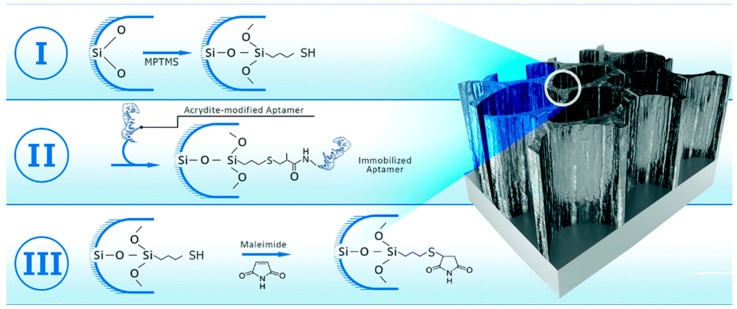
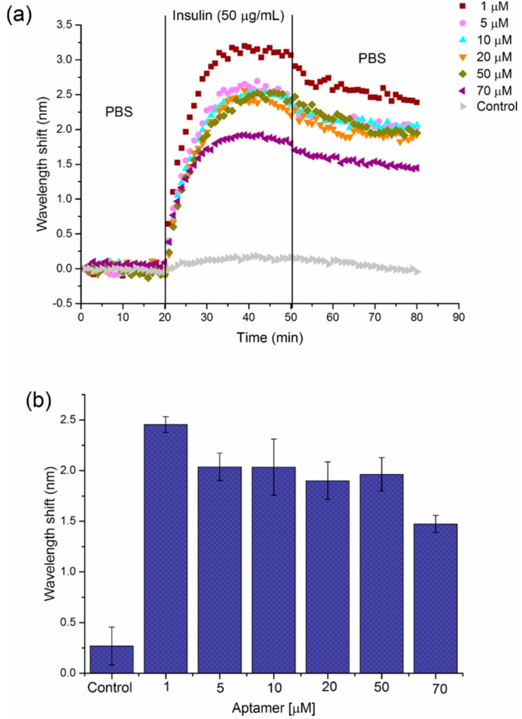
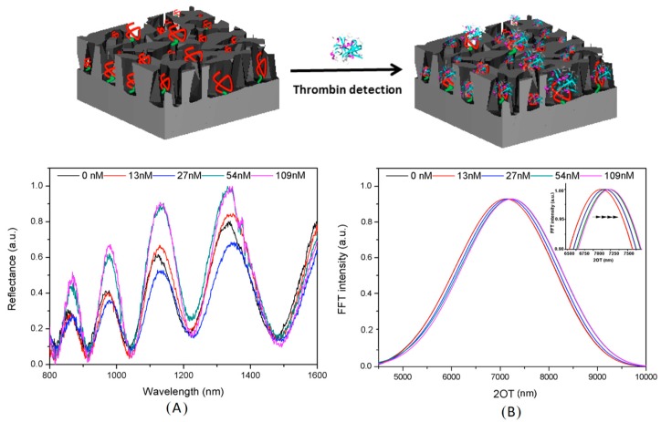
Aptamers are artificial nucleic acid ligands identified and obtained from combinatorial libraries of synthetic nucleic acids through the in vitro process SELEX (systematic evolution of ligands by exponential enrichment). Aptamers are able to bind an ample range of non-nucleic acid targets with great specificity and affinity. Devices based on aptamers as bio-recognition elements open up a new generation of biosensors called aptasensors. This review focuses on some recent achievements in the design of advanced label-free optical aptasensors using porous silicon (PSi) as a transducer surface for the detection of pathogenic microorganisms and diagnostic molecules with high sensitivity, reliability and low limit of detection (LoD).

Keywords: aptamer; aptasensor; optical label free-sensing; porous silicon; surface modification.


Conflict of interest statement

The authors declare no conflict of interest.

Figures

Figure 1
Figure 1
Three-step biofunctionalization process for aptamers immobilization onto PSiO2 device. (I) Silanization with MPTMS via a thioether bond, (II) reaction with acrydite-modified Hemag1P aptamer via a thioether bond and (III) blocking of residual thiol groups with maleimide. Reproduced with permission from [56].
Figure 2
Figure 2
(a) Relative intensity change of the Hemag1P-modified PSiO2 device upon exposure to L. acidophilus bacterial suspensions (107 cells per mL). (b) Microscope image taken after the biosensing experiment depicts L. acidophilus cells captured onto the aptamer-modified PSiO2 device. Reproduced with permission from [56].
Figure 3
Figure 3
Sensitivity enhancement of the PAA-functionalized PSiO2 biosensor. Averaged optical response (relative EOT) of the biosensor upon exposure to 1 M of: PA, IgG or both in a successive manner. Schematic illustration of biomolecules captured by the aptamers within the porous scaffold. Upper dashed line indicates the LoD value. Differences between both single exposures (of PA or IgG) and the sandwich assay are statistically significant (p < 0.05, n ≥ 3). Reproduced with permission from [58].
Figure 4
Figure 4
Sensorgram for the detection of insulin secreted by human islets upon stimulation with 20 mM glucose for 2 h in Krebs buffer using IRS. Reproduced with permission from [63].
Figure 5
Figure 5
(a) Sensograms showing the effect of IBA solution concentration used for modified-PSi device on the response to 50 μg/mL insulin. (b) Graph representing the average wavelength shift (nm) obtained for detection of 50 μg/mL insulin by PSi-5 cycles surface, which is modified with a range of aptamer concentrations (1–70 μM). Error bars correspond to the standard deviation from three individual experiments on PSi-devices prepared with each IBA concentration. Reproduced with permission from [64].
Figure 6
Figure 6
Reflectivity spectra (A) and corresponding Fourier transforms (B) of PSi-aptasensor after exposure to different thrombin concentrations (13, 27, 54 and 109 nM). Reproduced with permission from [71].

Similar articles

Cited by

References

    1. Lin V.S.Y., Motesharei K., Dancil K.P.S., Sailor M.J., Ghadiri M.R. A porous silicon-based optical interferometric biosensor. Science. 1997;278:840–843. doi: 10.1126/science.278.5339.840. - DOI - PubMed
    1. Jane A., Dronov R., Hodges A., Voelcker N.H. Porous silicon biosensors on the advance. Trends Biotechnol. 2009;27:230–239. doi: 10.1016/j.tibtech.2008.12.004. - DOI - PubMed
    1. Dancil K.P.S., Greiner D.P., Sailor M.J. A porous silicon optical biosensor: Detection of reversible binding of IgG to a protein A-modified surface. J. Am. Chem. Soc. 1999;121:7925–7930. doi: 10.1021/ja991421n. - DOI
    1. De Stefano L., Rea I., Giardina P., Armenante A., Rendina I. Protein-Modified Porous Silicon Nanostructures. Adv. Mat. 2008;20:1529–1533. doi: 10.1002/adma.200702454. - DOI
    1. Lehninger A.L., Nelson D.L., Cox M.M. The Molecular Basis of Cell Structure and Function, Biochemistry. 2nd ed. Worth Publishers; New York, NY, USA: 1975. pp. 71–77.

LinkOut - more resources