. 2019 Jun 14;38(1):257.
doi: 10.1186/s13046-019-1248-2.

Colorectal cancer-derived extracellular vesicles induce transformation of fibroblasts into colon carcinoma cells

Affiliations

Colorectal cancer-derived extracellular vesicles induce transformation of fibroblasts into colon carcinoma cells

Mohamed Abdouh et al. J Exp Clin Cancer Res. .

Abstract

Background: We reported that horizontal transfer of malignant traits to target cells is a potential pathway to explain cancer dissemination. Although these results were encouraging, they were never corroborated by data showing the molecular mechanisms responsible for the observed phenomenon.

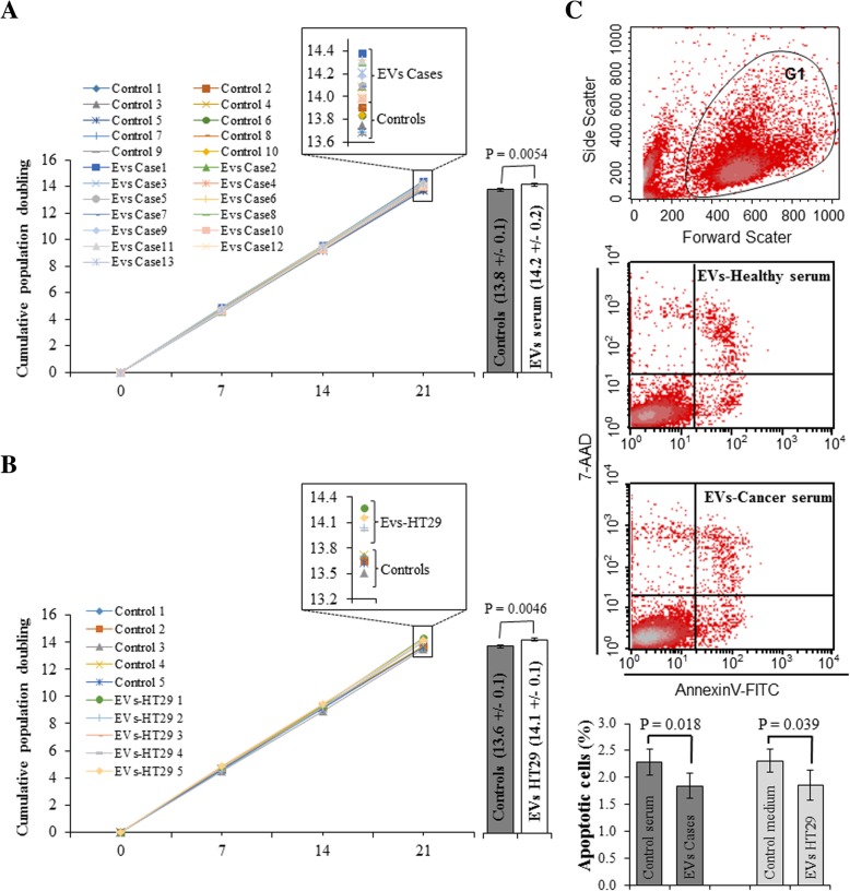
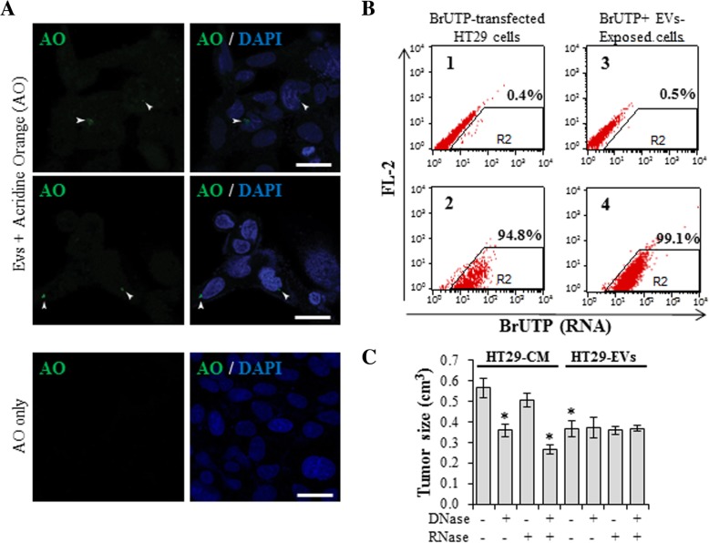
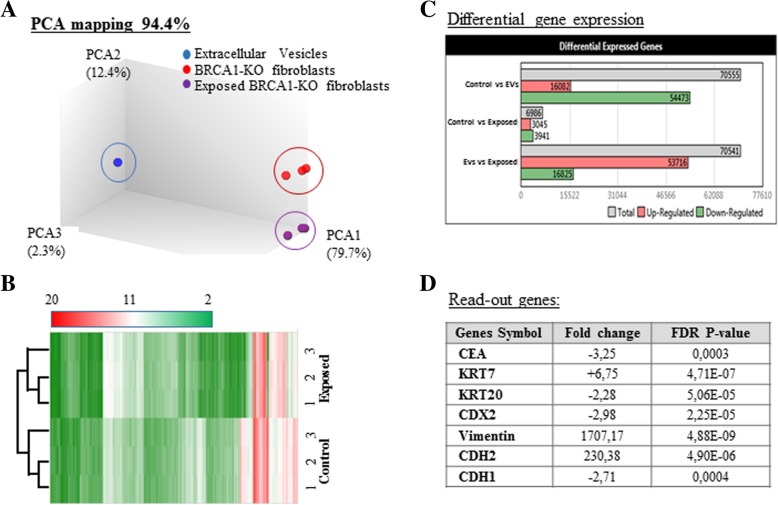
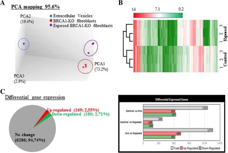
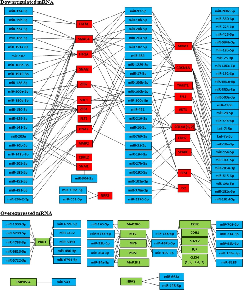
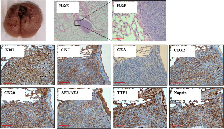
Methods: In the present study, we exposed BRCA1-KO fibroblasts to extracellular vesicles (EVs) isolated from a colon cancer cell line (HT29) and from sera of patients with colorectal cancer. Three weeks after exposure, fibroblasts were injected subcutaneously into NOD-SCID mice. Whole genome sequencing, transcriptome analysis and RNA sequencing of cancer EVs and fibroblasts prior and after exposure to cancer EVs were performed.

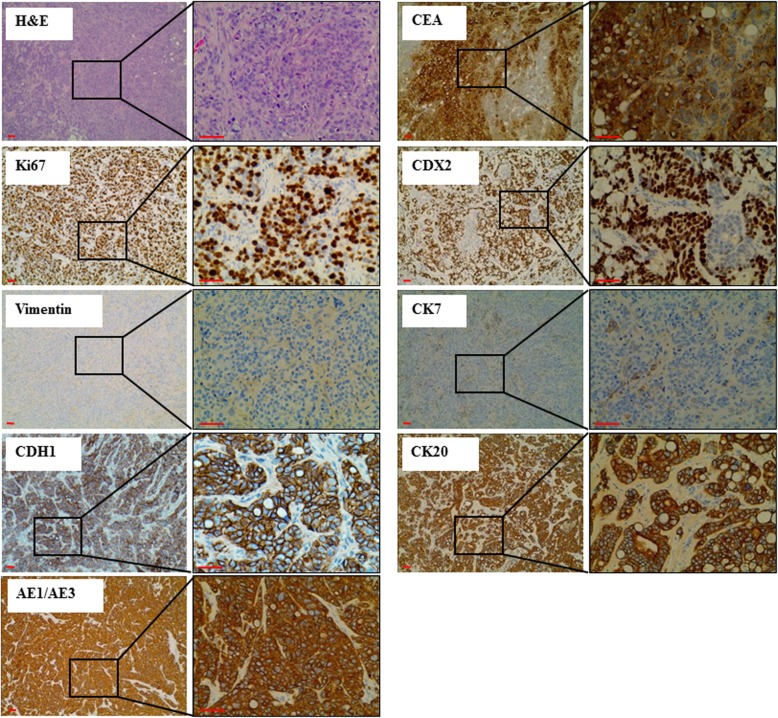
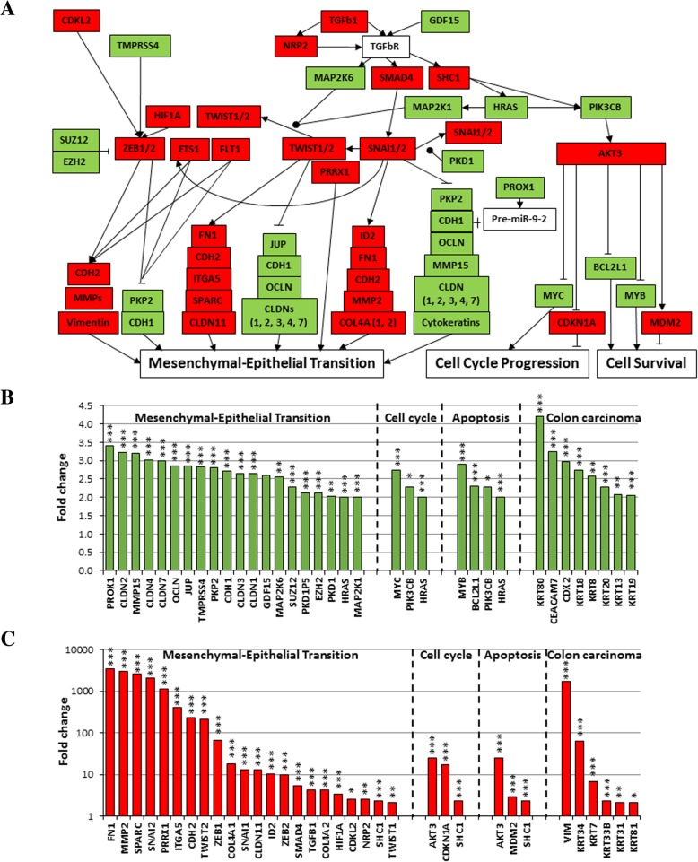
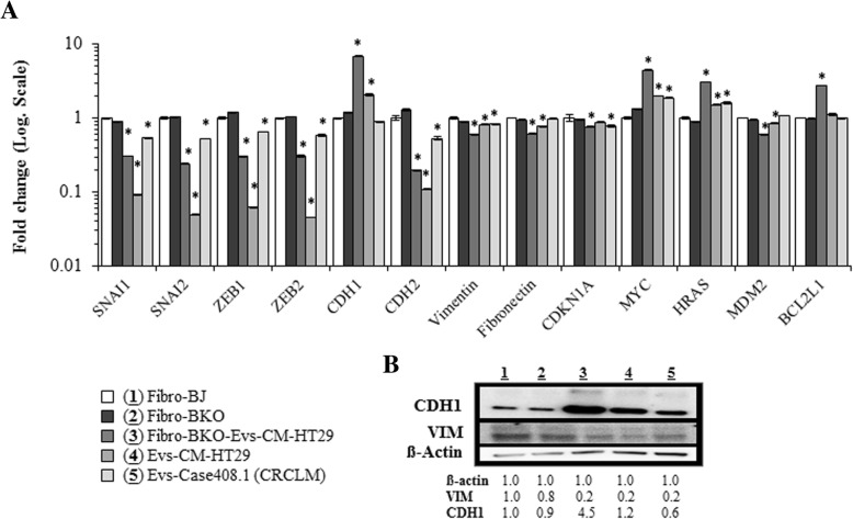
Results: Phenotypical transformation of the fibroblasts into colon cancer cells was confirmed by histopathological study of the xenotransplants. We observed that EV-mediated transfer of cancer microRNAs was responsible for the transition from a mesenchymal to an epithelial phenotype (MET) in the treated fibroblasts as well as activation of cell cycle progression and cell survival pathways. DNA and RNA sequencing suggested that cancer DNA was transferred and possibly transcribed in target cells. Furthermore, injection of colon cancer EVs in the tail vein of NOD-SCID mice determined neoplastic transformation and metastases in the lungs of the mice confirming for the first time the hypothesis that transfer of malignant epithelial cancer traits to distant target cells is a concept applicable to in vivo models.

Conclusions: These discoveries shed new light into the molecular mechanisms behind the horizontal transfer of malignant traits and confirm the notion that metastatic disease might be reproduced through transfer of circulating genetic material.

Keywords: Colorectal Cancer; Extracellular vesicles; Genetic material; Horizontal transfer; Metastasis.


Conflict of interest statement

The authors declare that they have no competing interests.

Figures

Fig. 1
Fig. 1
Colorectal cancer EVs increased BRCA1-KO fibroblasts proliferation and reduced their cell mortality. BRCA1-KO fibroblasts were cultured for 3 weeks in the presence of EVs isolated from control human sera, or cancer patient sera (a, c), and control medium or EVs isolated from HT-29 cell line conditioned medium (b, c). Cells were analyzed for their growth potential (a, b) and cell viability (c). a and b Population doublings capability was calculated at every passage. Data in inserts and column graphs represent cumulative population doublings at the end of the treatment periods. Column graphs represent pooled data from 10 control vs. 13 cancer patient sera (a), and from 5 control vs. 5 independent batches of HT29 conditioned media (b). Data are mean ± SD. P values are represented on the column graphs. (c) Treated cells were analyzed for cell viability using AnnexinV and 7-AAD staining followed by flow cytometry analyses. Cells were gated (G1) in a biparametric FSC/SSC graph. The percentage of apoptotic (AnnexinV positive and 7AAD negative) cells were plotted for comparison. Column graphs represent pooled data from 5 control vs. 5 cancer patient sera, and from 4 control vs. 4 independent batches of HT29 conditioned media. Data are mean ± SD. P values are represented on the column graphs
Fig. 2
Fig. 2
Cancer EVs transfer malignant trait to target cells in vitro. BRCA1-KO fibroblasts were treated for 3 weeks with EVs isolated from cancer cells conditioned medium, cancer patient sera or respective controls. Treated cells were injected subcutaneously into NOD/SCID mice that were followed for 4 weeks for tumors growth. Developing tumors were excised, fixed and embedded in paraffin. Tissue sections were processed for H&E staining or immunolabeled with antibodies to Ki67, vimentin, CDH1, CEA, CDX2, CK7, CK20 and AE1/AE3. Representative pictures are shown. Data are representative of five cell cultures exposed to EVs isolated from HT29 cells conditioned medium and thirteen cell cultures exposed to EVs derived from colorectal cancer sera. Scale bars: 50 μm
Fig. 3
Fig. 3
Cancer EVs transferred genetic material cargo to BRCA1-KO fibroblasts. a, EVs were tagged with acridine orange (AO) to label DNA and BRCA1-KO fibroblasts were exposed to these EVs for 24 h. Representative images are shown. Arrowheads point to EV-derived DNA (green) that is entering the nuclei (colored in blue with DAPI). Scale bars: 20 μm. b, HT29 cells were transfected with the BrUTP. Aliquotes of transfected (2) or untransfected (1) cells were analyzed for BrUTP incorporation in RNAs. Note that virtually all cells incorporated BrUTP in their RNA (sample 2). Afterwards, conditioned medium was collected from transfected cells for the isolation of EVs. BRCA1-KO fibroblasts were exposed (4) or not (3) to these EVs for 12 h. n = 2 independent experiments. c, HT29 conditioned medium or EVs were treated or left untreated either with DNase, RNase, or both. BRCA1-KO fibroblasts were treated for 3 weeks with these media, then treated cells were injected subcutaneously into NOD/SCID mice that were followed for 4 weeks for tumors growth. Data are mean +/− SD. n = 2–3 mice. *P < 0.05
Fig. 4
Fig. 4
Exposure of BRCA1-KO fibroblasts to cancer EVs changed their transcriptome profile. a and b, PCA mapping and hierarchical clustering showed that BRCA1-KO fibroblasts exposed to EVs clustered differently from non-exposed cells. In addition, EVs clustered far from cells. c, Differential gene expression between EVs-exposed and non-exposed cells. Data were obtained with 3 independent cell cultures. Filter criteria were set as follows: Fold change: > 2 or < − 2 and FDR P value < 0.05. d, Accuracy of the microarray data mining. List of read-out genes based on immunohistochemical labeling data (Fig. 1)
Fig. 5
Fig. 5
BRCA1-KO fibroblasts exposed to cancer cells EVs undergo Mesenchymal to Epithelial Transition (MET), increased proliferation and reduced apoptosis. BRCA1-KO fibroblasts were treated for 3 weeks with EVs isolated from HT29 cancer cells conditioned medium. RNA was isolated and processed for microarray analyses. a, Three major pathways involving genes implicated in MET, cell cycle progression and cell survival are shown. Green and Red highlight up-regulated and down-regulated genes in EVs-exposed cells, respectively. b and c, Up-regulated (b) and down-regulated (c) genes in EVs-exposed cells. Genes expression with a cut-off of − 2 and 2 and a FDR P value < 0.05 are shown. n = 3 independent cell cultures. ● phosphorylation. * P < 0.05, ** P < 0.01, *** P < 0.001
Fig. 6
Fig. 6
Validation of differential expression of genes involved in the MET process. a, Naïve fibroblasts (BJ) (1), BRCA1-KO fibroblasts untreated (2) or treated with EVs isolated from HT29 cancer cells conditioned medium (3), and EVs isolated from HT29 cells conditioned media (4) or colorectal cancer patient serum (4) were analyzed for the expression of mesenchymal (SNAI, ZEB, CDH2, Vimentin and Fibronectin), epithelial (CDH1), cell cycle regulators (CDKN1A, MYC and HRAS), and apoptosis regulators (MDM2 and BCL2L1) markers by qPCR. Results are representative of one experiment performed twice. Data are mean +/− SD. n = 2. *P < 0.05. Note that data are expressed in Log. Scale. b, The same samples as in (a) were analyzed by immunoblot for the expression of mesenchymal (Vimentin; VIM) and epithelial (CDH1) markers. β-actin is used as calibrator for proteins loading. The table shows proteins levels in the corresponding samples. n = 2 independent experiments
Fig. 7
Fig. 7
Exposure of BRCA1-KO fibroblasts to cancer EVs changed their pattern of miRNA expression. a and b, PCA mapping and hierarchical clustering showed that BRCA1-KO fibroblasts exposed to cancer EVs clustered differently from non-exposed cells. In addition, EVs clustered far from cells. c, Differential miRNA expression between EVs-exposed and non-exposed cells. Data are obtained with 3 independent cell cultures. Filter criteria were set as follows: Fold change: > 2 or < − 2 and FDR P value < 0.05
Fig. 8
Fig. 8
mRNA/miRNA interaction network derived from the microarray analyses describing the transcripts involved in MET process. Connections were retrieved from mirtarbase database (http://mirtarbase.mbc.nctu.edu.tw). Genes expression with a cut-off of 2 are shown. n = 3 independent cell cultures. See online Additional file 1: Tables S4 and S5 for the levels of expression
Fig. 9
Fig. 9
Cancer EVs transfer malignant traits in vivo. NOD-SCID mice were injected every other day in the tail vein with EVs isolated from MC38 cells conditioned medium for 5 weeks. Four weeks later, lungs were harvested photographed and analyzed. Tissue sections were processed for H&E staining or immunolabeled with antibodies to Ki67, CEA, CDX2, CK7, CK20, AE1/AE3, TTF1 and Napsin. Representative pictures are shown. Data are representative of staining perform on lungs harvested from three exposed mice. Scale bars: 50 μm

References

    1. Jemal A, Bray F, Center MM, Ferlay J, Ward E, Forman D. Global cancer statistics. CA Cancer J Clin. 2011;61:69–90. doi: 10.3322/caac.20107. - DOI - PubMed
    1. Yao D, Dai C, Pen S. Mechanism of the mesenchymal–epithelial transition and its relationship with metastatic tumor formation. Mol Cancer Res. 2011;9(12):1608–1620. doi: 10.1158/1541-7786.MCR-10-0568. - DOI - PubMed
    1. Kim MY, Oskarsson T, Acharyya S, Nguyen DX, Zhang XH, Norton L, et al. Tumor self-seeding by circulating cancer cells. Cell. 2009;139:1315–1326. doi: 10.1016/j.cell.2009.11.025. - DOI - PMC - PubMed
    1. Nguyen DX, Bos PD, Massagué J. Metastasis: from dissemination to organ specific colonization. Nat Rev Cancer. 2009;9:274–284. doi: 10.1038/nrc2622. - DOI - PubMed
    1. Hugo H, Ackland ML, Blick T, Lawrence MG, Clements JA, Williams ED, et al. Epithelial-mesenchymal and mesenchymal-epithelial transitions in carcinoma progression. J Cell Physiol. 2007;213:374–383. doi: 10.1002/jcp.21223. - DOI - PubMed

MeSH terms