. 2019 Jun 14;38(1):260.
doi: 10.1186/s13046-019-1199-7.

p65BTK is a novel potential actionable target in KRAS-mutated/EGFR-wild type lung adenocarcinoma

Affiliations

p65BTK is a novel potential actionable target in KRAS-mutated/EGFR-wild type lung adenocarcinoma

Federica Giordano et al. J Exp Clin Cancer Res. .

Abstract

Background: Lung cancer is still the main cause of cancer death worldwide despite the availability of targeted therapies and immune-checkpoint inhibitors combined with chemotherapy. Cancer cell heterogeneity and primary or acquired resistance mechanisms cause the elusive behaviour of this cancer and new biomarkers and active drugs are urgently needed to overcome these limitations. p65BTK, a novel isoform of the Bruton Tyrosine Kinase may represent a new actionable target in non-small cell lung cancer (NSCLC).

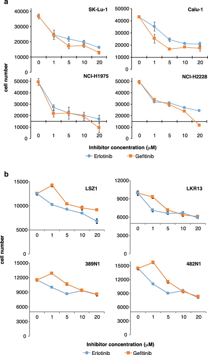
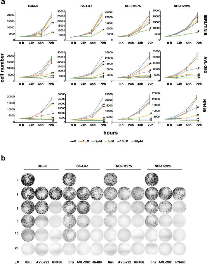
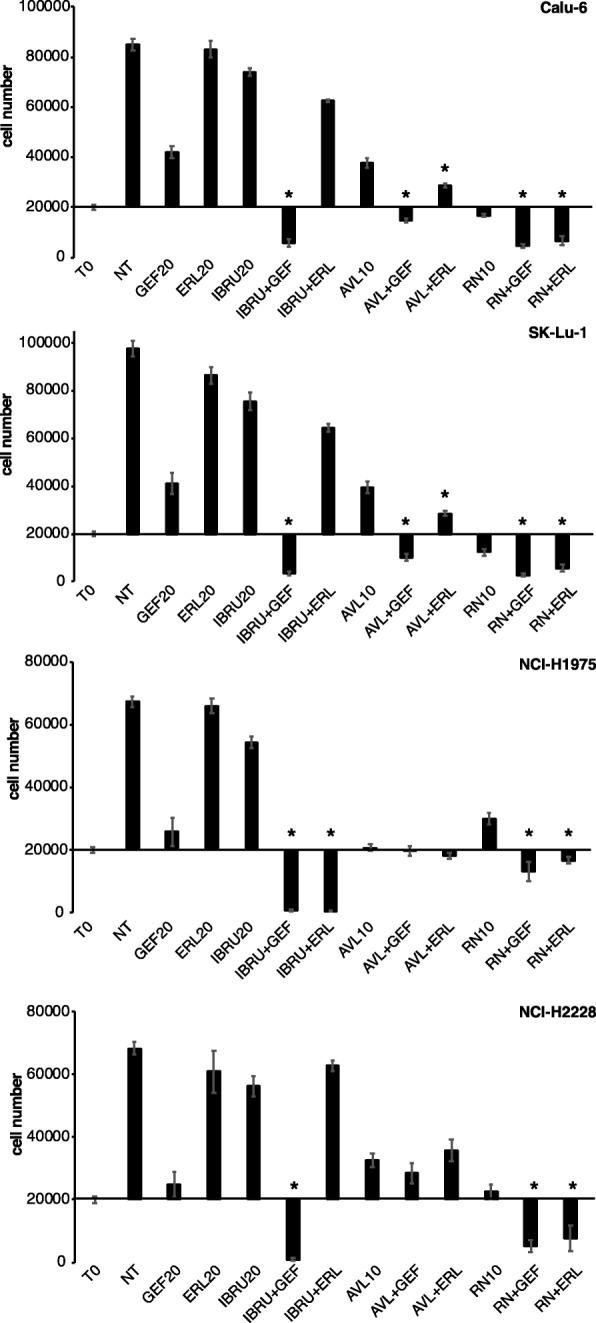
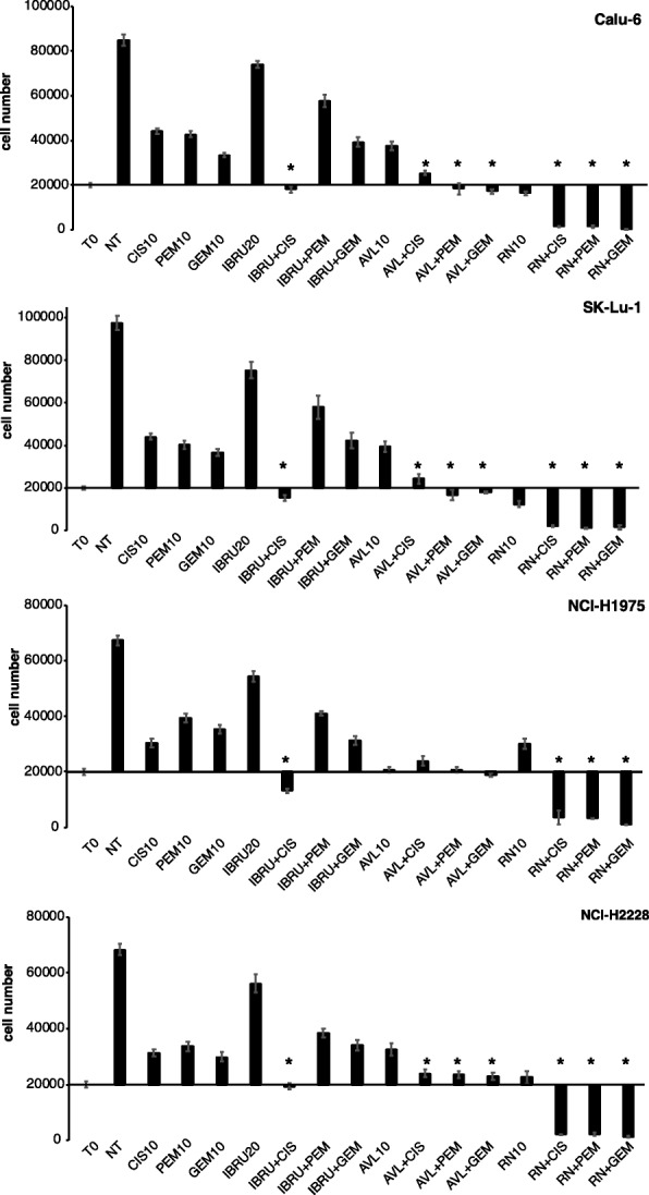
Methods: p65BTK expression was evaluated by immunohistochemistry in 382 NSCLC patients with complete clinico-pathological records including smoking habit, ALK and EGFR status, and in metastatic lymph nodes of 30 NSCLC patients. NSCLC cell lines mutated for p53 and/or a component of the RAS/MAPK pathway and primary lung cancer-derived cells from Kras/Trp53 null mice were used as a preclinical model. The effects of p65BTK inhibition by BTK Tyrosine Kinase Inhibitors (TKIs) (Ibrutinib, AVL-292, RN486) and first-generation EGFR-TKIs (Gefitinib, Erlotinib) on cell viability were evaluated by MTT. The effects of BTK-TKIs on cell growth and clonogenicity were assessed by crystal violet and colony assays, respectively. Cell toxicity assays were performed to study the effect of the combination of non-toxic concentrations of BTK-TKIs with EGFR-TKIs and standard-of-care (SOC) chemotherapy (Cisplatin, Gemcitabine, Pemetrexed).

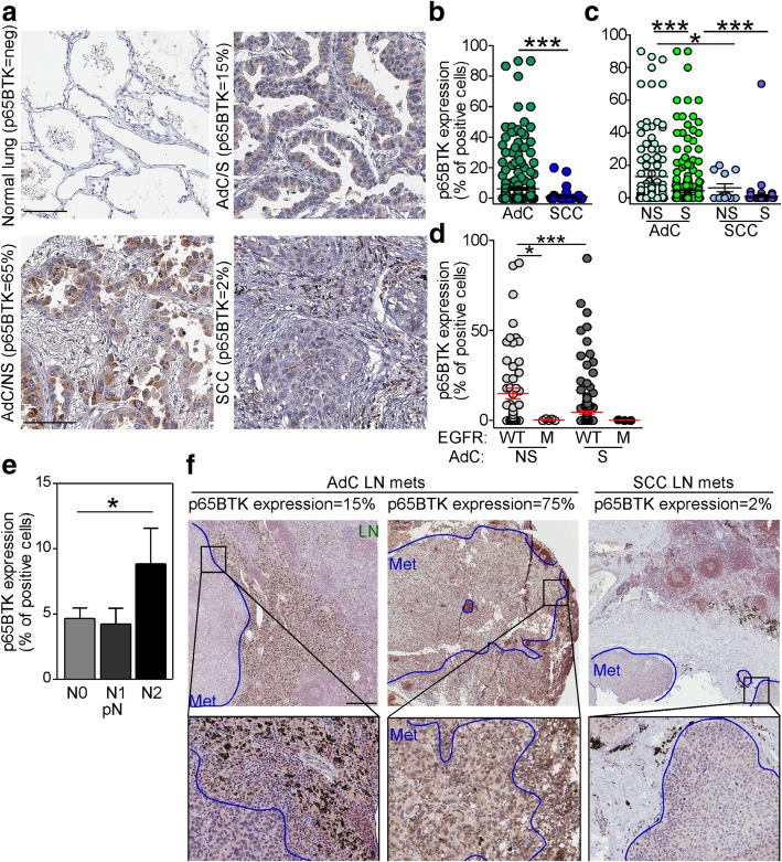
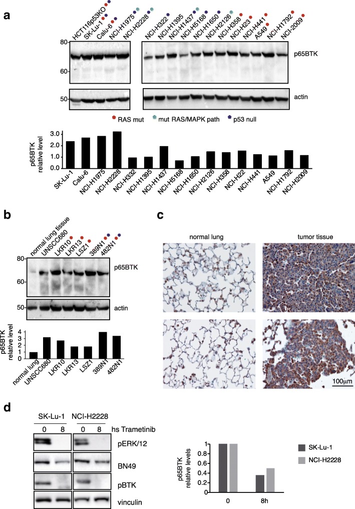
Results: p65BTK was significantly over-expressed in EGFR-wild type (wt) adenocarcinomas (AdC) from non-smoker patients and its expression was also preserved at the metastatic site. p65BTK was also over-expressed in cell lines mutated for KRAS or for a component of the RAS/MAPK pathway and in tumors from Kras/Trp53 null mice. BTK-TKIs were more effective than EGFR-TKIs in decreasing cancer cell viability and significantly impaired cell proliferation and clonogenicity. Moreover, non-toxic doses of BTK-TKIs re-sensitized drug-resistant NSCLC cell lines to both target- and SOC therapy, independently from EGFR/KRAS status.

Conclusions: p65BTK results as an emerging actionable target in non-smoking EGFR-wt AdC, also at advanced stages of disease. Notably, these patients are not eligible for EGFR-TKIs-based therapy due to a lack of EGFR mutation. The combination of BTK-TKIs with EGFR-TKIs is cytotoxic for EGFR-wt/KRAS-mutant/p53-null tumors and BTK-TKIs re-sensitizes drug-resistant NSCLC to SOC chemotherapy. Therefore, our data suggest that adding BTK-TKIs to SOC chemotherapy and EGFR-targeted therapy may open new avenues for clinical trials in currently untreatable NSCLC.

Keywords: BTK inhibitors; Chemotherapy; Drug resistance; EGFR; EGFR inhibitors; NSCLC; Targeted therapy; p65BTK.


Conflict of interest statement

The authors declare that they have no competing interests.

Figures

Fig. 1
Fig. 1
p65BTK is overexpressed in advanced lung adenocarcinomas with wild type EGFR from never-smoker patients. a IHC analysis of p65BTK in lung cancer tissue samples from a cohort of NSCLC patients using the BN30 antibody. Representative images of normal lung and lung cancer tissues are shown. SCC: squamous cell carcinoma; AdC/S: adenocarcinoma from smoker patient; AdC/NS: adenocarcinoma from non-smoker patient. Scale bar 100 μM. b Quantification of p65BTK expression in SCC and AdC patients. ***, p < 0.0001 by unpaired t test with Welch’s correction. c Quantification of p65BTK expression in smoker and non-smoker patients AdC and SCC patients. NS: non-smoker; S: smoker. Quantification of p65BTK expression. d Quantification of p65BTK expression in smoker and non-smoker AdC patients with either wild type (WT) or mutated (MT) EGFR. *, p = 0.04; ***, p < 0.0001 by non-parametric t test. e Quantification of p65BTK expression in primary NSCLC according to pN status. *, p = 0.04 by unpaired t test with Welch’s correction. f IHC analysis of p65BTK in metastatic lymph nodes of lung adenocarcinomas (AdC) or squamous cell carcinoma (SCC). Representative images show different expression levels of the kinase in the metastatic setting. Scale bars 500 μm (top panels) or 200 μm (lower panels)
Fig. 2
Fig. 2
NSCLC cells with activated KRAS express high levels of p65BTK. a Top: Western Blot analysis of p65BTK expression in NSCLC human cell lines with different mutations along the RAS/MAPK pathway and in p53. Lysate from HCT116p53KO colon cancer cells was loaded as a positive control. Bottom: fold change of p65BTK protein expression in NSCLC cell lines normalized to beta actin, setting as expression level = 1 NCI-H1935 which do not possess mutations in KRAS or in the RAS/MAP pathway not in the p53 gene. b Top: Western Blot analysis of p65BTK expression in primary lung cancer cells derived from KrasLSL-G12D (LKR10, LKR13, LSZ1) and KrasLSL-G12D;Trp53f/f (389 N1, 482 N1) mice. UNSCC680 is a primary cell line from a mouse squamous cell carcinoma. Bottom: fold change of p65BTK protein expression normalized to beta actin. In a and b p65BTK was detected by BN49 antibody [18] and beta actin was used as a loading control. c IHC analysis of p65BTK in normal and tumoral lung tissue samples from 2 different KrasLSLG12D; Trp53 f/f mice using BN30 antibody. d Left: Western Blot analysis of p65BTK expression in SK-Lu-1 and NCI-H2228 cells after treatment with the MEK inhibitor Trametinib (1 μM). Right: fold change of p65BTK protein expression normalized to vinculin
Fig. 3
Fig. 3
EGFR inhibition does not affect cell viability of NSCLC cell lines and tumor-derived primary cells with mutations along the EGFR/RAS/MAPK pathway. Dose-response curves of a human NSCLC cell lines (SK-Lu1, Calu-6, NCI-H1975 and NCI-H2228) and b primary lung cancer cell lines derived from KrasLSL-G12D (LSZ1, LKR13) and KrasLSL-G12D;Trp53f/f mice, (389 N1, 482 N1) treated with increasing concentrations of EGFR inhibitors (Erlotinib and Gefitinib). Cell viability was evaluated by MTT assay. X-axis crosses in correspondence of T0 values (before starting the treatment); 72 h values are then expressed as the variation relative to the initial cell number. Scale on Y-axis is adapted to the different growth rates shown by each cell line. Data are presented as mean ± SEM. n ≥ 3 independent experiments
Fig. 4
Fig. 4
p65BTK targeting affects cell viability of NSCLC cell lines and tumor-derived primary cells scarcely responsive to EGFR inhibition. Dose-response curves of a human NSCLC cell lines (SK-Lu1, Calu-6, NCI-H1975 and NCI-H2228) and b primary lung cancer cells derived from KrasLSL-G12D (LSZ1, LKR13) and KrasLSL-G12D;Trp53f/f mice (389 N1, 482 N1) treated with increasing concentrations of BTK inhibitors (Ibrutinib, AVL-292, RN486). Cell viability was evaluated by crystal violet staining. X-axis crosses in correspondence of T0 values (before starting the treatment); 72 h values are then expressed as the variation relative to the initial cell number. Scale on Y-axis is adapted to the different growth rates shown by each cell line. Data are presented as mean ± SEM. n ≥ 3 independent experiments
Fig. 5
Fig. 5
p65BTK inhibition strongly impairs proliferation and clonogenicity of NSCLC cell lines. a Growth curves of human p53-null NSCLC cell lines treated with increasing concentrations of BTK inhibitors; cell number was evaluated each 24 h by MTT assay. Scale on Y-axis is adapted to the different growth rates shown by each cell line. Data are presented as mean ± SEM. n ≥ 3 independent experiments. * indicates p < 0.05 vs untreated. b Clonogenicity assay of human p53-null NSCLC cell lines treated with increasing concentrations of BTK inhibitors for 10 days. A representative image of one experiment out of 3 is shown
Fig. 6
Fig. 6
p65BTK inhibition sensitizes NSCLC cell lines scarcely responsive to EGFR-targeted therapy. Cell viability of human p53-null NSCLC cell lines in response to different combinations of BTK and EGFR inhibitors (T0 = time 0; NT = untreated; GEF20 = Gefitinib 20 μM; ERL20 = Erlotinib 20 μM. IBRU20 = Ibrutinib 20 μM; AVL10 = AVL-292 10 μM; RN10 = RN486 10 μM). X-axis crosses in correspondence of T0 values (before starting the treatment); 72 h values are then expressed as the percentage variation relative to the initial cell number. Scale on Y-axis is adapted to the different growth rates shown by each cell line. Data are presented as mean ± SEM. n ≥ 3 independent experiments. * indicates p < 0.05 vs T0 values
Fig. 7
Fig. 7
p65BTK inhibition reverts resistance of NSCLC cell lines to chemotherapy. Cell viability of human p53-null NSCLC cell lines in response to different combinations of BTK and SOC chemotherapeutic agents (T0 = time 0; NT = untreated; CIS10 = Cisplatin 10 μM; PEM10 = Pemetrexed 10 μM; GEM10 = Gemcitabine 10 μM; IBRU20 = Ibrutinib 20 μM; AVL10 = AVL-292 10 μM; RN10 = RN486 10 μM). X-axis crosses in correspondence of T0 values (before starting the treatment); 72 h values are then expressed as the percentage variation relative to the initial cell number. Scale on Y-axis is adapted to the different growth rates shown by each cell line. Data are presented as mean ± SEM. n ≥ 3 independent experiments. * indicates p < 0.05 vs T0 values

References

    1. Wong MC, Lao XQ, Ho K-F, Goggins WB, Shelly L. Incidence and mortality of lung cancer: global trends and association with socioeconomic status. Sci Rep. 2017;7:14300. doi: 10.1038/s41598-017-14513-7. - DOI - PMC - PubMed
    1. Herbst RS, Heymach JV, Lippman SM. Lung cancer. N Engl J Med. 2008;359:1367. doi: 10.1056/NEJMra0802714. - DOI - PMC - PubMed
    1. Jones GS, Baldwin DR. Recent advances in the management of lung cancer. Clin Med. 2018;18:s41. doi: 10.7861/clinmedicine.18-2-s41. - DOI - PMC - PubMed
    1. Jemal A, Siegel R, Ward E, Hao Y, Xu J, Murray T, et al. Cancer statistics, 2008. CA-Cancer J Clin. 2008;58:71. doi: 10.3322/CA.2007.0010. - DOI - PubMed
    1. Ye Q, She Q-B. Integration of AKT and ERK signaling pathways in Cancer: biological and therapeutic implications. J Pharmacol Clin Toxicol. 2013;1(2):1009.

MeSH terms