. 2019 Jul 25;75(2):224-237.e5.
doi: 10.1016/j.molcel.2019.05.023. Epub 2019 Jun 11.

Sister DNA Entrapment between Juxtaposed Smc Heads and Kleisin of the Cohesin Complex

Affiliations

Sister DNA Entrapment between Juxtaposed Smc Heads and Kleisin of the Cohesin Complex

Christophe Chapard et al. Mol Cell. .

Abstract

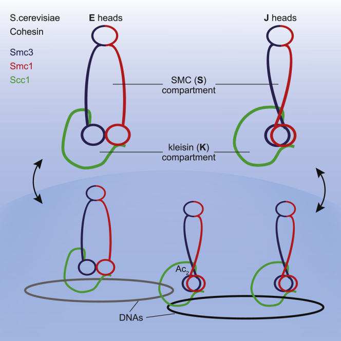
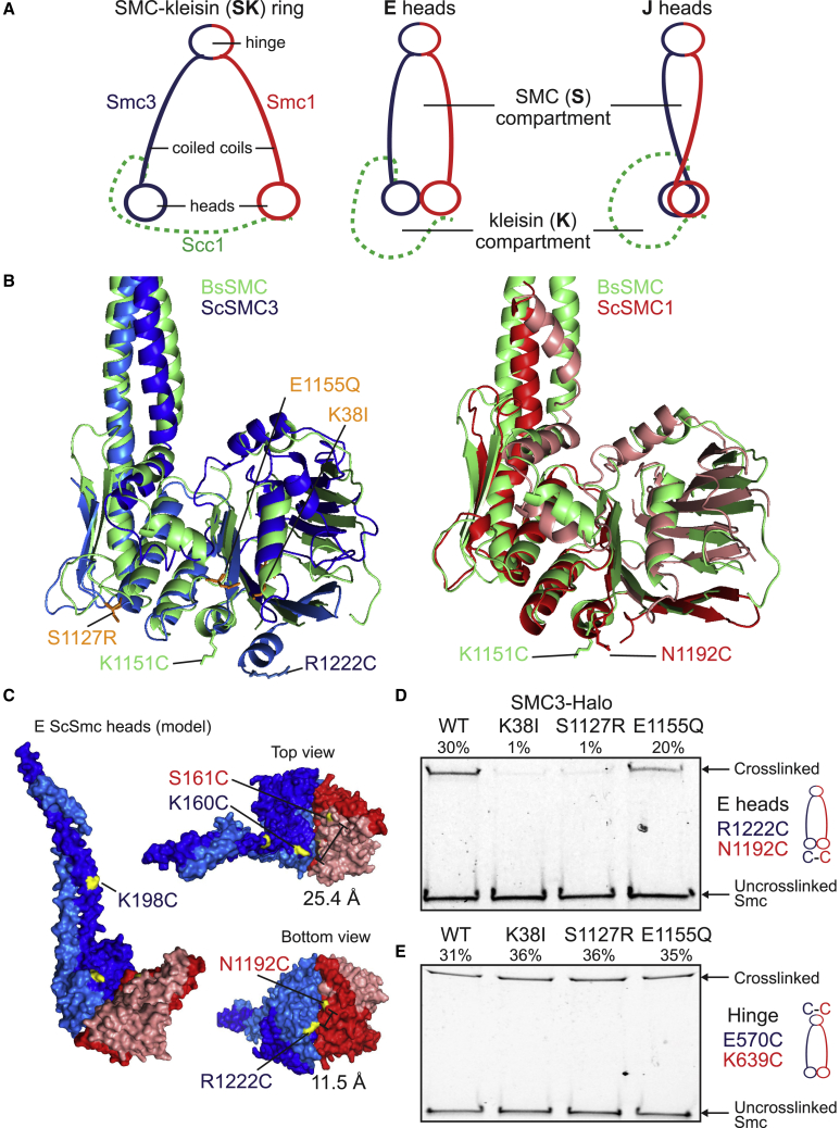
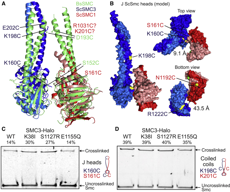
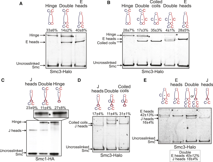
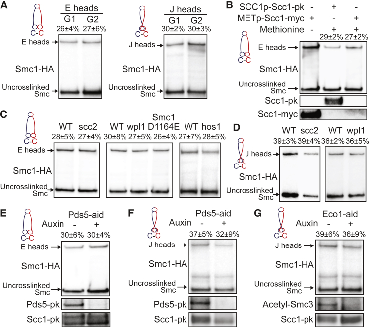
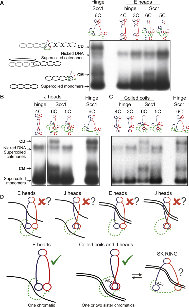
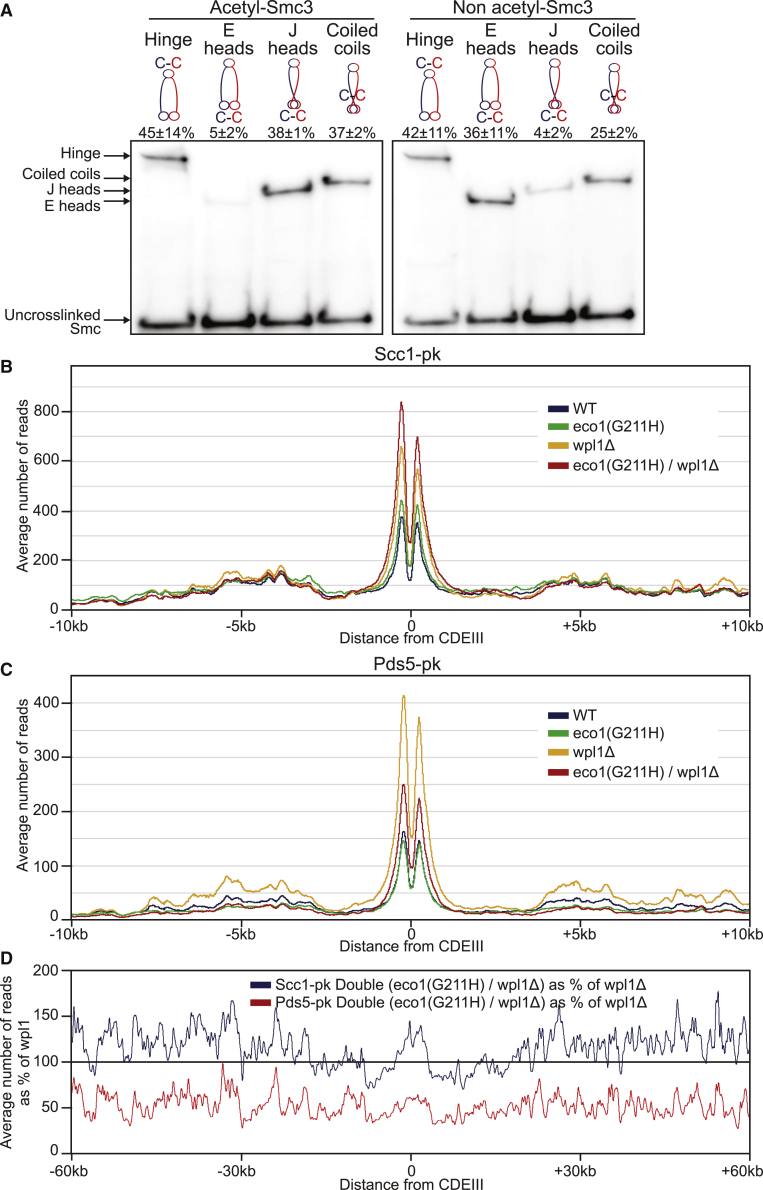
Cohesin entraps sister DNAs within tripartite rings created by pairwise interactions between Smc1, Smc3, and Scc1. Because Smc1/3 ATPase heads can also interact with each other, cohesin rings have the potential to form a variety of sub-compartments. Using in vivo cysteine cross-linking, we show that when Smc1 and Smc3 ATPases are engaged in the presence of ATP (E heads), cohesin rings generate a "SMC (S) compartment" between hinge and E heads and a "kleisin (K) compartment" between E heads and their associated kleisin subunit. Upon ATP hydrolysis, cohesin's heads associate in a different mode, in which their signature motifs and their coiled coils are closely juxtaposed (J heads), creating alternative S and K compartments. We show that K compartments of either E or J type can entrap single DNAs, that acetylation of Smc3 during S phase is associated with J heads, and that sister DNAs are entrapped in J-K compartments.

Keywords: DNA; S and K compartments; Scc1; Smc ATPase domains; acetylation; cohesin rings; engaged; entrapment; juxtaposed.


Conflict of interest statement

The authors declare no competing interests.

Figures

None
Graphical abstract
Figure 1
Figure 1
ATP-Dependent Head Engagement (E) State of Smc Head Domains (A) Schematic representation of the cohesin compartments. (B) Structure alignment of Sc Smc3 head (PDB: 4UX3, blue) and Sc Smc1 head (PDB: 1W1W, red) to Bs Smc head (PDB: 3ZGX, green). Selected residues displaying efficient cross-linking when mutated to cysteine are marked (for B. subtilis, see Diebold-Durand et al., 2017). Residues associated with the Smc3 ATP binding mutant (K38I), the signature motif mutant (S1127R), and the ATP hydrolysis mutant (E1155Q) are displayed in orange. (C) Model of ATP-engaged Smc3/Smc1 heads in surface representation in front (left) and top and bottom views (right). ATP-engaged head dimer is constructed by superimposition of Smc1 head to one of Smc3 head homodimer (Gligoris et al., 2014). Distance between selected residues is given. (D and E) In vivo cysteine cross-linking of Smc1 proteins with Halo-tagged wild-type and ATPase mutant Smc3. Cross-linking of Smc1(N1192C) and Smc3(R1222C) E head residues (C) or Smc1(K639C) and Smc3(E570C) hinge residues (D) was performed in vivo using BMOE. Cell extracts were labeled with HaloTag-TMR ligand. Smc-HaloTag species were separated by SDS-PAGE and quantified by in-gel fluorescence. Percentage of cross-link efficiency is indicated. See also Figure S1.
Figure 2
Figure 2
Signature Motif Juxtaposed (J) State of Smc Head Domains (A) Structure alignment of Sc Smc3 head (PDB: 4UX3, blue) and Sc Smc1 head (PDB: 1W1W, red) to disengaged Bs Smc heads (PDB: 3ZGX, green). Selected residues displaying efficient cross-linking when mutated to cysteine are marked (for B. subtilis, see Diebold-Durand et al., 2017). (B) Model of J Smc3/Smc1 heads in surface representation in front (left) and top and bottom views (right). J head dimer is constructed by superimposition of Smc1/3 heads onto the rod aligned Bs Smc head model as in (A) (Diebold-Durand et al., 2017). Distance between selected residues is given. (C and D) In vivo cysteine cross-linking of Smc1(Cys) proteins with Halo-tagged wild-type and mutant ATPase Smc3. Cross-linking of Smc1(S161C) and Smc3(K160C) J head residues (C), or Smc1(K201C) and Smc3(K198C) coiled coil residues (D) was performed in vivo using BMOE. Cell extracts were labeled with HaloTag-TMR ligand. Smc-HaloTag species were separated by SDS-PAGE and quantified by in-gel fluorescence. Percentage of cross-link efficiency is given. See also Figure S1.
Figure 3
Figure 3
S Compartments: Coiled Coils, E Head, and J Head Interactions in Cohesin Heterodimers (A) E state of Smc heads in heterodimers. Smc1 and Smc3-HaloTag proteins containing hinge and/or E heads cysteine pairs were cross-linked in vivo using BMOE. Complexes were immunoprecipitated on Scc1-PK6, labeled with TMR ligand, separated by SDS-PAGE, and quantified by in-gel fluorescence. Percentage (mean ± SD of 3 independent experiments) of cross-linking efficiency is given. Asterisk shows the location of the double cross-link. ICC1 = 0.74. (B) E Smc heads and coiled coils interactions in heterodimers. Smc1 and Smc3-HaloTag proteins containing hinge, E heads, and/or coiled coils cysteine pairs were analyzed as in (A). ICC1 = 0.84. (C) J state of Smc heads in heterodimers. Smc1-HA, Smc3, and Scc1-PK proteins containing hinge and/or J heads cysteine pairs were cross-linked in vivo, immunoprecipitated on Scc1-PK6, separated by SDS-PAGE, and analyzed by western blot. ICC1 = 0.37. (D) J heads interactions with Smc coiled coils. Smc1 and Smc3-HaloTag proteins containing coiled coils and/or alternative J heads cysteine pairs were analyzed as in (A). ICC1 = 0.84. (E) E and J states of Smc heads are mutually exclusive. Smc1 and Smc3-HaloTag proteins containing E heads and/or J heads cysteine pairs were analyzed as in (A). Percentage of the double cross-link efficiency is shown in box. Left lane: double cross-link of E heads with coiled coils shown for size indication. ICC1 = 0.52. See also Figure S2.
Figure 4
Figure 4
K Compartments: E and J Head Interactions with Scc1 (A) E heads interact with Scc1 at Smc3-Scc1 interface. Smc1-HA, Smc3, and Scc1-PK proteins containing E heads or Smc3-Scc1 interface cysteine were cross-linked in vivo, immunoprecipitated on Scc1-PK6, separated by SDS-PAGE, and semi-quantified by western blot. Percentage (mean ± SD of 3 independent experiments) of cross-link efficiency is given: E heads = 26 ± 4%, Smc3/Scc1 = 56 ± 17%, and Double = 15 ± 3%. Asterisk shows the location of the Smc3/Scc1 cross-link. ▼ indicates lane used for measurements. ICC1 = 0.72. (B) E heads interact with Scc1 at Smc1-Scc1 interface. Smc1-HA, Smc3, and Scc1-PK proteins containing E heads or Smc1-Scc1 interface cysteine were analyzed as in (A). Percentage (mean ± SD of 3 independent experiments) of cross-link efficiency is given: E heads = 21 ± 2%, Smc1/Scc1 = 52 ± 5%, and Double = 10 ± 2%. ▼ indicates lane used for measurements. ICC1 = 0.97. (C) J heads interact with Scc1 at Smc3-Scc1 interface. Smc1-HA, Smc3, and Scc1-PK proteins containing J heads or Smc3-Scc1 interface cysteine were analyzed as in (A). Asterisk shows the location of the Smc3/Scc1 cross-link. Percentage (mean ± SD of 3 independent experiments) of cross-link efficiency is given: J heads = 29 ± 6%, Smc3/Scc1 = 67 ± 17%, and Double = 18 ± 7%. ▼ indicates lane used for measurements. ICC1 = 0.77. (D) J heads interact with Scc1 at Smc1-Scc1 interface. Smc1-HA, Smc3, and Scc1-PK proteins containing J heads or Smc1-Scc1 interface cysteine were analyzed as in (A). Percentage (mean ± SD of 3 independent experiments) of cross-link efficiency is given: J heads = 24 ± 3%, Smc1/Scc1 = 54 ± 7%, and Double = 16 ± 5%. ▼ indicates lane used for measurements. ICC1 = 0.90. See also Figure S2.
Figure 5
Figure 5
Head Engagement and Juxtaposition Occur throughout the Cell Cycle (A) E and J heads over the cell cycle. Smc1-HA and Smc3 proteins containing E or J heads cysteine pairs were cross-linked in G1 (Gal-Sic1) or G2/M (nocodazole) arrested cells using BMOE. Complexes were immunoprecipitated on Scc1-PK, separated by SDS-PAGE, and analyzed by western blot. Percentage (mean ± SD of 3 independent experiments) of cross-link efficiency is given. (B) E heads upon absence of Scc1. Cells were pheromone arrested in early G1 prior to Scc1-myc transcription repression using an inducible methionine promoter and release into nocodazole. Smc1-HA and Smc3 proteins containing E heads cysteine pairs were cross-linked in vivo using BMOE. Protein extracts were analyzed by SDS-PAGE and western blot. Left lane: cycling cells expressing Scc1-myc under control of methionine promoter were analyzed in parallel. (C) E heads upon absence of functional cohesin loader Scc2 or releasing activity. Smc1-HA and Smc3 proteins containing E heads cysteine pairs were cross-linked in vivo using BMOE. Complexes were analyzed as in (A). Left panel: WT and scc2-45 strains were arrested in G1 with alpha factor at 25°C and released into nocodazole at 37°C. Middle panel: cycling WT, wpl1-deleted, and Smc1(D1164E, N1192C)-HA strains. Right panel: cycling WT and hos1-deleted strains. (D) J heads upon absence of functional cohesin loader Scc2 or releasing activity. Smc1-HA and Smc3 proteins containing J heads cysteine pairs were cross-linked in vivo using BMOE. Complexes were analyzed as in (A). Left panel: WT and scc2-45 strains were arrested in G1 with alpha factor at 25°C and released into nocodazole at 37°C. Right panel: cycling WT and wpl1-deleted strains. (E) E heads in the absence of Pds5. Pds5-aid strain was arrested in G1 with alpha factor and supplemented with auxin for 1 h prior to release into nocodazole-containing media supplemented with auxin. In vivo cross-linked proteins were analyzed as in (A). (F and G) J heads in the absence of Pds5 (F) or Eco1 (G). WT, Pds5-aid, or Eco1-aid strains were analyzed as in (E). See also Figure S3.
Figure 6
Figure 6
CMs and CDs in Core K compartments with E and J Heads (A) CMs and CDs in exponentially growing strains containing cysteines in the hinge, E heads, or Smc1/Scc1/Smc3 interfaces. Strains with cysteine pairs at interfaces (4C or 6C) and strains lacking just one of the cysteines (3C or 5C), carrying a 2.3-kb circular minichromosome, were treated with BMOE. DNAs associated with cohesion immunoprecipitates (Scc1-PK) were denatured with SDS and separated by agarose gel electrophoresis. Southern blotting reveals supercoiled monomers and nicked and supercoiled catenanes along with two forms of DNA unique to 6C cells, termed CMs and CDs (see Gligoris et al., 2014 for details). Full circularisation efficiencies (mean ± SD of 3 independent experiments): ES = 14 ± 2% (as measured by in-gel fluorescence) and 15% (as measured by western blot after engineered cleavage of Smc3, Figure S2A), EK = 7 ± 1%, and SK RING = 13 ± 6%. Representative of at least 3 independent experiments. See also Figure S4A. (B) CMs and CDs in exponentially growing strains containing cysteines in the hinge, J heads, or Smc1/Scc1/Smc3 interfaces. Full circularisation efficiencies (mean ± SD of 3 independent experiments): JS = 11 ± 4%, JK = 7 ± 2%, and SK RING = 13 ± 6%. Representative of at least 3 independent experiments. See also Figure S4B. The lane corresponding to hinge/J heads 3C control is not shown. (C) CMs and CDs in exponentially growing strains containing cysteines in the hinge, coiled coils, or Smc1/Scc1/Smc3 interfaces. Full circularisation efficiencies (mean ± SD of 3 independent experiments): CS = 22 ± 2%, CK = 12 ± 2%, and SK RING = 13 ± 6%. Representative of at least 3 independent experiments. (D) Schematic representation of DNA topological association with cohesin compartments (see discussion for details). See also Figure S4.
Figure 7
Figure 7
Acetylation-Mediated Control of Both J Head and Pds5 Chromatin Association (A) Smc3 acetylation of heterodimers with E and J heads. Smc1-HA and Smc3 proteins containing hinge, E heads, J heads, or coiled coils cysteine pairs were cross-linked in vivo with BMOE. Complexes were immunoprecipitated on Scc1-PK, separated by SDS-PAGE, and analyzed by western blot against acetylated-Smc3 or non-acetylated Smc3. Percentage (mean ± SD of 3 independent experiments) of the cross-link signal is given for each antibody. ICC1(acetyl) = 0.79, ICC1(non-acetyl) = 0.72. (B and C) Average calibrated ChIP-seq profiles of Scc1-PK (B) and Pds5-PK (C) in the presence or absence of Wpl1 and/or functional Eco1. Cells were arrested in G2 using nocodazole at restrictive temperature after release from pheromone arrest at permissive temperature. (D) Averaged calibrated ChIP-seq profiles 60 kb either side of CDEIII plotted as a percentage of the average number of reads of Scc1-PK and Pds5-PK obtained for wpl1Δ cells in (B) and (C) respectively. See also Figures S4, S5, and S6.

Comment in

  • Compartments in the Ring.
    Nishiyama T. Nishiyama T. Mol Cell. 2019 Jul 25;75(2):201-203. doi: 10.1016/j.molcel.2019.07.002. Mol Cell. 2019. PMID: 31348876

Similar articles

Cited by

References

    1. Arumugam P., Gruber S., Tanaka K., Haering C.H., Mechtler K., Nasmyth K. ATP hydrolysis is required for cohesin’s association with chromosomes. Curr. Biol. 2003;13:1941–1953. - PubMed
    1. Arumugam P., Nishino T., Haering C.H., Gruber S., Nasmyth K. Cohesin’s ATPase activity is stimulated by the C-terminal Winged-Helix domain of its kleisin subunit. Curr. Biol. 2006;16:1998–2008. - PubMed
    1. Beckouët F., Hu B., Roig M.B., Sutani T., Komata M., Uluocak P., Katis V.L., Shirahige K., Nasmyth K. An Smc3 acetylation cycle is essential for establishment of sister chromatid cohesion. Mol. Cell. 2010;39:689–699. - PMC - PubMed
    1. Beckouët F., Srinivasan M., Roig M.B., Chan K.L., Scheinost J.C., Batty P., Hu B., Petela N., Gligoris T., Smith A.C. Releasing activity disengages cohesin’s Smc3/Scc1 interface in a process blocked by acetylation. Mol. Cell. 2016;61:563–574. - PMC - PubMed
    1. Bürmann F., Lee B.-G., Than T., Sinn L., O’Reilly F.J., Yatskevich S., Rappsilber J., Hu B., Nasmyth K., Löwe J. A folded conformation of MukBEF and cohesin. Nat. Struct. Mol. Biol. 2019;26:227–236. - PMC - PubMed

Publication types

MeSH terms

Substances