. 2019 Jun 14;4(36):eaau6571.
doi: 10.1126/sciimmunol.aau6571.

IL-23-producing IL-10Rα-deficient gut macrophages elicit an IL-22-driven proinflammatory epithelial cell response

Affiliations

IL-23-producing IL-10Rα-deficient gut macrophages elicit an IL-22-driven proinflammatory epithelial cell response

Biana Bernshtein et al. Sci Immunol. .

Abstract

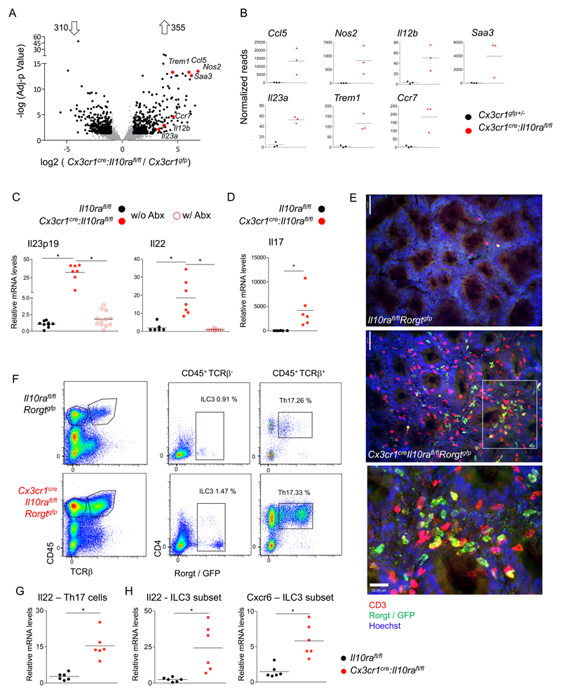
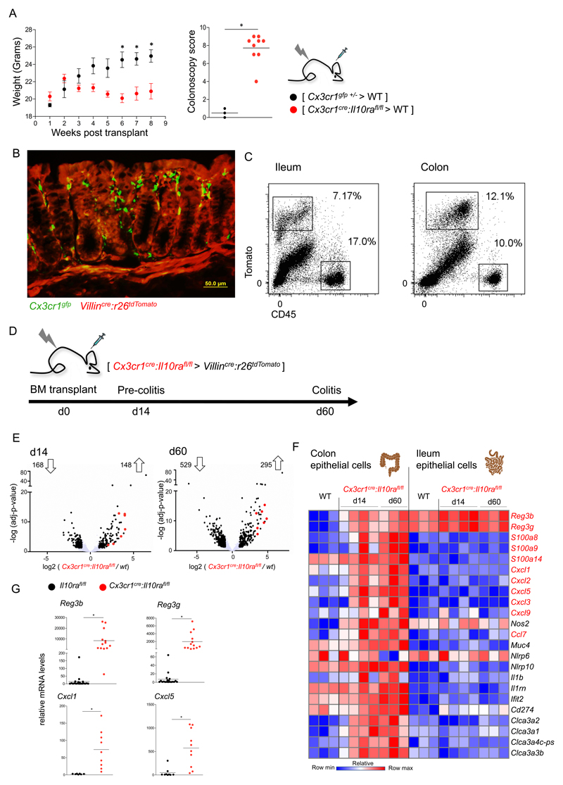
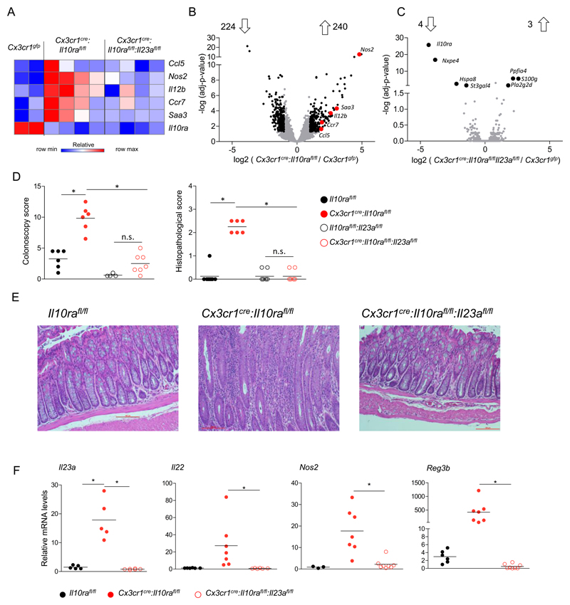
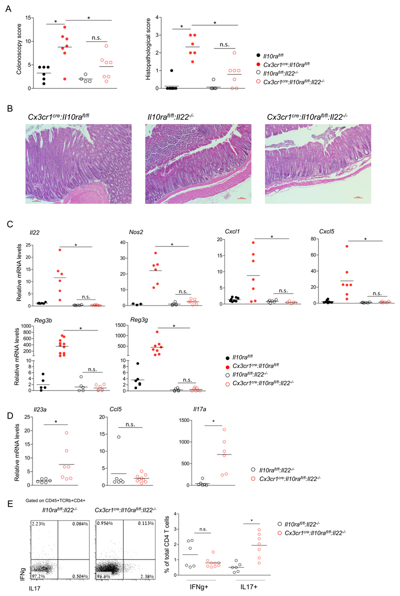
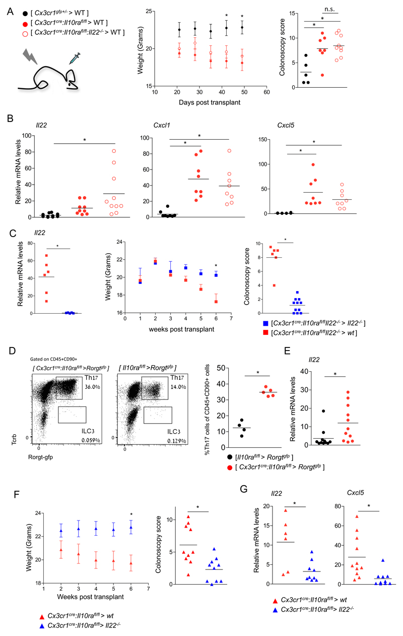
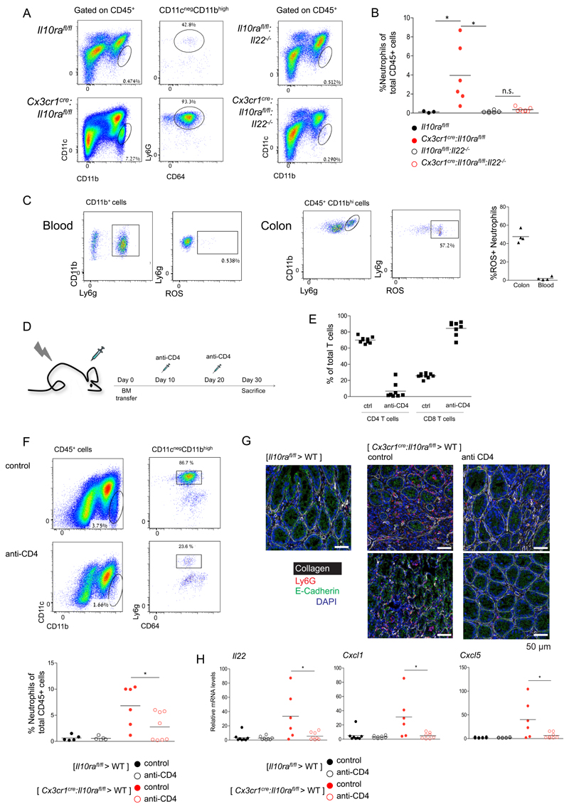
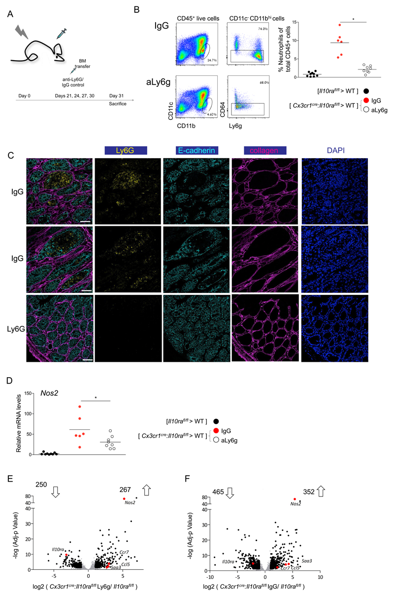
Cytokines maintain intestinal health, but precise intercellular communication networks remain poorly understood. Macrophages are immune sentinels of the intestinal tissue and are critical for gut homeostasis. Here, we show that in a murine inflammatory bowel disease (IBD) model based on macrophage-restricted interleukin-10 (IL-10) receptor deficiency (Cx3cr1Cre:Il10rafl/fl mice), proinflammatory mutant gut macrophages cause severe spontaneous colitis resembling the condition observed in children carrying IL-10R mutations. We establish macrophage-derived IL-23 as the driving factor of this pathology. Specifically, we report that Cx3cr1Cre:Il10rafl/fl:Il23afl/fl mice harboring macrophages deficient for both IL-10R and IL-23 are protected from colitis. By analyzing the epithelial response to proinflammatory macrophages, we provide evidence that T cells of colitic animals produce IL-22, which induces epithelial chemokine expression and detrimental neutrophil recruitment. Collectively, we define macrophage-specific contributions to the induction and pathogenesis of colitis, as manifested in mice harboring IL-10R deficiencies and human IBDs.


Conflict of interest statement

Competing interests: The authors have no competing interests.

Figures

Figure 1
Figure 1. IL-10 receptor-deficient macrophages secrete IL-23, inducing IL-22 secretion by ILC3 and Th17 cells.
(A) Volcano plot of statistical significance (-log10 p-value) against log 2 ratio of macrophages sorted from the colonic lamina propria of 6-7 weeks old Cx3cr1creIl10rafl/fl and Cx3cr1gfp/+ mice, based on RNA-seq data. Significantly up or down regulated genes (fold change>2, adj-p value<0.05) are in black, relevant pro inflammatory up-regulated genes are highlighted in red. Data are representative of two independent experiment, n>=3 for each group. (B) RNA-seq normalized read numbers for single genes of interest are plotted separately, each dot represents one mouse. (C) qRT-PCR analysis of il23p19 expression by sorted colonic MFs from 6-7 weeks old mice (left), or of il22 expression (right) in colonic whole tissue extracts of 6-7 weeks old mice. Data collected from two independent experiments, n>=3 in each. (D) qRT-PCR analysis of il17 expression in colonic whole tissue extracts of indicated mice. Data collected from two independent experiments, n>=3 in each. (E) Whole mount staining of colonic tissue of Cx3cr1cre:Il10rafl/fl:Rorgtgfp mice and Cre negative littermates (age 3-4 months), analysed by confocal microscope, GFP in green, CD3 in red. Indent in the bottom is in the white rectangle, Th17 marked with full arrow, ILC3 dashed arrow. Top scale=50μm, bottom scale= 20μm. (F) Representative picture of flow cytometry analysis and sorting strategy of Cx3cr1cre:Il10rafl/fl:Rorgtgfp mice and Cre negative littermates 3-4 months old. (G-H) qRT-PCR analysis of RNA extracted from sorted colonic Th17 cells (F) or ILC3 (G) for indicated genes. Data are collected from two independent experiments, n=3 in each.
Figure 2
Figure 2. The response of epithelial cells to pro-inflammatory macrophages.
(A) Weight and colonoscopic analysis of [ Cx3cr1cre:Il10rafl/fl > WT ] or [ Cx3cr1gfp/+ > WT ] BM chimeras. Colonoscopy was performed 7 weeks post-transplant. Data are representative of 3 independent experiments, n>=3 for each group. *p<0.05. (B) Representative picture of microscopic analysis of [Cx3cr1gfp>Villincre:R26-tdTomato] BM chimera. (C) Representative picture of flow cytometry analysis and sorting strategy of epithelial cells from [Cx3cr1gfp>Villincre:R26-tdTomato] BM chimera. (D) Description of BM chimera experiment and timeline of epithelial cell harvest. (E) Volcano plot of statistical significance (log10 p-value) against log 2 ratio of epithelial cells sorted from the colon of [Cx3cr1creil10rafl/lf >Villincre:R26-tdTomato] BM chimeras and Villincre:R26-tdTomato mice, based on RNA-seq data. Significantly up or down regulated genes (fold change>2, adj-p value<0.05) are in black, il22 induced genes are highlighted in red. Data are from one experiment, n>=3 for each group. (F) Heatmap of RNAseq data of colonic and ileal epithelial cells extracted from the same mouse. Normalized reads number were log transformed. Presented are genes of interest, significantly upregulated in colonic but not ileal epithelial cells in response to pro-inflammatory macrophages at both disease stages (d14 and d60 post-transplant). (G) qRT-PCR analysis of reg3b, reg3g, cxcl1 and cxcl5 expression in whole tissue extracts of colons of 6-7 weeks old mice. Data collected from two independent experiments, n>=3 in each.
Figure 3
Figure 3. IL-23 deficiency prevents the colitogenic activity of IL-10 receptor-mutant macrophages.
(A) Heatmap of RNAseq data of colonic macrophages sorted from 6-7 weeks old mice. Normalized reads number were log transformed. (B-C) Volcano plot of statistical significance (log10 p-value) against log 2 ratio of macrophages sorted from the colonic lamina propria of B - Cx3cr1cre:Il10rafl/fl and Cx3cr1gfp/+ mice or C - Cx3cr1cre:Il10rafl/fl:Il23afl/fl and Cx3cr1gfp/+ mice, based on RNA-seq data. Significantly up or down regulated genes (fold change>2, adj-p value<0.05) are in black, relevant pro inflammatory up-regulated genes are highlighted in red. In (B) Data are representative of two independent experiment (B) or from one experiment (C), in each experiment n>=3 for each group. (D) Colonoscopic (left) and histopathological (right) analysis of 3-4 months old mice. Data are collected from two independent experiments, n>=3 in each. (E) Representative images of histopathological analysis of indicated mouse strains. (F) qRT-PCR analysis of il23a expression by sorted colonic macrophages of indicated mouse strains (left), or il22, nos2 and reg3b expression in colonic whole tissue RNA extracts of indicated mouse strains. Data are collected from two independent experiments, n>=3 in each. All mice were age matched; each dot represents one mouse.
Figure 4
Figure 4. IL-22 is critical for colitis driven by IL-10 receptor-deficient macrophages.
(A) Colonoscopic (left) and histopathological (right) analysis of 3-4 months old mice. (B) Representative images of histopathological analysis of indicated mouse strains. (C) qRT-PCR analysis of il22, cxcl1, cxcl5, reg3b, reg3g and nos2 expression in colonic whole tissue extracts of indicated mouse strains. (D) qRT-PCR analysis of il23a and Ccl5 by sorted colonic macrophages of indicated mouse strains (left), and whole tissue il17a levels of indicated mouse strains (E) Flow cytometry analysis and quantification of lamina propria T cells extracted from the colons of indicated mouse strains. Data in A,C,D and E are collected from two independent experiments, n>=3 in each. All mice were age matched, each dot represents one mouse.
Figure 5
Figure 5. Definition of the cellular source of IL-22.
(A) Weight and colonoscopic analysis of [ Cx3cr1cre:Il10rafl/fl > WT ], [ Cx3cr1gfp/+ > WT ] or [ Cx3cr1cre:Il10rafl/fl:Il-22-/- > WT ] BM chimeras. Colonoscopy was performed 6 weeks post-transplant. Data are collected from 2 independent experiments, n>=3 in each. (B) qRT-PCR analysis of il22, cxcl1 and cxcl5 expression in whole tissue extracts of colons of indicated BM chimeric mice. Data are collected from 2 independent experiments, n=3-5 in each group. (C) qRT-PCR analysis of il22 expression in whole tissue extracts of colons of indicated BM chimeric mice. Data are from one representative experiment of two. (left) Weight and colonoscopic analysis of [ Cx3cr1cre:Il10rafl/fl:Il22-/- > WT ], [ Cx3cr1cre:Il10rafl/fl:Il22-/- > Il22-/- ] BM chimeras. Colonoscopy was performed 7 weeks post-transplant. Weight data are from one representative experiment of two, n=3-4 in each group, colonoscopy data are collected from two experiments, n=3-4 in each. (right) (D) Flow cytometry analysis of the lamina propria of [ Cx3cr1cre:Il10rafl/fl > Rorgtgfp ], [Il10rafl/fl> Rorgtgfp ] BM chimeras. Data are representative of three experiments, n=3-5 in each group. (E) qRT-PCR analysis of il22 expression by sorted Th17 cells from indicated BM chimeras. Data are collected from three independent experiments, n=3-5 in each group. (F) Weight and colonoscopic analysis of [ Cx3cr1cre:Il10rafl/fl > WT ] or [ Cx3cr1cre:Il10rafl/fl > Il- 22-/- ] BM chimeras. Colonoscopy was performed 6 weeks post-transplant. Data are collected from 2 independent experiments, n>=3 in each. (G) qRT-PCR analysis of il22 and cxcl5 expression in whole tissue extracts of colons of indicated BM chimeric mice. Data are collected from 2 independent experiments, n=3-5 in each group.
Figure 6
Figure 6. Neutrophil recruitment to the colonic lamina propria depends on CD4+ T cells.
(A) Representative plots of flow cytometry analysis of the colonic lamina propria of indicated mouse strains. (B) Quantification of flow cytometry analysis according to gating strategy indicated in A. (C) Representative plots of flow cytometry analysis of the colonic lamina propria of Cx3cr1cre:Il10rafl/fl mice (left). Quantification of flow cytometry analysis (right). (D) Schematic of T cell depletion protocol (E) Quantification of flow cytometry analysis of mesenteric LN indicating efficient ablation of CD4+ T cells, but not CD8+ T cells. (F) Representative plots of flow cytometry analysis of the colonic lamina propria of [ Cx3cr1cre:Il10rafl/fl > WT ] BM chimeras. (G) Representative immunofluorescence images of colon sections of [ Il10rafl/fl > WT ] BM chimeras, [ Cx3cr1cre:Il10rafl/fl > WT ] BM chimeras and [ Cx3cr1cre:Il10rafl/fl > WT ] BM chimeras treated with the anti-CD4 regimen. (H) qRT-PCR analysis of il22, cxcl1 and cxcl5 expression in whole tissue extracts of colons of indicated BM chimeric mice. Data are collected from two independent experiments, n>=3 in each
Figure 7
Figure 7. Neutrophil depletion ameliorates colitis in Cx3cr1cre:Il10rafl/fl BM chimeras
(A) Schematic of neutrophil depletion protocol (B) Representative plots of flow cytometry analysis of the colonic lamina propria of [ Cx3cr1cre:Il10rafl/fl > WT ] BM chimeras treated with IgG control or anti Ly6G antibody (left). Quantification of flow cytometry analysis (right). (C) Representative immunofluorescence images of the colonic tissue of [ Cx3cr1cre:Il10rafl/fl > WT ] BM chimeras treated with IgG control or anti Ly6G antibody. (D) qRT-PCR analysis of Nos2 expression in whole tissue extracts of colons of indicated BM chimeric mice. (E-F) Volcano plot of statistical significance (log10 p-value) against log 2 ratio of macrophages isolated from colonic tissue of [Cx3cr1cre:Il10rafl/fl > WT ] BM chimeras treated with IgG control (F) or anti Ly6G antibody (E) and [Il10rafl/fl > WT ] BM chimeras, based on RNA-seq data. Significantly up or down regulated genes (fold change>2, adj-p value<0.05) are in black, relevant pro inflammatory up-regulated genes are highlighted in red. Data in A-D are collected from two independent experiments, n>=3 in each

References

    1. Manolio TA, et al. Finding the missing heritability of complex diseases. Nature. 2009;461:747–753. - PMC - PubMed
    1. Huang H, et al. Fine-mapping inflammatory bowel disease loci to single-variant resolution. Nature. 2017;547:173–178. - PMC - PubMed
    1. Joeris T, Müller-Luda K, Agace WW, Mowat AM. Diversity and functions of intestinal mononuclear phagocytes. Mucosal Immunol. 2017;10:845–864. - PubMed
    1. Sonnenberg GF, Artis D. Innate lymphoid cells in the initiation, regulation and resolution of inflammation. Nat Med. 2015;21:698–708. - PMC - PubMed
    1. Mortha A, et al. Microbiota-dependent crosstalk between macrophages and ILC3 promotes intestinal homeostasis. Science. 2014;343:1249288–1249288. - PMC - PubMed

Publication types