Review
. 2019 Jul 1;218(7):2096-2112.
doi: 10.1083/jcb.201903090. Epub 2019 Jun 14.

The cell biology of the hepatocyte: A membrane trafficking machine

Affiliations
Review

The cell biology of the hepatocyte: A membrane trafficking machine

Ryan J Schulze et al. J Cell Biol. .

Abstract

The liver performs numerous vital functions, including the detoxification of blood before access to the brain while simultaneously secreting and internalizing scores of proteins and lipids to maintain appropriate blood chemistry. Furthermore, the liver also synthesizes and secretes bile to enable the digestion of food. These diverse attributes are all performed by hepatocytes, the parenchymal cells of the liver. As predicted, these cells possess a remarkably well-developed and complex membrane trafficking machinery that is dedicated to moving specific cargos to their correct cellular locations. Importantly, while most epithelial cells secrete nascent proteins directionally toward a single lumen, the hepatocyte secretes both proteins and bile concomitantly at its basolateral and apical domains, respectively. In this Beyond the Cell review, we will detail these central features of the hepatocyte and highlight how membrane transport processes play a key role in healthy liver function and how they are affected by disease.


Figures

Figure 1.
Figure 1.
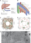
Liver and hepatocellular architecture. (A) Organization of the hepatic blood supply. The liver receives a mixture of nutrient-rich blood from the lower gastrointestinal tract via the portal vein (∼75%) as well as oxygenated blood from the heart via the hepatic artery (∼25%). Deoxygenated blood from the liver is released into the hepatic vein, while bile is released into the common bile duct for delivery to the gall bladder and gastrointestinal tract to aid in digestion. (B) Schematic of the hepatic sinusoid. Portal venous and hepatic arterial blood enters the hepatic sinusoid and flows along cords of hepatocytes to the central vein. Bile flows in the opposite direction through bile canaliculi before entering a bile ductule. (C) Drawing of the complexity of the crowded cytoplasm of the hepatocyte. This sketch represents a single hepatocyte flanked by four bile canaliculi (BC) and four sinusoids, representing the apical and basolateral membranes of the hepatocyte, respectively. Mitochondria, ER, and Golgi membranes are highlighted in red, orange, and green, respectively. Panel C is reprinted and modified with permission from Porter and Bonneville (1973). (D) Multiple microtubule-organizing centers are observed in a single hepatocyte, with the (−) end of microtubules extending outward from periapical domains with (+) ends oriented toward the basolateral domains. (E) Electron micrograph of the crowded cytoplasm from a pericentral hepatocyte (courtesy of Keith Porter, personal collection). Magnification of 8,100.
Figure 2.
Figure 2.
Distinct proteins of the hepatocyte sinusoidal and canalicular domains. (A) Left: Electron micrograph reveals the ultrastructure of the sinusoidal plasma membrane with numerous microvilli protruding into the perisinusoidal space (marked “Sin,” arrow) adjacent to sinusoidal endothelial cells (marked “E”). The sinusoidal domain contains abundant coated endocytic pits marked by arrowheads, highlighting the striking endocytic capacity of the hepatocyte. The corresponding cartoon (right) illustrates some of the prominent sinusoidal resident proteins that include a wide variety of receptors, signal transduction proteins, and transporters. (B) Left: The canalicular domain also contains microvilli that protrude into the bile canaliculus (marked “BC,” arrow), where bile transport and lysosomal secretion occurs (arrows denote electron-dense lysosomes). As shown in the cartoon (right), the canalicular domain is dominated by ectoenzymes and numerous transporters that shuttle water, bile acids, lipids, and other organic and inorganic molecules across the membrane and into the bile canaliculus. Electron micrographs (magnification of 9,100) are reprinted with permission from Schroeder and McNiven (2009). AQP, aquaporin; MATE-1, multidrug and extrusion protein 1; AP-N, alanine aminopeptidase.
Figure 3.
Figure 3.
Vesicle trafficking pathways prominent to the hepatocyte. Left: Hepatocyte ER–Golgi trafficking pathways deliver newly synthesized secreted soluble factors into the perisinusoidal space and deliver nascent transmembrane cargo directly to the canalicular or sinusoidal membrane. Some of the transmembrane cargoes at the sinusoidal plasma membrane may be destined for endocytosis into recycling or degradative pathways or by transcytosis for delivery to the bile canaliculus lumen. Middle: Transcytosis occurs by endocytosis of soluble factors or transmembrane cargo that are delivered either directly to the subapical compartment or indirectly via basolateral early endosomes before deposition into the bile canaliculus (BC). Right: During RME, receptor–ligand complexes are imported into early endosomes and sorted into recycling or degradative pathways. Recycling endosomes deliver receptors back to the plasma membrane, whereas degradation occurs within multivesicular bodies and late endosomes that may secrete their contents directly into the bile canaliculus for export. APN, aminopeptidase N; EE, early endosome; EGFR, EGF receptor; IR, insulin receptor; LE, late endosome; MVB, multivesicular body; PDE, phosphodiesterase; pIgA-R, polymeric IgA receptor; RE, recycling endosome; SAC, subapical compartment; Tf, transferrin.
Figure 4.
Figure 4.
Lipid accumulation and catabolism in hepatic disease. (A) Ultrastructure of a “ballooned” human hepatocyte, a hallmark of lipid-induced inflammatory liver diseases such as nonalcoholic steatohepatitis, showing the dilated lumen of the ER (labeled dER) and its intimate connection with LDs, as indicated by arrows, and mitochondria (M). Panel A is reprinted with permission from Caldwell et al. (2010). (B) Cartoon illustrates the synthesis of cytosolic LDs and lipoprotein particles and trafficking stages between the ER, cytosol, and Golgi apparatus. DGAT, diacylglycerol acyltransferase; MTP, microsomal triglyceride transfer protein. (C) Diseased rat hepatocyte showing the accumulation of lipoprotein particles within the secretory Golgi compartment (arrows) following alcohol-induced liver damage. Fig. 4 C was modified with permission from Ehrenreich et al. (1973). Magnification of 50,000. (D and E) Histology of human liver tissue shows the morphology of normal hepatocytes (D) versus the dramatic accumulation of LDs within steatotic hepatocytes (E). At the cellular level, LDs are catabolized in part by lysosomal digestion via lipophagy as shown in F and G. The fluorescence micrograph (F) displays BODIPY-stained LDs in red that intimately associate with lysosomes stained positive by LAMP1 immunofluorescence in green. The electron micrograph (G) illustrates the ultrastructure of this interaction and close proximity of lysosomes to LDs in hepatocytes.

Similar articles

Cited by

References

    1. Abul-Husn N.S., Cheng X., Li A.H., Xin Y., Schurmann C., Stevis P., Liu Y., Kozlitina J., Stender S., Wood G.C., et al. . 2018. A Protein-Truncating HSD17B13 Variant and Protection from Chronic Liver Disease. N. Engl. J. Med. 378:1096–1106. 10.1056/NEJMoa1712191 - DOI - PMC - PubMed
    1. Anderson G.J., and Frazer D.M.. 2017. Current understanding of iron homeostasis. Am. J. Clin. Nutr. 106(Suppl 6):1559S–1566S. 10.3945/ajcn.117.155804 - DOI - PMC - PubMed
    1. Arroyo V., García-Martinez R., and Salvatella X.. 2014. Human serum albumin, systemic inflammation, and cirrhosis. J. Hepatol. 61:396–407. 10.1016/j.jhep.2014.04.012 - DOI - PubMed
    1. Authier F., and Desbuquois B.. 2008. Glucagon receptors. Cell. Mol. Life Sci. 65:1880–1899. 10.1007/s00018-008-7479-6 - DOI - PMC - PubMed
    1. Banales J.M., Huebert R.C., Karlsen T., Strazzabosco M., LaRusso N.F., and Gores G.J.. 2019. Cholangiocyte pathobiology. Nat. Rev. Gastroenterol. Hepatol. 16:269–281. 10.1038/s41575-019-0125-y - DOI - PMC - PubMed

Publication types

Substances