. 2019 Jun 14;10(1):2631.
doi: 10.1038/s41467-019-10462-z.

XX sex chromosome complement promotes atherosclerosis in mice

Affiliations

XX sex chromosome complement promotes atherosclerosis in mice

Yasir AlSiraj et al. Nat Commun. .

Abstract

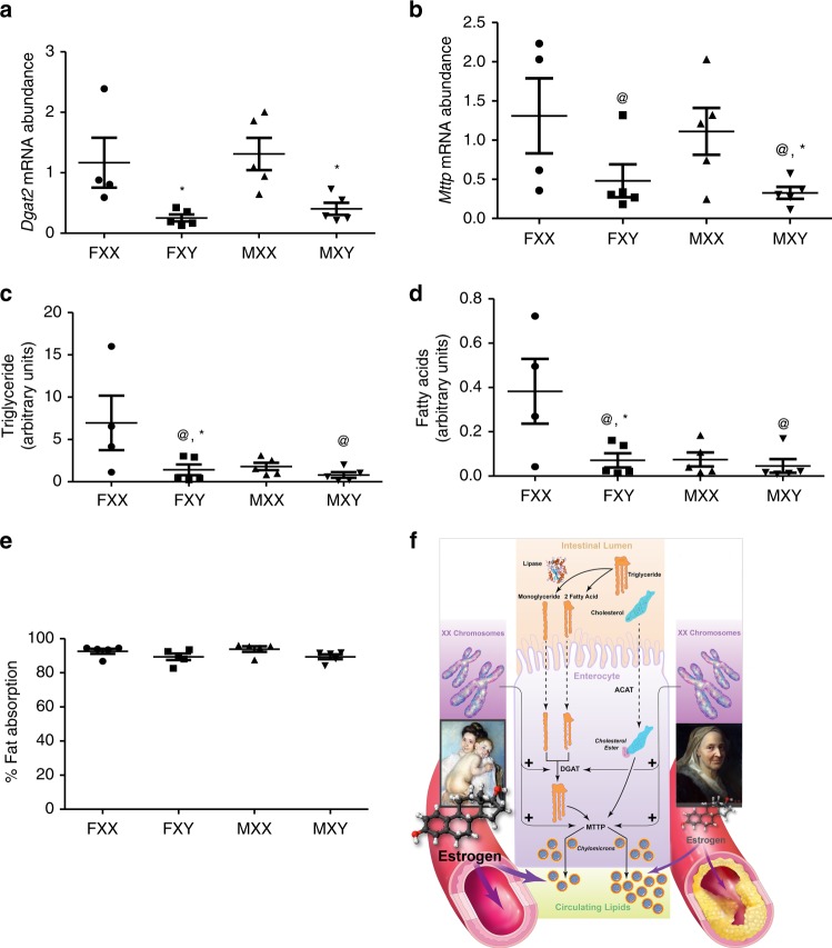
Men and women differ in circulating lipids and coronary artery disease (CAD). While sex hormones such as estrogens decrease CAD risk, hormone replacement therapy increases risk. Biological sex is determined by sex hormones and chromosomes, but effects of sex chromosomes on circulating lipids and atherosclerosis are unknown. Here, we use mouse models to separate effects of sex chromosomes and hormones on atherosclerosis, circulating lipids and intestinal fat metabolism. We assess atherosclerosis in multiple models and experimental paradigms that distinguish effects of sex chromosomes, and male or female gonads. Pro-atherogenic lipids and atherosclerosis are greater in XX than XY mice, indicating a primary effect of sex chromosomes. Small intestine expression of enzymes involved in lipid absorption and chylomicron assembly are greater in XX male and female mice with higher intestinal lipids. Together, our results show that an XX sex chromosome complement promotes the bioavailability of dietary fat to accelerate atherosclerosis.


Conflict of interest statement

The authors declare no competing interests.

Figures

Fig. 1
Fig. 1
XX male and female mice have higher body weight, fat mass and food intake. a Body weight of mice of each sex chromosome complement and gonadal sex at baseline (when fed standard murine diet) or after 1 week of consumption of a Western diet (WD). Lean (b) and fat (c) mass (gm). d Food intake, normalized to lean mass, of mice fed standard murine diet. e Physical activity, normalized to lean mass, of mice fed standard murine diet. f Energy expenditure, normalized to lean mass, of mice fed standard murine diet. g Food intake, normalized to lean mass, of mice fed a Western diet (1 week). h Physical activity, normalized to lean mass, of mice fed a Western diet. i Energy expenditure, normalized to lean mass, of mice fed a Western diet. Symbols represent individual mice per group (n = 5 mice/group) per measurement, with horizontal lines representing mean ± SEM. *P < 0.05 compared to XX within gonadal sex. #P < 0.05 compared to female within sex chromosome complement. $P < 0.05 compared to XX females. @P < 0.05 main effect of sex chromosome complement. Data were analyzed by 3-way ANOVA (A-C) with Holm–Sidak test, or by 2-way ANOVA (di) with Holm–Sidak test. Source data are available as a Source Data file
Fig. 2
Fig. 2
An XX sex chromosome complement promotes obesity and dyslipidemias. a Body weights (gm) of male and female intact and gonadectomized (GDX) mice of each genotype (Intact: FXX, n = 11; FXY, n = 14; MXX, n = 9; MXY, n = 11; GDX: FXX, n = 9; FXY, n = 8; MXX, n = 7; MXY, n = 7). b Representative pictures of mice from each group (left), with adipose tissue depots illustrated. c Representative pictures of serum from XX and XY male mice. d Total serum cholesterol concentrations (Intact: FXX, n = 10; FXY, n = 12; MXX, n = 9; MXY, n = 11; GDX: FXX, n = 6; FXY, n = 4; MXX, n = 6; MXY, n = 5). Concentrations of very low density lipoprotein (VLDL)-cholesterol (e) (Intact: FXX, n = 6; FXY, n = 6; MXX, n = 5; MXY, n = 6; GDX: FXX, n = 6; FXY, n = 4; MXX, n = 6; MXY, n = 5), low density lipoprotein (LDL)-cholesterol (f) (Intact: FXX, n = 6; FXY, n = 6; MXX, n = 5; MXY, n = 6; GDX: FXX, n = 6; FXY, n = 4; MXX, n = 6; MXY, n = 5) and high density lipoprotein (HDL)-cholesterol (g) (Intact: FXX, n = 6; FXY, n = 4; MXX, n = 6; MXY, n = 5; GDX: FXX, n = 6; FXY, n = 4; MXX, n = 6; MXY, n = 5) and TG (h) (Intact: FXX, n = 4; FXY, n = 6; MXX, n = 4; MXY, n = 5; GDX: FXX, n = 6; FXY, n = 4; MXX, n = 6; MXY, n = 5) in Ldlr−/− mice fed a Western diet for 4 months. Concentrations of total serum cholesterol (i) (FXX, n = 11; FXY, n = 10; MXX, n = 12; MXY, n = 8), LDL-cholesterol (j) (FXX, n = 11; FXY, n = 10; MXX, n = 12; MXY, n = 8), HDL-cholesterol (k) (FXX, n = 11; FXY, n = 10; MXX, n = 12; MXY, n = 8) and TG (l) (FXX, n = 11; FXY, n = 10; MXX, n = 12; MXY, n = 8) in Apoe−/− mice fed a standard murine diet. Symbols represent individual mice per group per measurement, with horizontal lines representing mean ± SEM. *P < 0.05 compared to XX within gonadal sex. #P < 0.05 compared to female within sex chromosome complement. $P < 0.05 compared to XX females. Data were analyzed by 3-way ANOVA (a, dh) with Holm–Sidak test, or by 2-way ANOVA (il) with Holm–Sidak test. Source data are available as a Source Data file. RPF retroperitoneal, EF epididymal, SubQ subcutaneous
Fig. 3
Fig. 3
An XX sex chromosome complement augments atherosclerosis in male and female mice. a Atherosclerotic lesion surface area, expressed as a percentage of the aortic arch, in aortic arches from male and female XX and XY intact or gonadectomized (GDX) Ldlr−/− mice. (Intact: FXX, n = 11; FXY, n = 12; MXX, n = 9; MXY, n = 11; GDX: FXX, n = 5; FXY, n = 4; MXX, n = 7; MXY, n = 7). b Representative aortic arch, stained with Oil Red O, from Ldlr−/− mice of each group. c Atherosclerotic lesion area, quantified as Oil Red O staining, in tissue sections from the aortic sinus of male and female XX and XY intact or GDX Ldlr−/− mice. (Intact: FXX, n = 4; FXY, n = 5; MXX, n = 4; MXY, n = 4; GDX: FXX, n = 4; FXY, n = 4; MXX, n = 5; MXY, n = 5). d Representative aortic sinus tissue sections, stained with Oil Red O, from Ldlr−/− mice of each group. e Atherosclerotic lesion area in tissue sections from XX and XY male and female Apoe−/− GDX mice. (FXX, n = 5; FXY, n = 4; MXX, n = 5; MXY, n = 4). f Representative aortic sinus tissue sections from Apoe−/− GDX mice of each group. g Atherosclerotic lesion area in tissue sections from aortic sinus of XX and XY male and female C57BL/6 GDX mice fed an atherogenic diet. Symbols represent individual mice per group per measurement, with horizontal lines representing mean ± SEM. (FXX, n = 8; FXY, n = 6; MXX, n = 10; MXY, n = 5). *P < 0.05 compared to XX within gonadal sex. #P < 0.05 compared to female within sex chromosome complement. $P < 0.05 compared to XX females. Data were analyzed by 3-way ANOVA (A,C) with Holm–Sidak test, or by 2-way ANOVA (e, g) with Holm–Sidak test. Scale bar = 200 μm. Source data are available as a Source Data file
Fig. 4
Fig. 4
Sex chromosome complement influences hepatic gene expression of Ldlr−/− gonadectomized (GDX) mice. a Total probe sets on the array were filtered to retain annotated transcript clusters with reliable signal intensity. The filtered data set was analyzed by two-way ANOVA. The number of genes whose expression was significantly changed (multiple testing corrected q value ≤ 0.01) by the main effects of gene status (XX vs XY), biological sex (male vs. female), as well as by interaction, are listed. Note that only genes significant by the chromosome effect survived multiple testing correction. b Fold change in gene expression (log 2 scale, x-axis) and statistical significance (p value, y-axis) for the main effect of chromosome are plotted. Genes labelled in blue exhibited significant increases in livers from XY compared to XX mice; genes labelled in red exhibited significant increases in livers from XX compared to XY mice. (FXX, n = 5; FXY, n = 5; MXX, n = 4; MXY, n = 5). Source data are available as a Source Data file
Fig. 5
Fig. 5
Intestinal gene expression, triglyceride content, and fat absorption are higher in XX than XY Ldlr−/− mice. mRNA abundance of Dgat2 (a) (FXX, n = 4; FXY, n = 5; MXX, n = 5; MXY, n = 5) and Mttp (b) (FXX, n = 4; FXY, n = 5; MXX, n = 5; MXY, n = 5) in small intestines from gonadectomized (GDX) male and female XX and XY mice. c Intestinal triglyceride content in GDX XX and XY male and female mice (FXX, n = 4; FXY, n = 5; MXX, n = 5; MXY, n = 5). d Fatty acids (Palmitic/palmitic/oleic acids) that are enriched in intestinal triglycerides from XX compared to XY GDX mice (FXX, n = 4; FXY, n = 5; MXX, n = 5; MXY, n = 5). e Absorption of dietary fat (%) in XX and XY male and female mice. Symbols represent individual mice per group per measurement, with horizontal lines representing mean ± SEM (n = 5 mice/genotype/sex). *P < 0.05 compared to XX within gonadal sex. #P < 0.05 compared to female within genotype. @P < 0.05 XX vs. XY. f Overall conclusions from this study. Data (ad) were analyzed by 2-way ANOVA with Holm–Sidak test. Dietary fat absorption data (e) were analyzed by Kruskal–Wallis. Source data are available as a Source Data file

Similar articles

Cited by

References

    1. Arnold AP, Cassis LA, Eghbali M, Reue K, Sandberg K. Sex hormones and sex chromosomes cause sex differences in the development of cardiovascular diseases. Arterioscler Thromb. Vasc. Biol. 2017;37:746–756. doi: 10.1161/ATVBAHA.116.307301. - DOI - PMC - PubMed
    1. Palmisano BT, Zhu L, Eckel RH, Stafford JM. Sex differences in lipid and lipoprotein metabolism. Mol. Metab. 2018;15:45–55. doi: 10.1016/j.molmet.2018.05.008. - DOI - PMC - PubMed
    1. Galman C, Angelin B, Rudling M. Pronounced variation in bile acid synthesis in humans is related to gender, hypertriglyceridaemia and circulating levels of fibroblast growth factor 19. J. Intern. Med. 2011;270:580–588. doi: 10.1111/j.1365-2796.2011.02466.x. - DOI - PubMed
    1. Middelberg RP, Spector TD, Swaminathan R, Snieder H. Genetic and environmental influences on lipids, lipoproteins, and apolipoproteins: effects of menopause. Arterioscler. Thromb. Vasc. Biol. 2002;22:1142–1147. doi: 10.1161/01.ATV.0000022889.85440.79. - DOI - PubMed
    1. Kannel WB, Hjortland MC, McNamara PM, Gordon T. Menopause and risk of cardiovascular disease: the Framingham study. Ann. Intern. Med. 1976;85:447–452. doi: 10.7326/0003-4819-85-4-447. - DOI - PubMed

Publication types

MeSH terms

Grants and funding