. 2019 Jun 14;10(1):2653.
doi: 10.1038/s41467-019-10568-4.

Convergent allostery in ribonucleotide reductase

Affiliations

Convergent allostery in ribonucleotide reductase

William C Thomas et al. Nat Commun. .

Abstract

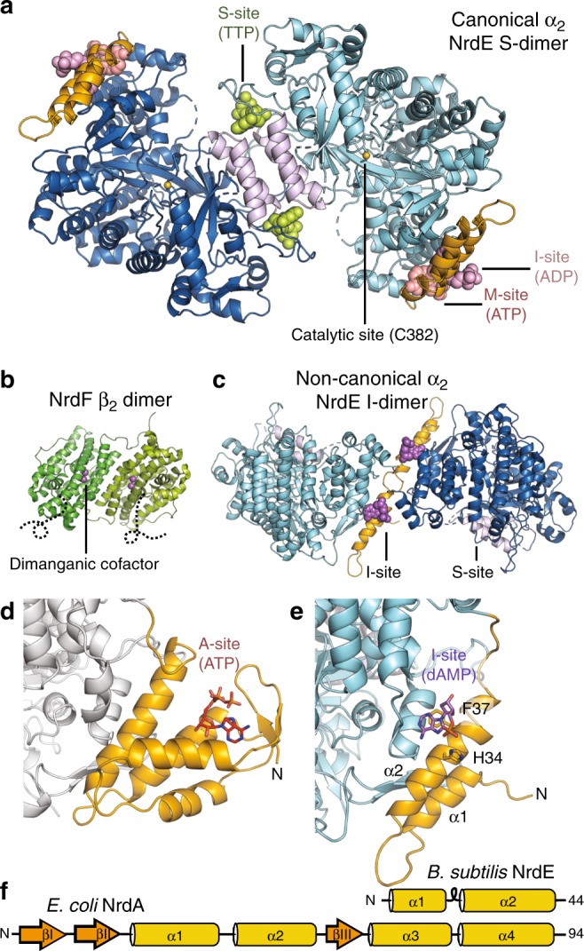
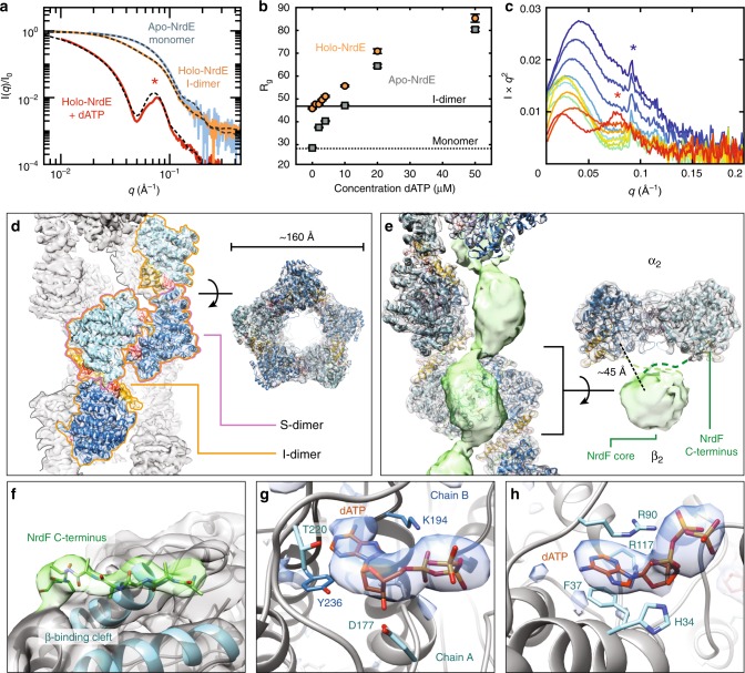
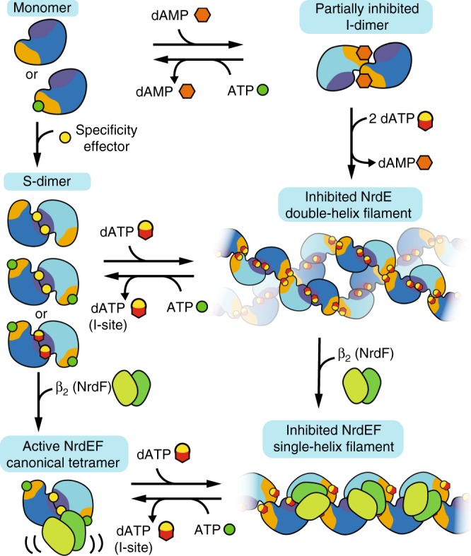
Ribonucleotide reductases (RNRs) use a conserved radical-based mechanism to catalyze the conversion of ribonucleotides to deoxyribonucleotides. Within the RNR family, class Ib RNRs are notable for being largely restricted to bacteria, including many pathogens, and for lacking an evolutionarily mobile ATP-cone domain that allosterically controls overall activity. In this study, we report the emergence of a distinct and unexpected mechanism of activity regulation in the sole RNR of the model organism Bacillus subtilis. Using a hypothesis-driven structural approach that combines the strengths of small-angle X-ray scattering (SAXS), crystallography, and cryo-electron microscopy (cryo-EM), we describe the reversible interconversion of six unique structures, including a flexible active tetramer and two inhibited helical filaments. These structures reveal the conformational gymnastics necessary for RNR activity and the molecular basis for its control via an evolutionarily convergent form of allostery.


Conflict of interest statement

The authors declare no competing interests.

Figures

Fig. 1
Fig. 1
Allosteric sites of the Bacillus subtilis class Ib ribonucleotide reductase. a A 2.50 Å crystal structure of B. subtilis NrdE (α subunit) obtained under activating conditions depicts an S-shaped dimer (“S-dimer”) interfacing at the “specificity” or S-site (lavender). A specificity effector TTP (green) is bound to the S-site, and activating nucleotides, ADP (pink) and ATP (salmon), are bound to two allosteric sites that evolved near the N-terminus of B. subtilis NrdE. A catalytically essential radical is generated at a central cysteine in the catalytic site, C382 (yellow sphere). b B. subtilis NrdF (β subunit) is dimeric and utilizes a dimanganic tyrosyl cofactor (purple spheres) to initiate radical chemistry (PDB: 4DR0). A disordered region of the NrdF C-terminus (black dotted lines) is critical for radical transfer. c A recent structure of B. subtilis NrdE co-crystallized with dAMP (purple) depicts a partially inhibited, non-canonical “I-dimer” with the interface formed by the truncated ATP-cone (orange) (PDB: 6CGL). d In class Ia RNRs, ATP or dATP binds to the “activity” or A-site in the ATP-cone domain (orange) to mediate changes in quaternary structure and tune overall activity (PDB: 3R1R). e Class Ib RNRs only contain partial ATP-cones (orange). B. subtilis NrdE is unusual in that it displays activity regulation and binds dAMP (purple) in the N-terminally located I-site (PDB: 6CGL). f The partial N-terminal cone of class Ib RNRs (top) is structurally homologous to the last two helices of the canonical ATP-cone found in many class Ia RNRs (bottom) but lacks A-site residues
Fig. 2
Fig. 2
The inhibitor dATP stabilizes an unusual helical NrdE filament. a EFA-separated SEC–SAXS profiles of apo-NrdE (blue), holo-NrdE (orange), and holo-NrdE + 100 µM dATP (red) agree with the theoretical scattering (dashed) of a monomer, I-dimer, and a 34-mer double-helix model derived from cryo-EM. b SAXS titrations of 0–50 μM dATP to 4 μM holo-NrdE (orange) and apo-NrdE (gray) display increasingly larger Rg values, suggestive of non-terminating oligomerization. Theoretical Rg values of the I-dimer (46 Å, solid line) and monomer (27 Å, dotted line) are shown for comparison. c A SAXS titration of 0–20 μM NrdF to 4 µM C382S holo-NrdE + 50 µM dATP (red to blue), plotted in Kratky representation, shows the loss of the NrdE double-helix (red star) with the formation of the NrdEF filament. This change is followed by an accumulation of excess NrdF and eventual appearance of ordered NrdEF filament assemblies (blue star, Supplementary Fig. 4b, d). d A 6 Å cryo-EM map of the dATP-induced NrdE filament (threshold = 4.45) reveals a double-helical structure with each helix formed by alternating S-dimer (pink outline) and I-dimer (orange outline) interfaces. e A 4.7 Å cryo-EM map of dATP-inhibited NrdEF displays strong density for a helical NrdE filament (gray, threshold = 1.14) and weaker density for NrdF forming a central column of beads (green, threshold = 0.45). Each ASU consists of a NrdF dimer centered on a NrdE S-dimer, resulting in an α2β2 with a prominent gap (right). f The NrdF C-terminus (green sticks/surface, threshold = 1.18) is observed at high occupancy in the β-tail-binding cleft of NrdE (blue cartoon, gray surface, threshold = 1.18). g and h Difference densities in the NrdEF cryo-EM map for (g) dATP in the S-site (threshold = 7.73) and (h) dATP in the I-site (threshold = 12.6). Corresponding σ levels are estimated in Supplementary Table 5. Source data are provided as a Source Data file
Fig. 3
Fig. 3
SAXS reveals a complex interplay of four distinct oligomerization states of NrdE. In all plots, the theoretical Rg values of the I-dimer (46 Å, solid line), S-dimer (39 Å, dashed line), and monomer (27 Å, dotted line) are shown for comparison. a Titration of 0–1 mM ATP to 4 μM holo-NrdE (orange circles) leads to a reduction in Rg, consistent with the dissociation of I-dimer to monomer and confirmed by SEC–SAXS (Supplementary Fig. 7a). In contrast, the Rg of 4 μM apo-NrdE (gray squares) remains constant up to 1 mM ATP. b Titration of 0–15 mM ATP to 4 μM holo-NrdE (red circles) or apo-NrdE (lavender squares) in the presence of 50 μM dATP leads to a decrease in Rg that converges to a value near the theoretical value of an S-dimer. c Titration of 0–500 μM TTP to 4 μM apo-NrdE (gray squares) leads to an increase in Rg that is suggestive of a monomer to S-dimer transition. A similar transition is observed with the addition of TTP to 4 μM holo-NrdE in the presence of 3 mM ATP (blue circles) and was further confirmed by SEC–SAXS (Supplementary Fig. 7b). d Titration of 0–500 μM TTP to 4 μM holo-NrdE in the absence of ATP leads to an increase in Rg and a final profile that resembles the dATP-inhibited filament (Supplementary Fig. 2b, c). Source data are provided as a Source Data file
Fig. 4
Fig. 4
Crystallographic insight into allosteric activation and re-reduction of the catalytic site. ac Shown in blue mesh are the mFODFC Polder omit maps for the ligands in the 2.50 Å dataset, contoured at 4.0σ. a TTP binds to the S-site and interacts with both chains of the S-dimer interface. b ADP binds to the I-site, displacing dAMP that was previously observed to bind this site in holo-NrdE. c ATP is bound to a newly identified M-site. d The M-site and I-site are both found in the N-terminus (orange) in close proximity to each other. Binding of ATP at the M-site induces F47 to flip inward (orange) relative to its position in the I-dimer (gray, PDB: 6CGL). e In our 2.50 Å structure, we observe R117 making H-bonds to the 2′-OH of the ADP ribose and E119. f With dAMP bound to the I-site, R117 instead H-bonds with N42 and T45 (PDB: 6CGL). In this position, R117 forms part of the I-dimer interface (Supplementary Fig. 9a). R117 is thus important for discrimination of ribonucleotides and deoxyribonucleotides at this site. g The NrdE C-terminus (purple sticks) was captured in the catalytic site of the 2.55 Å reduced dataset, revealing specific interactions between highly conserved residues. C698 on the C-terminal tail is captured in a conformation well poised to initiate re-reduction of a C170–C409 disulfide that forms in the catalytic site after each turnover (Supplementary Fig. 10c, d)
Fig. 5
Fig. 5
The active NrdEF structure is a flexible α2β2. a SEC–SAXS of a stoichiometric mixture of C382S holo-NrdE and NrdF under activating conditions (1 mM ATP, 250 µM TTP) revealed a predominant species (experimental profile shown in blue) with a molecular weight estimate of 246 kDa, consistent with an α2β2 species (actual MW of 243 kDa). Structural modeling was performed in AllosMod-FoXS using three different α2β2 starting models. Using a symmetric α2β2 docking model as a starting model underestimated the shoulder observed in the mid-q region (purple dash; χ2 = 20.43), suggesting that the solution structure is more open. In contrast, the expanded α2β2 starting model derived from the ASU of the NrdEF cryo-EM structure (Fig. 2e, right) overestimated the shoulder (orange dash; χ2 = 18.55), suggesting that the subunits are closer together in solution under activating conditions. An asymmetric α2β2 starting model based on an S. typhimurium structure (PDB: 2BQ1) yielded the best-fit conformer (black dash; χ2 = 2.25). SAXS profiles are shown in Kratky representation (q vs. Ixq2) to emphasize mid-q features. b Best-fit models of the asymmetric α2β2 (top) and symmetric α2β2 (bottom). Source data are provided as a Source Data file
Fig. 6
Fig. 6
A model for the overall allosteric regulation of B. subtilis RNR. Without nucleotides, NrdE is a monomer, but dAMP and dATP can bind the I-site and induce a partially inhibited I-dimer. Addition of specificity effectors instead induces the monomer to form an S-dimer. When specificity effectors (including dATP) bind to the I-dimer, they induce formation of an inhibited double-helical NrdE filament composed of alternating S-dimer and I-dimer interfaces. NrdF competes for the NrdE double-helical interface, and thus NrdF binding leads to the dissociation of the NrdE double-helix into individual helical structures. NrdF binds to the helical interior of the NrdE filament, leading to an inter-subunit gap and positional confinement that prevent NrdF from accessing NrdE for turnover. Both the NrdE and NrdEF filaments are reversible by addition of ATP, which can displace dATP from the I-site and induce dissociation of the I-dimer interface. Finally, addition of NrdF to the S-dimer leads to formation of an active but asymmetric α2β2 tetramer in which a hinge motion between the two subunits plays an important role in activity

Similar articles

Cited by

References

    1. Nordlund P, Reichard P. Ribonucleotide reductases. Annu. Rev. Biochem. 2006;75:681–706. doi: 10.1146/annurev.biochem.75.103004.142443. - DOI - PubMed
    1. Stubbe J. Ribonucleotide reductases in the twenty-first century. Proc. Natl Acad. Sci. USA. 1998;95:2723–2724. doi: 10.1073/pnas.95.6.2723. - DOI - PMC - PubMed
    1. Hofer A, Crona M, Logan DT, Sjöberg BM. DNA building blocks: keeping control of manufacture. Crit. Rev. Biochem. Mol. Biol. 2012;47:50–63. doi: 10.3109/10409238.2011.630372. - DOI - PMC - PubMed
    1. Mathews CK. The most interesting enzyme in the world. Structure. 2016;24:843–844. doi: 10.1016/j.str.2016.05.006. - DOI - PubMed
    1. Meisburger SP, Thomas WC, Watkins MB, Ando N. X-ray scattering studies of protein structural dynamics. Chem. Rev. 2017;117:7615–7672. doi: 10.1021/acs.chemrev.6b00790. - DOI - PMC - PubMed

Publication types

MeSH terms