Review
. 2019 Nov;44(11):3559-3568.
doi: 10.1007/s00261-019-02089-x.

The use of PET/MRI for imaging rectal cancer

Affiliations
Review

The use of PET/MRI for imaging rectal cancer

Thomas A Hope et al. Abdom Radiol (NY). 2019 Nov.

Abstract

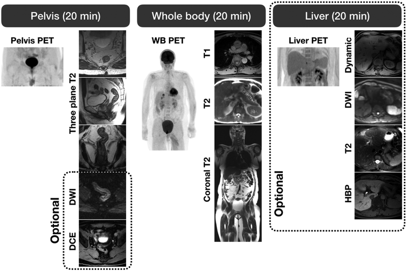
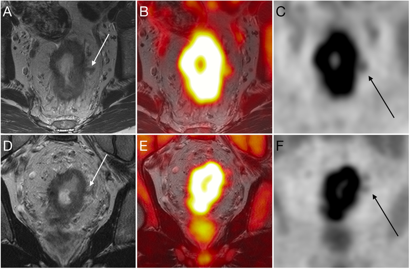
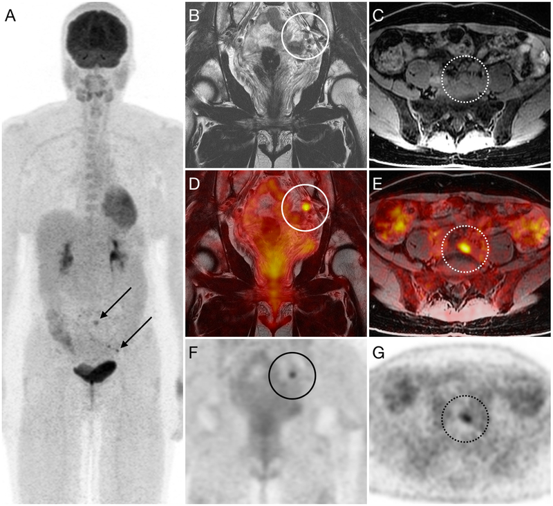
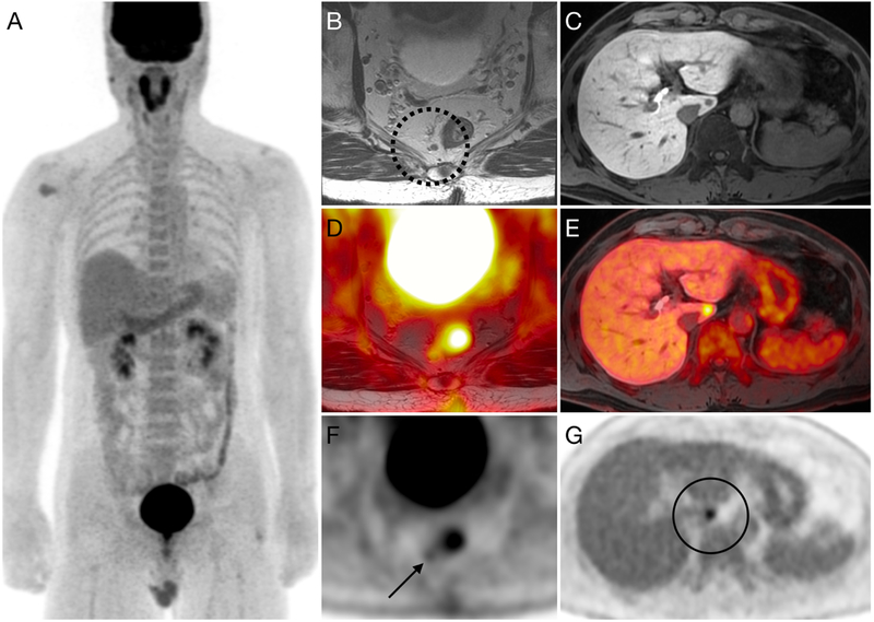
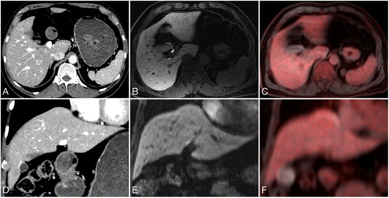
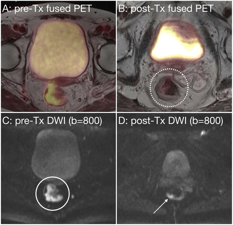
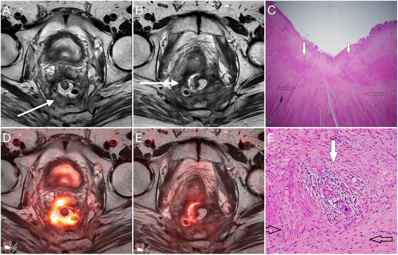
Combined PET/MRI is a proposed imaging modality for rectal cancer, leveraging the advantages of MRI and 18F-fluorodeoxyglucose PET. Rectal cancer PET/MRI protocols typically include dedicated pelvis bed positions utilizing small field-of-view T2-weighted imaging. For staging of the primary tumor, PET/MRI can help delineate the extent of tumor better as well as the extent of tumor beyond the muscularis propria. PET uptake may help characterize small lymph nodes, and the use of hepatobiliary phase imaging can improve the detection of small hepatic metastases. The most beneficial aspect of PET/MRI may be in treatment response, although current data are limited on how to combine PET and MRI data in this setting. Limitations of PET/MRI include the inability to detect small pulmonary nodules and issues related to attenuation correction, although the development of new attenuation correction techniques may address this issue. Overall PET/MRI can improve the staging of rectal cancer, although this potential has yet to be fulfilled.

Keywords: PET/MRI; Rectal cancer; Staging; Treatment response.


Figures

Figure 1:
Figure 1:
Overview of components of a rectal cancer PET/MRI protocol. DWI and DCE acquisitions in the pelvis may be considered optional, while a dedicated liver MRI using hepatobiliary agents is also optional.
Figure 2:
Figure 2:
43-year-old man with newly diagnosed rectal adenocarcinoma. PET/MRI demonstrates a circumferential hypermetabolic rectal mass. On oblique axial (A-C) and coronal (D-F) T2-weighted and PET images through the tumor soft tissue extending from the primary tumor into the mesorectal fat is noted consistent with extramural vascular invasion (white and black arrows).
Figure 3:
Figure 3:
53-year-old female with newly diagnosed rectal adenocarcinoma initially staged using PET/MRI. PET imaging demonstrated a 6-mm hypermetabolic left internal iliac node (B, D and F, solid circle) and an 8-mm left common iliac node (C, E and G, dotted circle).
Figure 4:
Figure 4:
52-year-old man with a newly diagnosed rectal adenocarcinoma. A whole-body PET/MRI with dedicated rectal and liver bed positions was performed (A). In the rectal bed position, a 5-mm right perirectal node is seen which demonstrates hypermetabolism consistent with nodal metastatic disease (B circle, D and F black arrow). In the liver, a 6-mm caudate lobe lesion was visualized on hepatobiliary phase imaging (C), which demonstrated hypermetabolism (E and G, black circle) consistent with hepatic metastatic disease.
Figure 5:
Figure 5:
65-year-old male status post abdominal-peritoneal resection with a nonspecific lesion on CT (A and D). At hepatobiliary phase MRI (B and E) and FDG PET (C and F), no suspicious features are identified. This case demonstrates how PET/MRI provides improved characterizations of liver lesions compared to PET/CT.
Figure 6:
Figure 6:
72-year-old female with rectal adenocarcinoma before (A) and after chemoradiation (B). Post-therapy images suggest residual tumor on T2 weighted images, given that there was only a 24% reduction in tumor volume (B, white dotted circle). FDG-PET shows a marked reduction in metabolic activity, with a reduction of the SUVmax from 13.7 to 4.5 (a 67% reduction). DWI images show a decrease in tumor that demonstrates restricted diffusion (C, white circle and D, white arrow). MRI indicated partial response on T2 weighted imaging, while PET imaging suggests potentially a complete response. At pathology this was confirmed a complete response.
Figure 7:
Figure 7:
PET/MRI performed in a 53-year-old man three years after surgery with a rising CEA. Axial T2 (A) demonstrates circumferential soft tissue thickening with heterogeneously mixed hyperintense and hypointense signal intensity within the surgical bed (arrow). Fused axial PET image (C) demonstrates intense FDG uptake within this circumferential soft tissue mass consistent with local recurrence. Inferiorly in the same patient, the MRI appearance is similar with semicircular T2 signal hypointensity (B, arrow), but FDG PET demonstrates an absence of hypermetabolism (D) consistent with fibrosis. At pathology, one can see both recurrent tumor (C and F, white arrows) as well as fibrosis surrounding the region of local recurrence (C and F, open arrows). The addition of FDG PET increases reader confidence and sensitivity for local recurrence.

References

    1. Horvat N, Carlos Tavares Rocha C, Clemente de Oliveira B, Petkovska I, Gollub MJ. MRI of Rectal Cancer: Tumor Staging, Imaging Techniques, and Management. Radiographics. 2019;:180114. - PMC - PubMed
    1. Jeong JH, Cho IH, Chun KA, Kong EJ, Kwon SD, Kim JH. Correlation Between Apparent Diffusion Coefficients and Standardized Uptake Values in Hybrid (18)F-FDG PET/MR: Preliminary Results in Rectal Cancer Nucl Med Mol Imaging. Springer Berlin Heidelberg; 2016;50:150–156. - PMC - PubMed
    1. Buchbender C, Heusner TA, Lauenstein TC, Bockisch A, Antoch G. Oncologic PET/MRI, part 1: tumors of the brain, head and neck, chest, abdomen, and pelvis. J Nucl Med. 2012;53:928–938. - PubMed
    1. Park MJ, Kim SH, Lee SJ, Jang KM, Rhim H. Locally advanced rectal cancer: added value of diffusion-weighted MR imaging for predicting tumor clearance of the mesorectal fascia after neoadjuvant chemotherapy and radiation therapy. Radiology. 2011;260:771–780. - PubMed
    1. Rosenkrantz AB, Friedman K, Chandarana H, et al. Current Status of Hybrid PET/MRI in Oncologic Imaging. American Journal of Roentgenology. 2016;206:162–172. - PMC - PubMed

Publication types

MeSH terms

Substances