

The value of radiographic markers in the diagnostic work-up of rotator cuff tears, an arthroscopic correlated study
- PMID: 31201467
- PMCID: PMC6858903
- DOI: 10.1007/s00256-019-03251-8
The value of radiographic markers in the diagnostic work-up of rotator cuff tears, an arthroscopic correlated study
Abstract
Objective: To evaluate the value of radiographs during the diagnostic work-up of rotator cuff tears, using arthroscopy as reference standard.
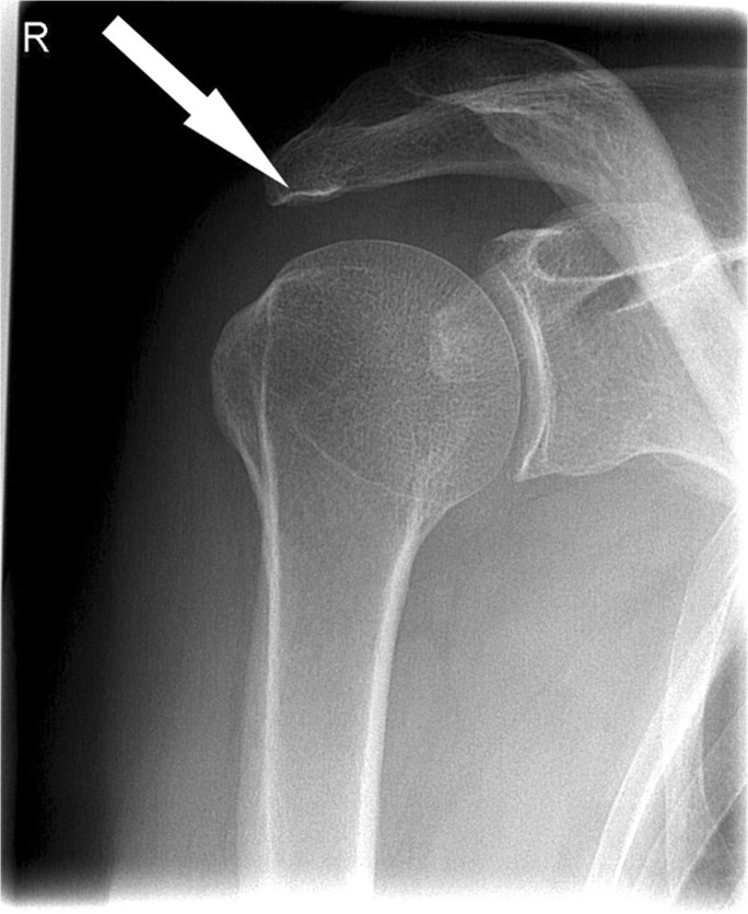
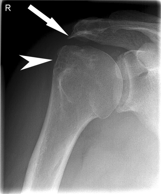
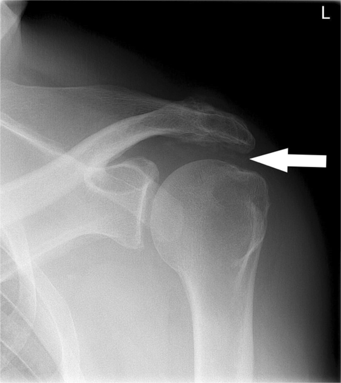
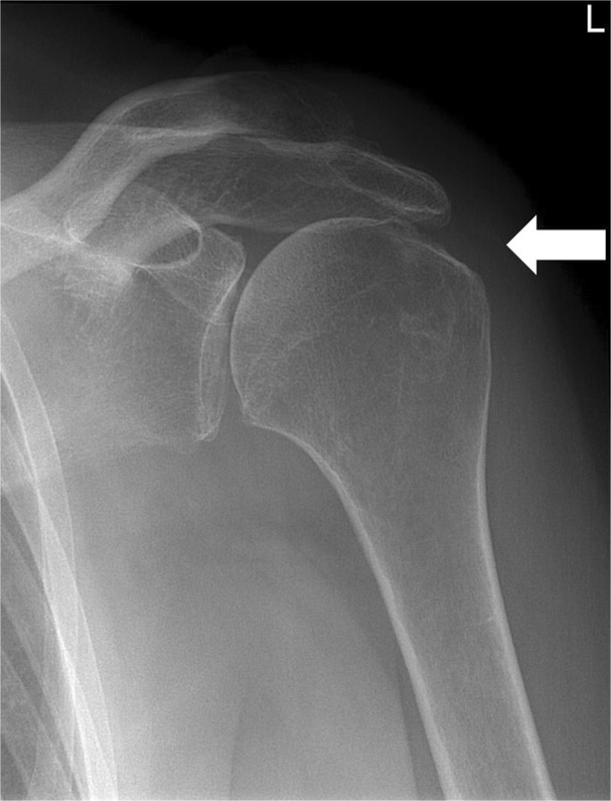
Materials and methods: This retrospective study included 236 shoulders of 236 patients. All radiographs were evaluated for inferior cortical acromial sclerosis, lateral acromial spur, superior migration of the humeral head, greater tubercle cysts, and subacromial space calcifications. Predictive value of these radiographic signs in predicting rotator cuff tears was determined with arthroscopy as reference standard.
Results: According to arthroscopy, 131 shoulders were diagnosed with rotator cuff tears. Seventy-two out of 131 shoulders (55%) had inferior cortical acromial sclerosis, 37 (28%) lateral acromial spur, 21 (16%) superior migration of the humeral head, 7 (5%) greater tubercle cysts and 15 subacromial space calcifications (11%). Inferior cortical acromial sclerosis (P = 0.001), lateral spur (P = 0.001), superior migration (P = 0.002), and cysts (P = 0.03) were significantly and independently associated with rotator cuff tears, whereas subacromial calcifications (p = 0.21) was not. Inferior cortical acromial sclerosis, superior migration, lateral acromial spur, and cysts combined have a positive predictive value of 78%.
Conclusions: The combination of inferior cortical acromial sclerosis, lateral acromial spur, superior migration of the humeral head, and greater tubercle cysts has a high positive predictive value for the presence of full-thickness rotator cuff tears. In patients with a high suspicion for having a rotator cuff tear based on radiographic findings, MRI can be performed directly without the delay and costs caused by an additional ultrasound exam.
Keywords: Arthroscopy; Diagnostic test; Predictive value; Radiographic markers; Rotator cuff tear; Shoulder pathology.
Conflict of interest statement
Jeroen J. van der Reijden, Syert L. Nienhuis, Matthijs P. Somford, Job N. Doornberg, Esther van’t Riet, Maaike P.J. van den Borne declare that they have no conflicts of interest.
Michel P.J. van den Bekerom reports a grant from Smith and Nephew and a grant from Wright/Tornier, during the conduct of the study.
Figures
Similar articles
-
The Correlation between Various Shoulder Anatomical Indices on X-Ray and Subacromial Impingement and Morphology of Rotator Cuff Tears.Orthop Surg. 2023 Aug;15(8):1997-2006. doi: 10.1111/os.13610. Epub 2022 Dec 26. Orthop Surg. 2023. PMID: 36573272 Free PMC article.
-
Classification and clinical significance of acromial spur in rotator cuff tear: heel-type spur and rotator cuff tear.Clin Orthop Relat Res. 2010 Jun;468(6):1542-50. doi: 10.1007/s11999-009-1058-5. Epub 2009 Sep 4. Clin Orthop Relat Res. 2010. PMID: 19760471 Free PMC article.
-
Effectiveness Of Plain Shoulder Radiograph In Detecting Degenerate Rotator Cuff Tears.J Ayub Med Coll Abbottabad. 2018 Jan-Mar;30(1):8-11. J Ayub Med Coll Abbottabad. 2018. PMID: 29504320
-
The keeled acromion: an aggressive acromial variant--a series of 20 patients with associated rotator cuff tears.Arthroscopy. 2004 Sep;20(7):744-53. doi: 10.1016/j.arthro.2004.06.018. Arthroscopy. 2004. PMID: 15346116 Review.
-
[Results of the surgical repair of the rotator cuff. Radio-clinical correlation].Rev Chir Orthop Reparatrice Appar Mot. 1994;80(7):582-94. Rev Chir Orthop Reparatrice Appar Mot. 1994. PMID: 7638384 Review. French.
Cited by
-
Capsular remnant in the rotator cuff footprint is a novel arthroscopic finding may indicate the etiology of the tear.Knee Surg Sports Traumatol Arthrosc. 2023 Aug;31(8):3559-3564. doi: 10.1007/s00167-023-07413-z. Epub 2023 Apr 10. Knee Surg Sports Traumatol Arthrosc. 2023. PMID: 37038018
-
Muscle Health & Fatty Infiltration with Advanced Rotator Cuff Pathology.Curr Rev Musculoskelet Med. 2025 Apr;18(4):160-172. doi: 10.1007/s12178-025-09955-w. Epub 2025 Feb 26. Curr Rev Musculoskelet Med. 2025. PMID: 40009348 Free PMC article. Review.
-
Evaluation of Radiographic Changes 5 Years After Arthroscopic Rotator Cuff Repair.Orthop J Sports Med. 2022 Sep 30;10(9):23259671221126095. doi: 10.1177/23259671221126095. eCollection 2022 Sep. Orthop J Sports Med. 2022. PMID: 36199829 Free PMC article.
-
Relationship between the morphology of the greater tuberosity and radiological and clinical outcomes after arthroscopic rotator cuff repair.JSES Int. 2021 Jan 25;5(3):493-499. doi: 10.1016/j.jseint.2020.11.009. eCollection 2021 May. JSES Int. 2021. PMID: 34136860 Free PMC article.
-
Rotator Cuff Tears and Mid-Term Shoulder Outcomes after Intramedullary Nail Fixation for Humeral Shaft Fracture: A Minimum Five-year Follow-up Study.Malays Orthop J. 2024 Nov;18(3):59-65. doi: 10.5704/MOJ.2411.008. Malays Orthop J. 2024. PMID: 39691577 Free PMC article.
References
-
- Smith C, Dattani R, Deans V, Drew S. The sourcil sign: a useful finding on plain X-ray? Should Elb. 2010;2:9–12. doi: 10.1111/j.1758-5740.2009.00034.x. - DOI
-
- Hamid N, Omid R, Yamaguchi K, Steger-May K, Stobbs G, Keener JD. Relationship of radiographic acromial characteristics and rotator cuff disease: a prospective investigation of clinical, radiographic, and sonographic findings. J Shoulder Elb Surg. 2012;21:1289–1298. doi: 10.1016/j.jse.2011.09.028. - DOI - PMC - PubMed
MeSH terms
LinkOut - more resources
Full Text Sources
Medical
