. 2019 Oct;71(4):719-730.
doi: 10.1016/j.jhep.2019.05.029. Epub 2019 Jun 13.

Activation of YAP attenuates hepatic damage and fibrosis in liver ischemia-reperfusion injury

Affiliations

Activation of YAP attenuates hepatic damage and fibrosis in liver ischemia-reperfusion injury

Yuan Liu et al. J Hepatol. 2019 Oct.

Abstract

Background & aims: Hepatic ischemia-reperfusion injury (IRI) is a major complication of hemorrhagic shock, liver resection and transplantation. YAP, a key downstream effector of the Hippo pathway, is essential for determining cell fate and maintaining homeostasis in the liver. We aimed to elucidate its role in IRI.

Methods: The role of YAP/Hippo signaling was systematically studied in biopsy specimens from 60 patients after orthotopic liver transplantation (OLT), and in a mouse model of liver warm IRI. Human biopsy specimens were collected after 2-10 h of cold storage and 3 h post-reperfusion, before being screened by western blot. In the mouse model, the role of YAP was probed by activating or inhibiting YAP prior to ischemia-reperfusion.

Results: In human biopsies, high post-OLT YAP expression was correlated with well-preserved histology and improved hepatocellular function at postoperative day 1-7. In mice, the ischemia insult (90 min) triggered intrinsic hepatic YAP expression, which peaked at 1-6 h of reperfusion. Activation of YAP protected the liver against IR-stress, by promoting regenerative and anti-oxidative gene induction, while diminishing oxidative stress, necrosis/apoptosis and the innate inflammatory response. Inhibition of YAP aggravated hepatic IRI and suppressed repair/anti-oxidative genes. In mouse hepatocyte cultures, activating YAP prevented hypoxia-reoxygenation induced stress. Interestingly, YAP activation suppressed extracellular matrix synthesis and diminished hepatic stellate cell (HSC) activation, whereas YAP inhibition significantly delayed hepatic repair, potentiated HSC activation, and enhanced liver fibrosis at 7 days post-IRI. Notably, YAP activation failed to protect Nrf2-deficient livers against IR-mediated damage, leading to extensive fibrosis.

Conclusion: Our novel findings document the crucial role of YAP in IR-mediated hepatocellular damage and liver fibrogenesis, providing evidence of a potential therapeutic target for the management of sterile liver inflammation in transplant recipients.

Lay summary: In the clinical arm, graft YAP expression negatively correlated with liver function and tissue damage after human liver transplantation. YAP activation attenuated hepatocellular oxidative stress and diminished the innate immune response in mouse livers following ischemia-reperfusion injury. In the mouse model, YAP inhibited hepatic stellate cell activation, and abolished injury-mediated fibrogenesis up to 7 days after the ischemic insult.

Keywords: Fibrogenesis; Hippo; Immune response; Inflammation; Liver ischemia-reperfusion injury; Orthotopic liver transplantation; YAP.


Conflict of interest statement

Conflict of interest statement: The authors declare no conflicts of interest that pertain to this work.

Figures

Fig.1
Fig.1
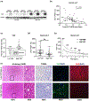
Peri-operative hepatic YAP expression and its correlation with liver damage in human OLT (n=60). (a)Western blot-assisted pre- and post-OLT YAP expression profiles in OLT biopsies (representative 6 cases). (b)The ratio of post-OLT YAP/β-actin correlated negatively with sALT at POD1. (c)Sixty human OLTs were divided into low ratio of post-OLT YAP/β-actin group (Low YAP: n=33) and high ratio of post-OLT YAP/β-actin group (High YAP: n=27) by using post-OLT YAP/β-actin ratio=0.95 as threshold. (d)sALT values in both Low YAP and High YAP groups in OLT recipients at POD1. (e)sALT decreasing curve in Low YAP and High YAP groups in OLT recipients (POD1-7). (f)Histology (H&E staining), TUNEL staining, YAP staining, DHE staining in OLT biopsies (representative 2 cases for Low YAP and High YAP groups) (magnification x100, x200 and x400; **p<0.01, *p<0.001 by Student’s t test).
Fig.2
Fig.2
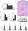
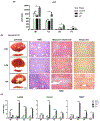
(a)Kinetics of hepatic YAP gene expression in murine liver IRI. Liver samples harvested from B6 mice that were sham-operated or subjected to 90min of partial warm ischemia, followed by various lengths of reperfusion. WT mice were pre-treated with PBS (Ctrl, control), LPA (1-oleoyl lysophosphatidic acid, YAP activator) or VP (verteporfin, YAP inhibitor) at 1h before ischemia. Liver and serum samples were collected at 6h post reperfusion for: (b)YAP gene induction, (c)sALT levels, (d)liver histology (representative H&E staining; magnification x100 and x400), (e)YAP and p-YAP(Ser127) protein induction and (f)YAP downstream gene expression (*p<0.001 by Student’s t test; n=10-12/group).
Fig.3
Fig.3
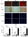
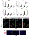
YAP activation prevented hepatocyte death and suppressed pro-inflammatory response in liver IRI. Mice pre-treated with PBS, LPA or VP were subjected to 90min of warm ischemia, and analyzed at 6h of reperfusion for: (a)ROS-sensing dye dihydroethidium (DHE) staining (red fluorescence indicating ROS production). (b)TUNEL-assisted detection of hepatic necrosis/apoptosis (red arrows) in ischemic liver lobes. (c)CD68+ macrophages and Ly6G+ neutrophils in IR liver lobes (white arrow indicated positive staining). (d)qRT-PCR assisted detection of TNF-α, IL-1β and IL-6 (magnification x200, *p<0.001 by Student’s t test; n=4-6/group).
Fig.4
Fig.4
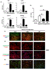
YAP activation protected primary hepatocytes against hypoxia-reoxygenation insult in vitro. WT mouse hepatocyte cultures were supplemented with PBS, LPA or VP. After 6h of hypoxia and 8h of reoxygenation, the cell cultures were analyzed for: (a)YAP downstream gene expressions. (b)Supernatant ALT levels. (c)Hepatocellular YAP staining (red fluorescence indicating YAP accumulation in nucleus). (d)Caspase-3/7 detection (green fluorescence indicating positive caspase-3/7 in nucleus). (e)JC-1 staining (green fluorescence indicating mitochondria depolarization, red fluorescence indicating intact mitochondria membrane) (magnification ×200, *p<0.001 by Student’s t test; n=4-6/group).
Fig.5
Fig.5
YAP activation diminished fibrogenesis development after liver IRI. Mice pre-treated with PBS, LPA or VP were subjected to 90min of warm ischemia, followed by various lengths of reperfusion. Serum and liver samples were collected for: (a)ALT. (b)Histology (7d after reperfusion): gross morphology (fibrotic change); H&E staining; Masson’s trichrome staining (blue color indicating collagen deposition); and Sirius red staining (red color indicating collagen deposition). (c)Fibrosis-related gene expression. (magnification x100, N.S. represented no significance, **p<0.01, *p<0.001 by Student’s ttest; n=4-6/group).
Fig.6
Fig.6
YAP inhibition exacerbated inflammation and activated HSCs in liver IRI. Mice pre-treated with PBS, LPA or VP were subjected to 90min of warm ischemia, followed by various lengths of reperfusion. (a)Liver samples were harvested serially between 6h to 7d after reperfusion to measure pro-inflammatory cytokine gene inductions. (b)HSCs were isolated from IR-livers at 7d after reperfusion, and stained for YAP (green fluorescence) and α-SMA (red fluorescence). (c)IR-livers at 7d after reperfusion stained for α-SMA (red fluorescence) (magnification x200, **p<0.01, *p<0.001 by Student’s ttest; n=4-6/group).
Fig.7
Fig.7
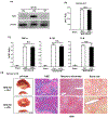
YAP activation failed to protect Nrf2-deficient livers against IR-damage. WT and Nrf2-deficient mice pre-treated with PBS, LPA or VP were subjected to 90min of warm ischemia followed by 6h or 7d of reperfusion. Liver and serum samples were collected for analysis. (a)Western blot-assisted Nrf2 expression in WT liver after IRI. In Nrf2-deficient mice: (b)sALT levels. (c)qRT-PCR detected gene induction of TNF-α, IL-1β, IL-6. (d)Liver histology (7d after reperfusion): gross morphology (fibrotic change); H&E staining; Masson’s trichrome staining (blue color indicating collagen deposition); and Sirius red staining (red color indicating collagen deposition). (magnification x100, N.S. represented no significance by Student’s t test; n=4-6/group).
Fig.8
Fig.8
YAP activation failed to protect Nrf2-deficient primary hepatocytes against hypoxia-reoxygenation insult in vitro, and failed to suppress Nrf2-deficient HSCs in liver IRI. Nrf2-deficient mouse hepatocyte cultures were supplemented with PBS or LPA. After 6h of hypoxia and 8h of reoxygenation, the cell cultures were analyzed for: (a)Supernatant ALT levels. (b)Caspase-3/7 detection (green fluorescence indicating positive caspase-3/7 in nucleus). (c)YAP downstream gene expressions. (d)HSCs were isolated from PBS or LPA treated IR-Nrf2-deficient livers at 7d after reperfusion, and stained for YAP (green fluorescence) and α-SMA (red fluorescence). (magnification x200, N.S. represented no significance by Student’s t test: n=4-6/qroup).

Comment in

References

    1. Ji H, Shen X, Gao F et al. Programmed death-1/B7-H1 negative costimulation protects mouse liver against ischemia and reperfusion injury. Hepatology 2010;52:1380–1389. - PMC - PubMed
    1. Ji H, Zhang Y, Shen X et al. Neuropeptide PACAP in mouse liver ischemia and reperfusion injury: Immunomodulation by the cAMP-PKA pathway. Hepatology 2013;57:1225–1237. - PMC - PubMed
    1. Ji H, Liu Y, Zhang Y et al. T-cell immunoglobulin and mucin domain 4 (TIM-4) signaling in innate immune-mediated liver ischemia-reperfusion injury. Hepatology 2014;60:2052–2064. - PMC - PubMed
    1. Duffy JP, Kao K, Ko CY et al. Long-term patient outcome and quality of life after liver transplantation: analysis of 20-year survivors. Ann Surg 2010;252:652–661. - PubMed
    1. Evans HM, Kelly DA, McKiernan PJ et al. Progressive histological damage in liver allografts following pediatric liver transplantation. Hepatology 2006;43:1109–1117. - PubMed

Publication types

MeSH terms