Review
. 2019 Jun:36:56-66.
doi: 10.1016/j.coviro.2019.05.003. Epub 2019 Jun 13.

Glycosylation of viral surface proteins probed by mass spectrometry

Affiliations
Review

Glycosylation of viral surface proteins probed by mass spectrometry

Audra A Hargett et al. Curr Opin Virol. 2019 Jun.

Abstract

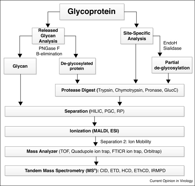
Glycosylation is a common and biologically significant post-translational modification that is found on numerous virus surface proteins (VSPs). Many of these glycans affect virulence through modulating virus receptor binding, masking antigenic sites, or by stimulating the host immune response. Mass spectrometry (MS) has arisen as a pivotal technique for the characterization of VSP glycosylation. This review will cover how MS-based analyses, such as released glycan profiles, glycan site localization, site-occupancy, and site-specific heterogeneity, are being utilized to map VSP glycosylation. Furthermore, this review will provide information on how MS glycoprofiling data are being used in conjunction with molecular and structural experiments to provide a better understanding of the role of specific glycans in VSP function.


Figures

Figure 1
Figure 1
Viral Fusion Protein Structures and N-glycosylation Sites (NGS). Viral fusion proteins are typically trimers of heterodimers with a globular head domain and a stalk/transmembrane domain. A GlucNAc1 or GlucNac2 are modeled at each NGS (Blue). (a) Lassa virus (LASV) glycoprotein (GP) complex contains 11 (NGS) with 7 in GP1 and 4 in GP2 (PDB: 5VK2) [27]. (b) Human immunodeficiency virus (HIV-1) Envelope (Env) GP contains around 29 NGS with 25 in gp120 the outer Env domain and 4 in gp41 the transmembrane domain (PDB: 5FYK) [49••]. (c) Ebola virus (EBOV) GP has 17 NGS with 15 on GP1 and 2 on GP2 (PDB: 6G9B) [101]. (d) Coronavirus (CoV) spike GP has 26 NGS with 15 in subunit 1 and 11 in subunit 2 (PDB: 6BFU) [89]. (e) Influenza virus hemagglutinin is a homotrimer with 11 NGS (PDB: 4FNK) [92].
Figure 2
Figure 2
Workflow diagram of glycoprotein analysis commonly used to characterize viral surface protein glycosylation.
Figure 3
Figure 3
Types of N-glycan profiling data. (a) Released glycan profile for HIV-1 gp120 produced in 293 T cells and treated with neuraminidase. (b) Site Occupancy data for Flu HA N-glycosylation sites [25]. (c) Tandem MS spectra confirming site localization for an HIV-1 gp120 glycopeptide after EndoH digestion. (d) Site-specific heterogeneity profile for a HIV-1 gp120 N-glycosylation site. (e) Tandem Mass spectra confirming peptide sequence in the MS1 spectra shown in panel d.
Figure 4
Figure 4
Examples of site-specific glycan profile outputs. All data obtained from publicly available HIV-1 recombinant gp120 data sets [24••,94••]. Each panel demonstrates how single-site glycan heterogeneity for 3–5 individual HIV-1 Env N-glycosylation sites are commonly represented in the literature. (a) Table of 5 NGS from three gp120 trimers indicating their predominant N-glycan type observed from site-specific MS data similar to reports from Go et al. [46]. (b) Quantitative site-specific N-glycosylation bar graph of the same NGSs categorized as oligomannose series (M5–M9), hybrids (H), and fucosylated hybrids (FH), and also by the number of branching antennae (a) of complex type glycans. A summary pie chart that compares the amount of high mannose (green) to processed glycans (pink) is provided next to each bar graph similar to data reported by Behrens et al. [94••] (c) Three of the same NGS presented as a weighted distribution graph of every oligosaccharide observed organized based on how N-glycans are processed (high-mannose glycans (yellow), hybrid glycans (green), complex glycans (blue)). A darker shading is used to indicate oligosaccharides that contain a sialic acid. (d) Relative abundance bar graph of every oligosaccharide observed at the N463 site of the WEAU.d16 and WEAU.d391from a recombinant gp120 trimer organized in the same order as the weighted distribution graph similar to Hargett et al. [24••].

Similar articles

Cited by

References

    1. Buchmann J.P., Holmes E.C. Cell walls and the convergent evolution of the viral envelope. Microbiol Mol Biol Rev. 2015;79:403–418. - PMC - PubMed
    1. Behrens A.J., Vasiljevic S., Pritchard L.K., Harvey D.J., Andev R.S., Krumm S.A., Struwe W.B., Cupo A., Kumar A., Zitzmann N. Composition and antigenic effects of individual glycan sites of a trimeric HIV-1 envelope glycoprotein. Cell Rep. 2016;14:2695–2706. - PMC - PubMed
    1. Francica J.R., Varela-Rohena A., Medvec A., Plesa G., Riley J.L., Bates P. Steric shielding of surface epitopes and impaired immune recognition induced by the Ebola virus glycoprotein. PLoS Pathog. 2010;6 - PMC - PubMed
    1. Helle F., Duverlie G., Dubuisson J. The hepatitis C virus glycan shield and evasion of the humoral immune response. Viruses. 2011;3:1909–1932. - PMC - PubMed
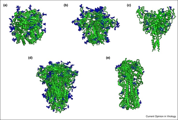
    1. Walls A.C., Tortorici M.A., Frenz B., Snijder J., Li W., Rey F.A., DiMaio F., Bosch B.J., Veesler D. Glycan shield and epitope masking of a coronavirus spike protein observed by cryo-electron microscopy. Nat Struct Mol Biol. 2016;23:899–905. - PMC - PubMed
    2. This article demonstrates how mass spectrometry N-glycan analysis can be combined with cryo-election microscopy data to characterize the human CoV spike glycoprotein trimer.

Publication types

LinkOut - more resources