Review
. 2020 Jan;13(1 Pt 2):245-257.
doi: 10.1016/j.jcmg.2018.12.034. Epub 2019 Jun 12.

Diastolic Dysfunction and Heart Failure With Preserved Ejection Fraction: Understanding Mechanisms by Using Noninvasive Methods

Affiliations
Review

Diastolic Dysfunction and Heart Failure With Preserved Ejection Fraction: Understanding Mechanisms by Using Noninvasive Methods

Masaru Obokata et al. JACC Cardiovasc Imaging. 2020 Jan.

Abstract

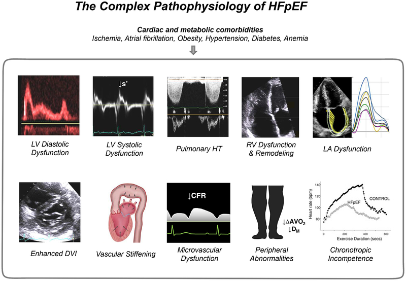
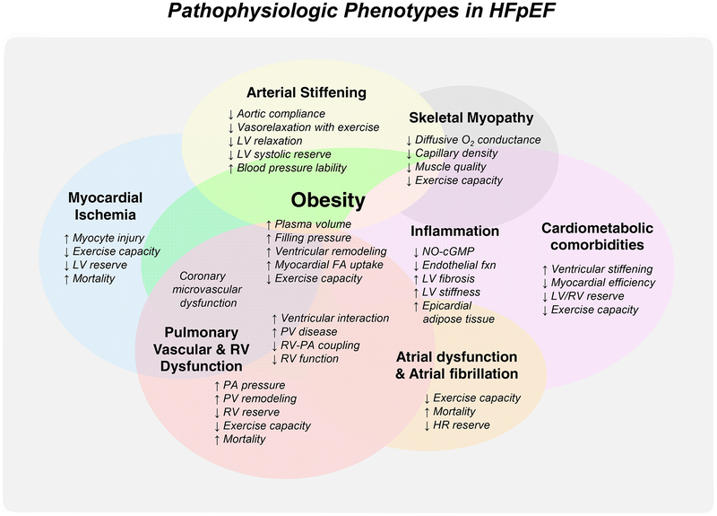
Research in the last decade has substantially advanced our understanding of the pathophysiology of heart failure with preserved ejection fraction (HFpEF). However, treatment options remain limited as clinical trials have largely failed to identify effective therapies. Part of this failure may be related to mechanistic heterogeneity. It is speculated that categorizing HFpEF patients based upon underlying pathophysiological phenotypes may represent the key next step in delivering the right therapies to the right patients. Echocardiography may provide valuable insight into both the pathophysiology and underlying phenotypes in HFpEF. Echocardiography also plays a key role in the evaluation of patients with unexplained dyspnea, where HFpEF is suspected but the diagnosis remains unknown. The combination of the E/e' ratio and right ventricular systolic pressure has recently been shown to add independent value to the diagnostic evaluation of patients suspected of having HFpEF. Finally, echocardiography enables identification of the different causes that mimic HFpEF but are treated differently, such as valvular heart disease, pericardial constriction, and high-output heart failure or infiltrative myopathies such as cardiac amyloid. This review summarizes the current understanding of the pathophysiology and phenotyping of HFpEF with particular attention to the role of echocardiography in this context.

Keywords: diagnosis; diastolic function; echocardiography; filling pressure; heart failure; noninvasive.


Figures

Figure 1.
Figure 1.. The Complex Pathophysiology of HFpEF:
Patients with HFpEF display impairments beyond diastolic dysfunction. The primary involvement in any one component may vary between patients. See text for details. Portions of this figure were adapted with permission from references and . ΔAVO2, arterial-venous oxygen content difference; CFR, coronary flow reserve; DM, diffusive oxygen conductance; DVI, diastolic ventricular interdependence; LA, left atrial; LV, left ventricular; s’, systolic mitral annular tissue velocity; RV, right ventricular; and HT, hypertension.
Figure 2.
Figure 2.. HFpEF with Pulmonary Vascular Disease:
Invasive pressure tracings and right ventricular outflow pulse-wave Doppler imaging in a HFpEF patient with severe pulmonary vascular disease. (A) There is severe elevation in mean pulmonary artery pressure [PAP] due to marked elevation in pulmonary capillary wedge pressure [PCWP] but also coexisting elevation in pulmonary vascular resistance (PVR 11.0 WU). Clinical right heart failure is also present, as evidenced by high right atrial pressure [RAP] (20 mmHg). (B) Right ventricular outflow pulse-wave Doppler in this patient demonstrates a distinct mid-systolic notch, presumably caused by backward traveling compression wave, with abbreviation of acceleration time. Abbreviations as in Figure 1.
Figure 3.
Figure 3.. Obesity-related HFpEF with Enhanced Pericardial Restraint:
(A) Diastolic pressure-volume relationships of patients with HFpEF (mean BMI 30.2) at rest (black) and with exercise (red). With exercise, the diastolic pressure-volume relationship (DPVR) curve shifts upward. While chamber stiffness (linearized slope of the diastolic pressure-volume relationship) increases significantly with exercise in HFpEF, the majority of the increase in LV end diastolic pressure is related to parallel shift upward in the DPVR, suggesting increased external forces from the right heart and pericardium. Adapted with permission from reference . (B) Parasternal short axis-views at end-diastole in a patient with obese HFpEF (body mass index 42 kg/m2) demonstrating worsening pericardial restraint from rest to exercise. Note the D-shaped septum during 20 watts supine ergometer exercise with increase in the LV eccentricity index. Abbreviations as in Figure 1.
Figure 4.
Figure 4.. The H2FPEF score to Facilitate Diagnostic Evaluation in HFpEF:
In this score, the echocardiographic parameters that were independently predictive for HFpEF (E/e’ >9 and right ventricular systolic pressure >35mmHg) are incorporated with clinical characteristics to determine the probability that HFpEF is present in patients presenting with unexplained dyspnea. Adapted with permission from reference . Abbreviations as in Figure 1.
Central illustration.
Central illustration.. The Phenotypes of HFpEF:
There are key clinical phenotypes that demonstrate distinct pathophysiologic features compared to “garden variety of HFpEF”. FA, fatty acid; fxn, function; HFpEF, heart failure with preserved ejection fraction; HR, heart rate; LA, left atrial; LV, left ventricular; NO-cGMP, nitric oxide-cyclic guanosine monophosphate signaling; O2, oxygen; PA, pulmonary artery; PH, pulmonary hypertension; PV, pulmonary vascular; and RV, right ventricular.

Comment in

  • Diastolic Stress Testing: Have You Considered Isometric Handgrip Echocardiography?
    Samuel TJ, Haykowsky MJF, Sarma S, Nelson MD. Samuel TJ, et al. JACC Cardiovasc Imaging. 2019 Oct;12(10):2095-2097. doi: 10.1016/j.jcmg.2019.07.023. JACC Cardiovasc Imaging. 2019. PMID: 31601382 Free PMC article. No abstract available.
  • The Authors Reply.
    Obokata M, Reddy YNV, Borlaug BA. Obokata M, et al. JACC Cardiovasc Imaging. 2019 Oct;12(10):2098-2099. doi: 10.1016/j.jcmg.2019.07.024. JACC Cardiovasc Imaging. 2019. PMID: 31601384 No abstract available.

References

    1. Borlaug BA. The pathophysiology of heart failure with preserved ejection fraction. Nat Rev Cardiol. 2014;11:507–15. - PubMed
    1. Shah SJ, Kitzman DW, Borlaug BA, et al. Phenotype-Specific Treatment of Heart Failure With Preserved Ejection Fraction: A Multiorgan Roadmap. Circulation. 2016;134:73–90. - PMC - PubMed
    1. Nagueh SF, Smiseth OA, Appleton CP, et al. Recommendations for the Evaluation of Left Ventricular Diastolic Function by Echocardiography: An Update from the American Society of Echocardiography and the European Association of Cardiovascular Imaging. J Am Soc Echocardiogr. 2016;29:277–314. - PubMed
    1. Brutsaert DL, Sys SU, Gillebert TC. Diastolic failure: pathophysiology and therapeutic implications. J Am Coll Cardiol. 1993;22:318–25. - PubMed
    1. Gaasch WH, Zile MR. Left ventricular diastolic dysfunction and diastolic heart failure. Annu Rev Med. 2004;55:373–94. - PubMed

Publication types