

An expanded proteome of cardiac t-tubules
- PMID: 31202980
- PMCID: PMC6732025
- DOI: 10.1016/j.carpath.2019.05.001
An expanded proteome of cardiac t-tubules
Abstract
Background: Transverse tubules (t-tubules) are important structural elements, derived from sarcolemma, found on all striated myocytes. These specialized organelles create a scaffold for many proteins crucial to the effective propagation of signal in cardiac excitation-contraction coupling. The full protein composition of this region is unknown.
Methods: We characterized the t-tubule subproteome using 52,033 immunohistochemical images covering 13,203 proteins from the Human Protein Atlas (HPA) cardiac tissue microarrays. We used HPASubC, a suite of Python tools, to rapidly review and classify each image for a specific t-tubule staining pattern. The tools Gene Cards, String 11, and Gene Ontology Consortium as well as literature searches were used to understand pathways and relationships between the proteins.
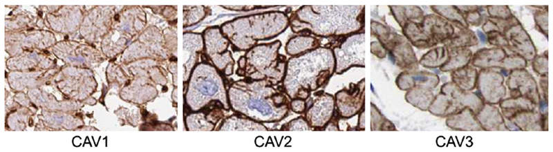
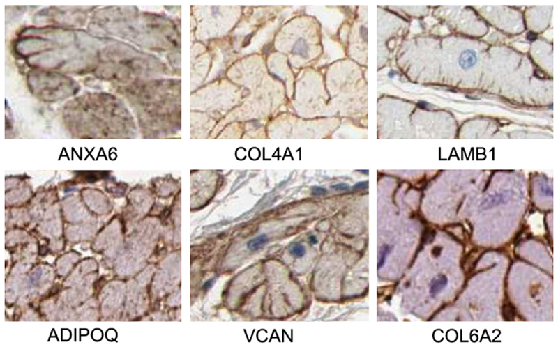
Results: There were 96 likely t-tubule proteins identified by HPASubC. Of these, 12 were matrisome proteins and 3 were mitochondrial proteins. A separate literature search identified 50 known t-tubule proteins. A comparison of the 2 lists revealed only 17 proteins in common, including 8 of the matrisome proteins. String11 revealed that 94 of 127 combined t-tubule proteins generated a single interconnected network.
Conclusion: Using HPASubC and the HPA, we identified 78 novel, putative t-tubule proteins and validated 17 within the literature. This expands and improves our knowledge of this important subcellular structure of the cardiac myocyte. This information can be used to identify new structural targets involved in excitation-contraction coupling that may be altered in disease.
Keywords: Caveolin; Proteomics; T-tubule.
Copyright © 2019 Elsevier Inc. All rights reserved.
Conflict of interest statement
Conflict of interest
The authors declare no conflict of interest.
Figures

References
-
- Lindner E [Submicroscopic morphology of the cardiac muscle]. Zeitschrift fur Zellforschung und mikroskopische Anatomie. 1957;45:702–46. - PubMed
MeSH terms
Substances
Grants and funding
LinkOut - more resources
Full Text Sources
