

When you're strange: Unusual features of the MUTYH glycosylase and implications in cancer
- PMID: 31203172
- PMCID: PMC6812671
- DOI: 10.1016/j.dnarep.2019.05.005
When you're strange: Unusual features of the MUTYH glycosylase and implications in cancer
Abstract
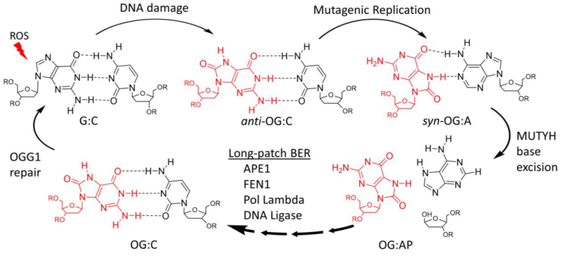
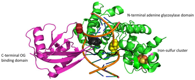
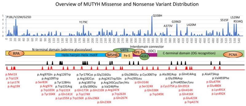
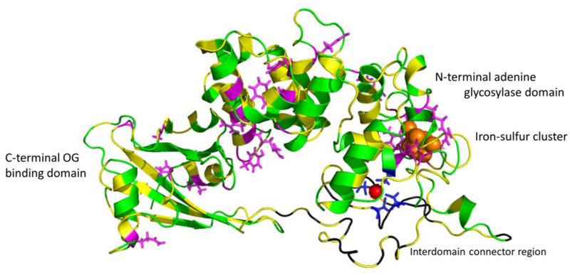
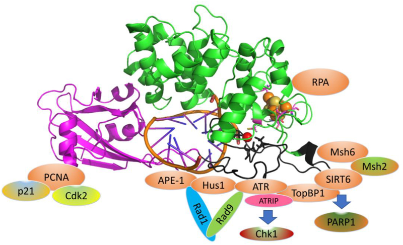
MUTYH is a base-excision repair glycosylase that removes adenine opposite 8-oxoguanine (OG). Variants of MUTYH defective in functional activity lead to MUTYH-associated polyposis (MAP), which progresses to cancer with very high penetrance. Whole genome and whole exome sequencing studies have found MUTYH deficiencies in an increasing number of cancer types. While the canonical OG:A repair activity of MUTYH is well characterized and similar to bacterial MutY, here we review more recent evidence that MUTYH has activities independent of OG:A repair and appear centered on the interdomain connector (IDC) region of MUTYH. We summarize evidence that MUTYH is involved in rapid DNA damage response (DDR) signaling, including PARP activation, 9-1-1 and ATR signaling, and SIRT6 activity. MUTYH alters survival and DDR to a wide variety of DNA damaging agents in a time course that is not consistent with the formation of OG:A mispairs. Studies that suggest MUTYH inhibits the repair of alkyl-DNA damage and cyclopyrimidine dimers (CPDs) is reviewed, and evidence of a synthetic lethal interaction with mismatch repair (MMR) is summarized. Based on these studies we suggest that MUTYH has evolved from an OG:A mispair glycosylase to a multifunctional scaffold for DNA damage response signaling.
Keywords: Base excision repair; DNA damage response; Glycosylase; MUTYH; MutY; Oxidative DNA damage.
Copyright © 2019 Elsevier B.V. All rights reserved.
Conflict of interest statement
Figures
Similar articles
-
FSHing for DNA Damage: Key Features of MutY Detection of 8-Oxoguanine:Adenine Mismatches.Acc Chem Res. 2024 Apr 2;57(7):1019-1031. doi: 10.1021/acs.accounts.3c00759. Epub 2024 Mar 12. Acc Chem Res. 2024. PMID: 38471078 Free PMC article. Review.
-
Structure-Activity Relationships Reveal Key Features of 8-Oxoguanine: A Mismatch Detection by the MutY Glycosylase.ACS Chem Biol. 2017 Sep 15;12(9):2335-2344. doi: 10.1021/acschembio.7b00389. Epub 2017 Aug 8. ACS Chem Biol. 2017. PMID: 28723094 Free PMC article.
-
Genomic 8-oxoguanine modulates gene transcription independent of its repair by DNA glycosylases OGG1 and MUTYH.Redox Biol. 2025 Feb;79:103461. doi: 10.1016/j.redox.2024.103461. Epub 2024 Dec 5. Redox Biol. 2025. PMID: 39662289 Free PMC article.
-
Distinct functional consequences of MUTYH variants associated with colorectal cancer: Damaged DNA affinity, glycosylase activity and interaction with PCNA and Hus1.DNA Repair (Amst). 2015 Oct;34:39-51. doi: 10.1016/j.dnarep.2015.08.001. Epub 2015 Aug 12. DNA Repair (Amst). 2015. PMID: 26377631 Free PMC article.
-
Repair of 8-oxoG:A mismatches by the MUTYH glycosylase: Mechanism, metals and medicine.Free Radic Biol Med. 2017 Jun;107:202-215. doi: 10.1016/j.freeradbiomed.2017.01.008. Epub 2017 Jan 10. Free Radic Biol Med. 2017. PMID: 28087410 Free PMC article. Review.
Cited by
-
FSHing for DNA Damage: Key Features of MutY Detection of 8-Oxoguanine:Adenine Mismatches.Acc Chem Res. 2024 Apr 2;57(7):1019-1031. doi: 10.1021/acs.accounts.3c00759. Epub 2024 Mar 12. Acc Chem Res. 2024. PMID: 38471078 Free PMC article. Review.
-
Saturation mapping of MUTYH variant effects using DNA repair reporters.bioRxiv [Preprint]. 2025 Mar 6:2025.03.01.640912. doi: 10.1101/2025.03.01.640912. bioRxiv. 2025. Update in: Am J Hum Genet. 2025 Jul 24:S0002-9297(25)00275-7. doi: 10.1016/j.ajhg.2025.07.005. PMID: 40093110 Free PMC article. Updated. Preprint.
-
Interplay of Guanine Oxidation and G-Quadruplex Folding in Gene Promoters.J Am Chem Soc. 2020 Jan 22;142(3):1115-1136. doi: 10.1021/jacs.9b11050. Epub 2020 Jan 9. J Am Chem Soc. 2020. PMID: 31880930 Free PMC article.
-
OGG1 and MUTYH repair activities promote telomeric 8-oxoguanine induced senescence in human fibroblasts.Nat Commun. 2025 Jan 21;16(1):893. doi: 10.1038/s41467-024-55638-4. Nat Commun. 2025. PMID: 39837827 Free PMC article.
-
Overview of Wnt/β-Catenin Pathway and DNA Damage/Repair in Cancer.Biology (Basel). 2025 Feb 11;14(2):185. doi: 10.3390/biology14020185. Biology (Basel). 2025. PMID: 40001953 Free PMC article. Review.
References
-
- David SS and Williams SD, Chemistry of Glycosylases and Endonucleases Involved in Base-Excision Repair. Chem Rev, 1998. 98(3): p. 1221–1262 DOI: cr980321h [pii]. - PubMed
-
- Steenken S and Jovanovic SV, How Easily Oxidizable Is DNA? One-Electron Reduction Potentials of Adenosine and Guanosine Radicals in Aqueous Solution. Journal of the American Chemical Society, 1997. 119(3): p. 617–618.
Publication types
MeSH terms
Substances
Grants and funding
LinkOut - more resources
Full Text Sources
Research Materials
Miscellaneous
