

Follicular dendritic cells display microvesicle-associated LMP1 in reactive germinal centers of EBV+ classic Hodgkin lymphoma
- PMID: 31203443
- PMCID: PMC6647529
- DOI: 10.1007/s00428-019-02605-w
Follicular dendritic cells display microvesicle-associated LMP1 in reactive germinal centers of EBV+ classic Hodgkin lymphoma
Abstract
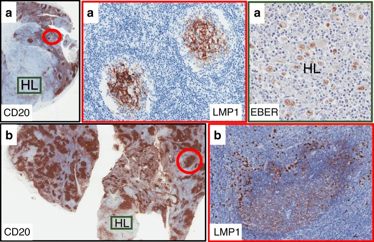
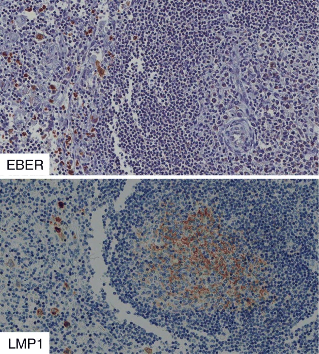
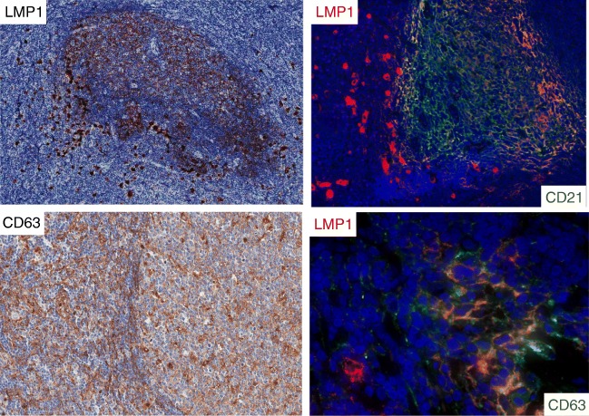
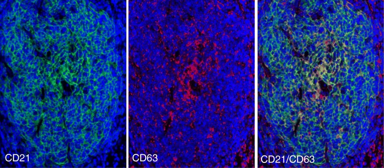
Expression of the latent membrane protein-1 (LMP1) of Epstein-Barr virus (EBV) was investigated in 153 cases of EBV+ classic Hodgkin lymphoma (cHL); 120 cases were pediatric patients (< 14 years of age) from Iraq, and 33 cases were adult patients from Italy. We describe for the first time the presence of LMP1 protein in EBV-encoded RNA (EBER)-negative follicular dendritic cells (FDCs) of reactive germinal centers (GC) associated with EBV+ cHL. Presence of LMP1+ GCs was independent of geographic region and age of patients. Variable numbers of reactive GCs were present in 22.2% of cases (34 of 153), whereas LMP1 staining of FDCs was present in about a third of cases (10 of 34) with reactive GC. Most cases with LMP1+ GC were mixed-cellularity (MC) subtype, but some nodular sclerosis (NS) was also present. GC cells with LMP1+ FDCs were surrounded by numerous EBV-infected cells which were positive for EBER, LMP1, and CD30. Double immunolocalization analysis revealed that LMP1 was associated with CD63, an exosomal marker, and with CD21. The possibility is discussed that peri-follicular EBV-infected cells release LMP1 protein, perhaps through exosomes, and that the protein is then captured by FDCs and is presented to EBER-negative GC B cells.
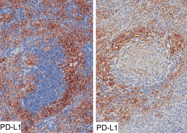
Keywords: Classic Hodgkin lymphoma (cHL); Epstein-Barr virus (EBV); Exosomes and microvesicles; Follicular dendritic cells (FDCs); Latent membrane protein-1 (LMP1); Programmed death ligand 1 (PD-L1).
Conflict of interest statement
All authors declare that they have no conflict of interest.
Figures
Similar articles
-
Epstein-Barr virus latent membrane protein-1 upregulates autophagy and promotes viability in Hodgkin lymphoma: Implications for targeted therapy.Cancer Sci. 2021 Apr;112(4):1589-1602. doi: 10.1111/cas.14833. Epub 2021 Feb 16. Cancer Sci. 2021. PMID: 33525055 Free PMC article.
-
Latent membrane protein 1 (LMP1) expression in Hodgkin lymphoma and its correlation with clinical and histologic parameters.World J Surg Oncol. 2017 Apr 20;15(1):89. doi: 10.1186/s12957-017-1147-y. World J Surg Oncol. 2017. PMID: 28427406 Free PMC article.
-
[Clinicopathologic features and association with Epstein-Barr virus infection in 235 cases of Hodgkin lymphoma from northern China].Zhonghua Bing Li Xue Za Zhi. 2015 Feb;44(2):84-9. Zhonghua Bing Li Xue Za Zhi. 2015. PMID: 25916637 Chinese.
-
LMP1 and Dynamic Progressive Telomere Dysfunction: A Major Culprit in EBV-Associated Hodgkin's Lymphoma.Viruses. 2017 Jun 27;9(7):164. doi: 10.3390/v9070164. Viruses. 2017. PMID: 28654015 Free PMC article. Review.
-
Effect of latent membrane protein 1 expression on overall survival in Epstein-Barr virus-associated cancers: a literature-based meta-analysis.Oncotarget. 2015 Oct 6;6(30):29311-23. doi: 10.18632/oncotarget.4906. Oncotarget. 2015. PMID: 26336130 Free PMC article. Review.
Cited by
-
CD169+ subcapsular sinus macrophage-derived microvesicles are associated with light zone follicular dendritic cells.Eur J Immunol. 2022 Oct;52(10):1581-1594. doi: 10.1002/eji.202249879. Epub 2022 Aug 10. Eur J Immunol. 2022. PMID: 35907260 Free PMC article.
-
Role of Exosomes in Pharyngucutaneous Fistula After Total Laryngectomy.Int J Nanomedicine. 2022 Sep 12;17:4119-4135. doi: 10.2147/IJN.S372042. eCollection 2022. Int J Nanomedicine. 2022. PMID: 36118178 Free PMC article. Review.
-
Epstein-Barr Functional Mimicry: Pathogenicity of Oncogenic Latent Membrane Protein-1 in Systemic Lupus Erythematosus and Autoimmunity.Front Immunol. 2021 Feb 3;11:606936. doi: 10.3389/fimmu.2020.606936. eCollection 2020. Front Immunol. 2021. PMID: 33613527 Free PMC article.
-
Comprehensive global collaboration in the care of 1182 pediatric oncology patients over 12 years: The Iraqi-Italian experience.Cancer Med. 2023 Jan;12(1):256-265. doi: 10.1002/cam4.4892. Epub 2022 Jun 3. Cancer Med. 2023. PMID: 35661436 Free PMC article.
-
Epstein-Barr Virus-Associated Malignancies and Immune Escape: The Role of the Tumor Microenvironment and Tumor Cell Evasion Strategies.Cancers (Basel). 2021 Oct 16;13(20):5189. doi: 10.3390/cancers13205189. Cancers (Basel). 2021. PMID: 34680337 Free PMC article. Review.
References
-
- Stein H, Pileri SA, Weiss LM, Poppema S, Gascoyne RD, Jaffe ES, et al. Hodgkin lymphomas: introduction. In: Swerdlow SH, Campo E, Harris NL, et al., editors. WHO classification of tumours of haematopoietic and lymphoid tissues, revised. 4. Lyon: IARC; 2017. pp. 424–430.
MeSH terms
Substances
LinkOut - more resources
Full Text Sources
Medical
Research Materials
Miscellaneous
