. 2019 Jun 1;75(Pt 6):405-411.
doi: 10.1107/S2053230X19006046. Epub 2019 May 10.

Structures of soluble rabbit neprilysin complexed with phosphoramidon or thiorphan

Affiliations

Structures of soluble rabbit neprilysin complexed with phosphoramidon or thiorphan

Shaunivan L Labiuk et al. Acta Crystallogr F Struct Biol Commun. .

Abstract

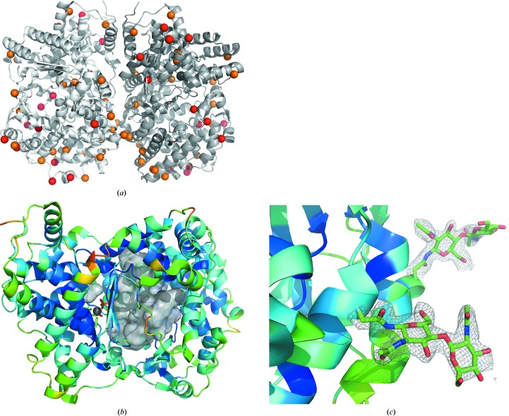
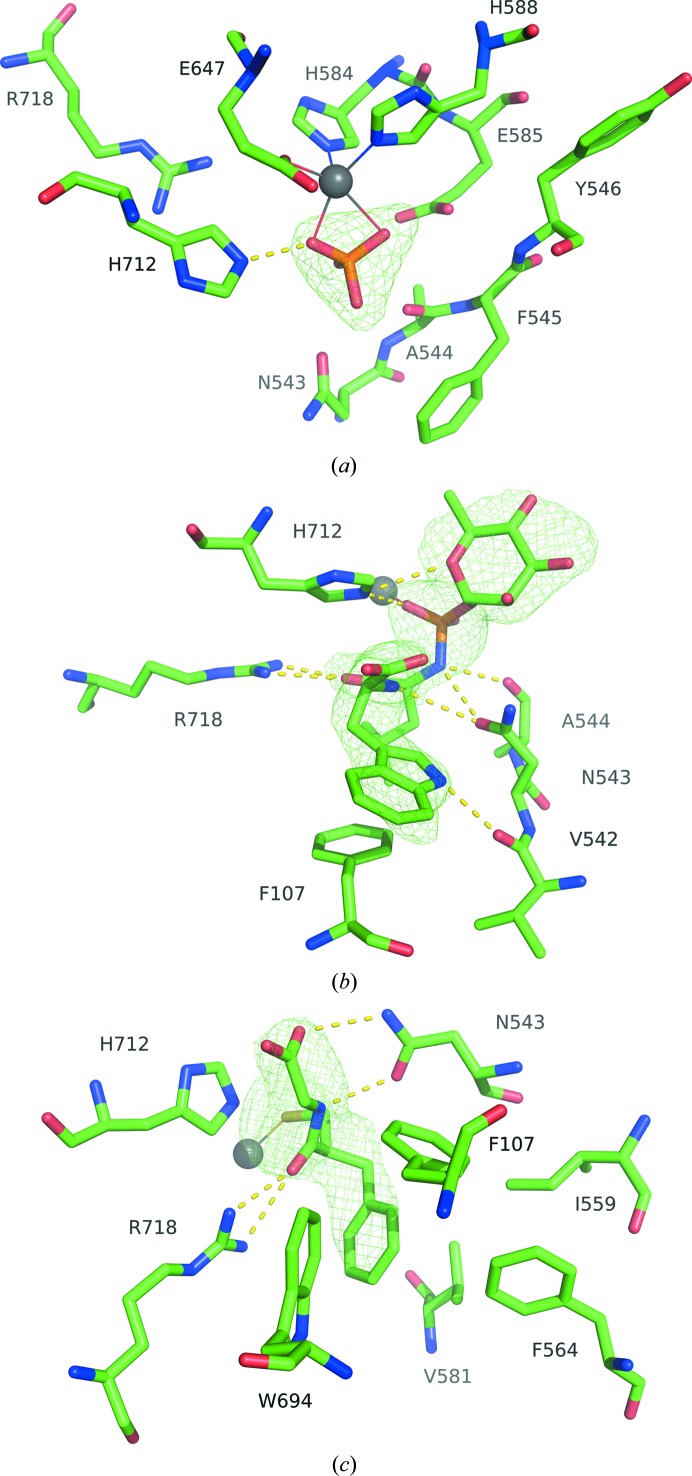
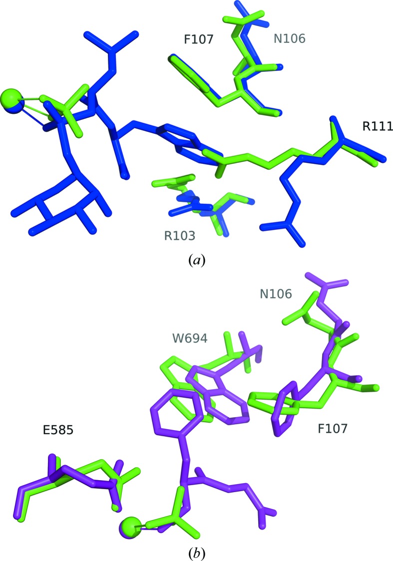
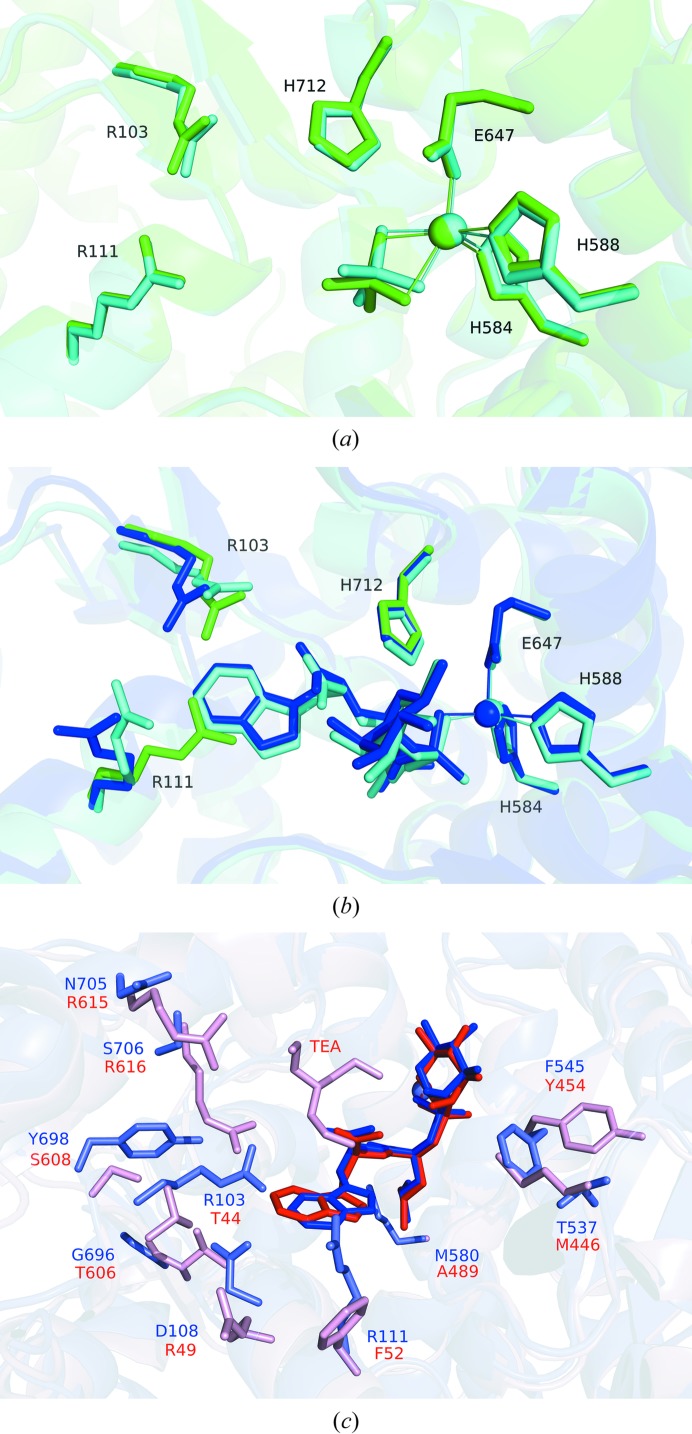
Neutral endopeptidase (neprilysin; NEP) is a proteinase that cleaves a wide variety of peptides and has been implicated in Alzheimer's disease, cardiovascular conditions, arthritis and other inflammatory diseases. The structure of the soluble extracellular domain (residues 55-750) of rabbit neprilysin was solved both in its native form at 2.1 Å resolution, and bound to the inhibitors phosphoramidon and thiorphan at 2.8 and 3.0 Å resolution, respectively. Consistent with the extracellular domain of human neprilysin, the structure reveals a large central cavity which contains the active site and the location for inhibitor binding.

Keywords: neprilysin; neutral endopeptidase; phosphoramidon; thiorphan.


Figures

Figure 1
Figure 1
(a) Ribbon representation of the asymmetric unit of native rabbit neprilysin. The moiety on the left (light grey) is designated as chain A, while chain B is on the right (dark grey). Red spheres represent the locations of residues that differ from human neprilysin, whereas orange spheres represent residues that are similar but non-identical. The active-site zinc is represented by a silver sphere. (b) Ribbon diagram of chain A coloured according to α-carbon B factors. The large central cavity is depicted using a grey surface representation. The disordered loop region is situated on the front surface of the cavity depiction, from the perspective of the viewer, and represents residues near 530–537. Zinc is represented by a silver sphere at the active site, bound to phosphate. (c) Examples of the quality of 2F oF c maps (grey) contoured at 1σ around the N-linked acetylglucosamine residues at sites Asn285 (foreground) and Asn311 (background).
Figure 2
Figure 2
(a) The active site of native neprilysin, with zinc represented as a silver sphere coordinated to His584, His588, Glu647 and phosphate ion. (b) Active site of neprilysin bound to phosphoramidon. The His584, His588 and Glu647 coordinations of Zn2+ are omitted for clarity. (c) Active site of the thiorphan complex. Ligand-omit F oF c density maps are contoured at 3σ (green).
Figure 3
Figure 3
(a) Comparison of the phosphoramidon-bound structure (blue) with the native structure (green). The ligand environment is very similar in both cases, with the notable exceptions of residues Arg111 and Arg103, which are displaced in order to accommodate the large ring of phosphoramidon. (b) Comparison of the thiorphan-bound structure (purple) with the native structure. In this case, there are adjustments in Trp694 and Phe107 to accommodate the aromatic ring of thiorphan. Spheres represent zinc. Alignments were performed against the core amino-acid residues coordinated to the metal ion using the align algorithm in the PyMOL molecular-graphics system (v.1.8; Schrödinger), which was used to produce the figures.
Figure 4
Figure 4
(a) Overlay of the native rabbit structure (green) with that of its human counterpart (cyan; PDB entry 6gid; Moss et al., 2018 ▸). (b) Overlay of the phosphoramidon-bound rabbit structure (dark blue) with that of its human counterpart (cyan; PDB entry 1dmt; Oefner et al., 2000 ▸). Residues Arg111, Arg103 and His712 from the native phosphate-bound rabbit structure are depicted (green) to highlight the differences in side-chain orientations. (c) Overlay of the phosphoramidon-bound rabbit neprilysin structure (blue) with Zmp1 from M. tuberculosis (red; PDB entry 3zuk; Ferraris et al., 2011 ▸). Phosphoramidon is coloured a darker blue or red, respectively, for clarity. TEA represents N,N′,N′′-triethanolamine in the Zmp1 structure.

References

    1. Adams, P. D., Afonine, P. V., Bunkóczi, G., Chen, V. B., Davis, I. W., Echols, N., Headd, J. J., Hung, L.-W., Kapral, G. J., Grosse-Kunstleve, R. W., McCoy, A. J., Moriarty, N. W., Oeffner, R., Read, R. J., Richardson, D. C., Richardson, J. S., Terwilliger, T. C. & Zwart, P. H. (2010). Acta Cryst. D66, 213–221. - PMC - PubMed
    1. Afonine, P. V., Grosse-Kunstleve, R. W., Echols, N., Headd, J. J., Moriarty, N. W., Mustyakimov, M., Terwilliger, T. C., Urzhumtsev, A., Zwart, P. H. & Adams, P. D. (2012). Acta Cryst. D68, 352–367. - PMC - PubMed
    1. Birner, C., Ulucan, C., Bratfisch, M., Götz, T., Dietl, A., Schweda, F., Riegger, G. A. & Luchner, A. (2012). Naunyn Schmiedebergs Arch. Pharmacol. 385, 1117–1125. - PubMed
    1. Caragea, C., Sinapov, J., Silvescu, A., Dobbs, D. & Honavar, V. (2007). BMC Bioinformatics, 8, 438. - PMC - PubMed
    1. Converse, P. J., Dannenberg, A. M. Jr, Estep, J. E., Sugisaki, K., Abe, Y., Schofield, B. H. & Pitt, M. L. (1996). Infect. Immun. 64, 4776–4787. - PMC - PubMed