. 2019 Jun 17;129(7):2775-2791.
doi: 10.1172/JCI123825.

Shear stress-induced endothelial adrenomedullin signaling regulates vascular tone and blood pressure

Affiliations

Shear stress-induced endothelial adrenomedullin signaling regulates vascular tone and blood pressure

Andras Iring et al. J Clin Invest. .

Abstract

Hypertension is a primary risk factor for cardiovascular diseases including myocardial infarction and stroke. Major determinants of blood pressure are vasodilatory factors such as nitric oxide (NO) released from the endothelium under the influence of fluid shear stress exerted by the flowing blood. Several endothelial signaling processes mediating fluid shear stress-induced formation and release of vasodilatory factors have been described. It is, however, still poorly understood how fluid shear stress induces these endothelial responses. Here we show that the endothelial mechanosensitive cation channel PIEZO1 mediated fluid shear stress-induced release of adrenomedullin, which in turn activated its Gs-coupled receptor. The subsequent increase in cAMP levels promoted the phosphorylation of endothelial NO synthase (eNOS) at serine 633 through protein kinase A (PKA), leading to the activation of the enzyme. This Gs/PKA-mediated pathway synergized with the AKT-mediated pathways leading to eNOS phosphorylation at serine 1177. Mice with endothelium-specific deficiency of adrenomedullin, the adrenomedullin receptor, or Gαs showed reduced flow-induced eNOS activation and vasodilation and developed hypertension. Our data identify fluid shear stress-induced PIEZO1 activation as a central regulator of endothelial adrenomedullin release and establish the adrenomedullin receptor and subsequent Gs-mediated formation of cAMP as a critical endothelial mechanosignaling pathway regulating basal endothelial NO formation, vascular tone, and blood pressure.

Keywords: G-protein coupled receptors; Hypertension; Vascular Biology; endothelial cells.


Conflict of interest statement

Conflict of interest: The authors have declared that no conflict of interest exists.

Figures

Figure 1
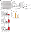
Figure 1. Gs mediates flow-induced cAMP formation and specific eNOS phosphorylation at serine 635 in BAECs.
(A) BAECs were pretreated with the PKA inhibitor PKI (1 μM) or solvent (control) for 20 minutes and were then kept under static conditions or were exposed to flow (15 dyn/cm2) for 30 minutes. Total and phosphorylated eNOS was determined by immunoblotting. Graphs show the densitometric evaluation (n = 3). (BG) BAECs were transfected with scrambled (control) siRNA or siRNA directed against Gαs as indicated and were exposed to flow for 30 minutes or the indicated time periods (15 dyn/cm2 in BE) or were incubated with isoproterenol (Isopr., 10 μM, 10 minutes) or VEGF (50 ng/ml, 10 minutes) (F and G). (B) Intracellular cAMP levels were determined (n = 4, control; n = 6, Gαs). (C) Phosphorylation of various eNOS sites was determined by LC-MS/MS under static conditions and after application of shear stress (15 dyn/cm2) for 30 minutes. Intensity values of the phoshpo-sites shown were normalized to eNOS intensity and thus protein abundance in the proteome. Bar diagrams show the fold change of the corrected ratios of flow versus static conditions (n = 2–4). Logarithmization of the y axis emphasizes the directional deviation from the static condition. (DG) Total and phosphorylated eNOS and AKT were analyzed by immunoblotting. Graphs and bar diagrams show the densitometric evaluation (n = 3). Analysis of Gαs and GAPDH expression served as control (D). Data represent the mean ± SEM; *P ≤ 0.05, **P ≤ 0.01, ***P ≤ 0.001, 1-way ANOVA with Tukey’s post hoc test (A, F, and G), 2-way ANOVA with Bonferroni’s post hoc test (B and E).
Figure 2
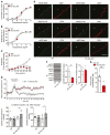
Figure 2. Gs and Gq/G11 additively regulate endothelial eNOS activity and NO formation.
BAECs were transfected with control siRNA (control) or siRNA directed against Gαs or Gαq and Gα11 as indicated. (A and B) Cells were exposed to 15 dyn/cm2 for the indicated time periods, and phosphorylation of eNOS and AKT was determined by immunoblotting (A). Bar diagrams show the densitometric evaluation (n = 3). (B) Nitrate and nitrite concentration in the cell culture medium (n = 9, control; n = 6, Gαq/Gα11; n = 7, Gαs; n = 5, Gαq/Gα11 + Gαs) (left), and percentage contribution of Gs and Gq/G11 to the maximal nitrate and nitrite formation (right). (C) HAECs were transfected with siRNA directed against eNOS, and eNOS WT or the eNOS phospho-site mutants S1177A and S633A were expressed by lentiviral transduction. Thereafter, the effect of flow (15 dyn/cm2 for 30 minutes) on nitrate/nitrite concentration in the cell culture medium was determined (n = 3). Data represent the mean ± SEM; *,P ≤ 0.05, **P ≤ 0.01, ***,###,‡‡‡P ≤ 0.001, 2-way ANOVA with Bonferroni’s post hoc test; * and #, compared with control; , compared with siRNA against Gαs; compared with siRNA against Gαq/Gα11.
Figure 3
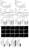
Figure 3. Endothelial Gαs is required for flow-induced vasodilation and blood pressure control.
(A and B) Aorta segments from WT or induced EC-Gαs-KO mice were incubated with increasing concentrations of phenylephrine (A) or were precontracted with 10 μM phenylephrine and exposed to acetylcholine (B), and vascular tension was determined (n = 5). (C) Mesenteric arteries isolated from tamoxifen-treated WT or EC-Gαs-KO mice were precontracted with 100 nM U46619 and exposed to stepwise increases in perfusion flow. Shown is the flow-induced vasodilation as a percentage of the passive vessel diameter (n = 11, WT; n = 7, EC-Gαs-KO). (D) Mean arterial blood pressure measured in conscious, freely moving WT (n = 11) and EC-Gαs-KO mice (n = 7) before, during, and after tamoxifen treatment. Bar diagrams show systolic and diastolic arterial blood pressure during 4 days before tamoxifen treatment and during the second week after induction. (E) Immuno-confocal microscopy images of aortae isolated from WT or EC-Gαs-KO mice stained with antibodies directed against eNOS, phosphorylated eNOS (S632 or S1176, green), and the endothelial marker CD31 (red). Shown is 1 of 2 independent experiments. Scale bar: 25 μm. (F) Phosphorylation of eNOS at serine 632 and 1176 in lysates from mesenteric arteries prepared from tamoxifen-treated WT and EC-Gαs-KO mice. Graphs show the densitometric evaluation (n = 3). (G) Plasma nitrate and nitrite (NOx) levels in WT (n = 8) and EC-Gαs-KO mice (n = 8) 10 days after induction. Data represent the mean ± SEM; *P ≤ 0.05, **P ≤ 0.01, ***P ≤ 0.001, 2-way ANOVA with Bonferroni’s post hoc test (AC), paired 2-tailed Student’s t test (D), or unpaired 2-tailed Student’s t test (F and G).
Figure 4
Figure 4. The adrenomedullin receptor CALCRL mediates endothelial fluid shear stress response in vitro.
(A) BAECs were transfected with siRNAs against Gαs (GNAS) and the indicated GPCRs and were then exposed to flow (15 dyn/cm2) for 30 minutes. Shown is the ratio of flow-induced eNOS S635 phosphorylation in cells transfected with a control siRNA and with a siRNA against a GPCR. The plot shows the ranked average ratios of 3 independent experiments. (BE) BAECs were transfected with control siRNA, siRNA directed against Gαs, or an alternative siRNA against CALCRL and GPR146 (B, D, and E) or were pretreated with the adrenomedullin receptor antagonist AM22–52 (1 μM) (C) and were exposed to flow (15 dyn/cm2) for 30 minutes (B and C) or to VEGF (50 ng/ml, 10 minutes) (D) or to isoproterenol (Isopr., 10 μM, 10 minutes) (E). Total and phosphorylated eNOS and AKT were determined by immunoblotting. Graphs show the densitometric evaluation of blots (n = 3). Data represent the mean ± SEM; *P ≤ 0.05, **P ≤ 0.01, ***P ≤ 0.001, 1-way ANOVA with Dunnett’s post hoc test (A), 2-way ANOVA with Bonferroni’s post hoc test (B), or 1-way ANOVA with Tukey’s post hoc test (CE).
Figure 5
Figure 5. Role of adrenomedullin in flow-induced eNOS regulation.
(A) BAECs were treated with adrenomedullin (ADM, 10 nM, 5 minutes), calcitonin gene–related peptide (CGRP; 10 nM, 10 minutes), or adrenomedullin-2 (ADM2, 1 nM, 3 minutes), and phosphorylation of eNOS S635 was determined by immunoblotting. Bar diagram shows the densitometric evaluation (n = 3). (BD and FH) BAECs (B, C, and FH) or HAECs (D) were transfected with scrambled (control) siRNA or siRNA directed against Gαs, CALCRL, eNOS, or ADM as indicated. In D, eNOS WT or the eNOS phospho-site mutants S1177A and S633A were expressed by lentiviral transduction. Cells were treated with adrenomedullin (ADM, 10 nM, 5 minutes [B] or 30 minutes [D]) or adrenomedullin-2 (ADM2, 1 nM, 3 minutes, C) or were exposed to 15 dyn/cm2 for 30 minutes or for the indicated time periods (FH). Phosphorylation of eNOS at serine 635 and serine 1179 was determined by immunoblotting (B, C, and F). Intracellular cAMP concentration (n = 7, control; n = 6, CALCRL; n = 8, ADM) (G) or nitrate and nitrite concentration in the cell culture medium (n = 6 [D]; n = 13, control; n = 4, CALCRL; n = 5, ADM [H]) was determined. Bar diagrams in B, C, and F show densitometric evaluation of immunoblots (n = 3). (E) Expression of ADM, CGRP (CALCA), ADM2, and RAMP1–3 RNA in BAECs and HUVECs (n = 4). Data represent the mean ± SEM; *P ≤ 0.05, **P ≤ 0.01, ***P ≤ 0.00, 2-way ANOVA with Bonferroni’s post hoc test (AD, G, and H) or 1-way ANOVA with Tukey’s post hoc test (F).
Figure 6
Figure 6. Mechanism of flow-induced adrenomedullin release from endothelial cells.
(AF) BAECs (AC, E, and F) or HAECs (D) were transfected with scrambled (control) siRNA or siRNA directed against Gαs, PIEZO1, or eNOS as indicated (AD and F) or were pretreated in the absence or presence of BAPTA-AM (100 μM) for 20 minutes (E). In D, eNOS WT or the eNOS phospho-site mutants S1177A and S633A were expressed by lentiviral transduction. Cells were then exposed to flow (15 dyn/cm2) (A, B, and E) for the indicated time periods or were incubated with Yoda1 (1 μM) for 30 minutes (C, D, and F). Thereafter the adrenomedullin concentration in the cell culture medium was determined (A, E, and F), phosphorylation of eNOS serine 635 and serine 1179 was determined by immunoblotting (B and C), or the nitrate and nitrite concentration in the cell culture medium was determined (D). Bar diagrams show adrenomedullin concentration (n = 4 in A, n = 5 in E, and n = 3 in F) or the densitometric evaluation (B and C; n = 3) or the nitrate/nitrite concentration (n = 6 in D). (G) Plasma adrenomedullin concentration in WT (n = 4) and EC-Piezo1-KO mice (n = 5) 10 days after induction. Data represent the mean ± SEM; *P ≤ 0.05, **P ≤ 0.01, ***P ≤ 0.001, 1-way ANOVA with Tukey’s post hoc test (A, B, E, and F), 2-way ANOVA with Bonferroni’s post hoc test (C and D), or 2-tailed Student’s t test (G).
Figure 7
Figure 7. Endothelial adrenomedullin and CALCRL control vascular tone and blood pressure.
(A and B) Effect of increasing perfusion flow on the diameter of mesenteric arteries precontracted with 100 nM U46619 from EC-Calcrl-KO (n = 8) and WT animals (n = 7) as well as EC-Adm-KO (n = 6) and corresponding WT mice (n = 6). (C and D) Blood pressure in WT (n = 11; C and D), EC-Calcrl-KO mice (n = 8; C), and EC-Adm-KO animals (n = 4; D) before, during, and after induction of tamoxifen. Average blood pressure 3 days before induction was set to 100%. Bar diagrams show systolic and diastolic arterial blood pressure 3 days before tamoxifen treatment and during 3 days within the second week after induction. (E) Immuno-confocal microscopy images of aortae isolated from WT or EC-Gαs-KO mice stained with antibodies directed against eNOS, phosphorylated eNOS (S632 or S1176, green), and the endothelial marker CD31 (red). Shown is 1 of 2 independent experiments. Scale bar: 25 μm. (F and G) Plasma nitrite/nitrate (F) and adrenomedullin levels (G) in WT (n = 7) and EC-Calcrl-KO animals (n = 5) and EC-Adm-KO mice (n = 4) measured 10 days after induction. Data presented are the mean ± SEM; *P ≤ 0.05, **P ≤ 0.01, ***P ≤ 0.001, 2-way ANOVA with Bonferroni’s post hoc test (A and B), paired 2-tailed Student’s t test (C and D), or unpaired Student’s t test (F and G).
Figure 8
Figure 8. Model of the role of the adrenomedullin receptor and its downstream signaling pathway in flow-induced phosphorylation of eNOS.
Laminar flow activates the mechanosensitive cation channel PIEZO1, resulting in the release of adrenomedullin (ADM), which in an autocrine or paracrine fashion activates its receptor consisting of CALCRL and RAMP2, which then through activation of the heterotrimeric G protein Gs results in the stimulatory regulation of adenylyl cyclase (AC). The increase in cAMP levels leads to activation of protein kinase A (PKA), which phosphorylates murine eNOS at serine 632, resulting in the activation of the enzyme and increased NO formation. This pathway acts synergistically with Gq/G11-mediated activation of the mechanosensitive complex consisting of PECAM-1, VE-cadherin, and VEGFR2, which phosphorylates eNOS at serine 1176 via PI3K and AKT. The latter pathway is also induced by laminar flow through PIEZO1 activation and ATP release acting on the Gq/G11-coupled purinergic P2Y2 receptor. PDE, phosphodiesterase.

References

    1. Baeyens N, Bandyopadhyay C, Coon BG, Yun S, Schwartz MA. Endothelial fluid shear stress sensing in vascular health and disease. J Clin Invest. 2016;126(3):821–828. doi: 10.1172/JCI83083. - DOI - PMC - PubMed
    1. Félétou M, Köhler R, Vanhoutte PM. Endothelium-derived vasoactive factors and hypertension: possible roles in pathogenesis and as treatment targets. Curr Hypertens Rep. 2010;12(4):267–275. doi: 10.1007/s11906-010-0118-2. - DOI - PMC - PubMed
    1. Fleming I. Molecular mechanisms underlying the activation of eNOS. Pflugers Arch. 2010;459(6):793–806. doi: 10.1007/s00424-009-0767-7. - DOI - PubMed
    1. Balligand JL, Feron O, Dessy C. eNOS activation by physical forces: from short-term regulation of contraction to chronic remodeling of cardiovascular tissues. Physiol Rev. 2009;89(2):481–534. doi: 10.1152/physrev.00042.2007. - DOI - PubMed
    1. Garcia V, Sessa WC. Endothelial NOS: perspective and recent developments. Br J Pharmacol. 2019;176(2):189–196. doi: 10.1111/bph.14522. - DOI - PMC - PubMed

Publication types

MeSH terms