

Image Content Enhancement Through Salient Regions Segmentation for People With Color Vision Deficiencies
- PMID: 31205319
- PMCID: PMC6537263
- DOI: 10.1177/2041669519841073
Image Content Enhancement Through Salient Regions Segmentation for People With Color Vision Deficiencies
Abstract
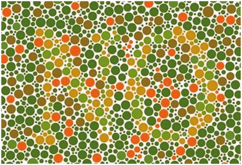
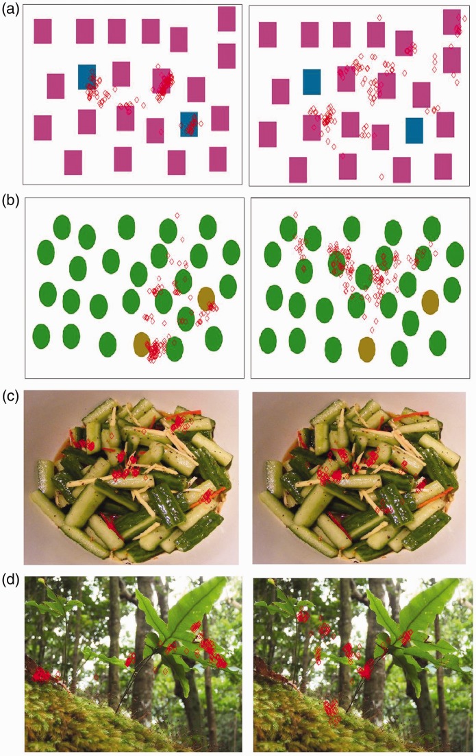
Color vision deficiencies affect visual perception of colors and, more generally, color images. Several sciences such as genetics, biology, medicine, and computer vision are involved in studying and analyzing vision deficiencies. As we know from visual saliency findings, human visual system tends to fix some specific points and regions of the image in the first seconds of observation summing up the most important and meaningful parts of the scene. In this article, we provide some studies about human visual system behavior differences between normal and color vision-deficient visual systems. We eye-tracked the human fixations in first 3 seconds of observation of color images to build real fixation point maps. One of our contributions is to detect the main differences between the aforementioned human visual systems related to color vision deficiencies by analyzing real fixation maps among people with and without color vision deficiencies. Another contribution is to provide a method to enhance color regions of the image by using a detailed color mapping of the segmented salient regions of the given image. The segmentation is performed by using the difference between the original input image and the corresponding color blind altered image. A second eye-tracking of color blind people with the images enhanced by using recoloring of segmented salient regions reveals that the real fixation points are then more coherent (up to 10%) with the normal visual system. The eye-tracking data collected during our experiments are in a publicly available dataset called Eye-Tracking of Color Vision Deficiencies.
Keywords: color vision deficiencies; eye movements; eye-tracking; image enhancement; image segmentation; imagery; visual saliency.
Figures


Similar articles
-
Bio-driven visual saliency detection with color factor.Front Bioeng Biotechnol. 2022 Aug 4;10:946084. doi: 10.3389/fbioe.2022.946084. eCollection 2022. Front Bioeng Biotechnol. 2022. PMID: 35992342 Free PMC article.
-
Visual Search in the Real World: Color Vision Deficiency Affects Peripheral Guidance, but Leaves Foveal Verification Largely Unaffected.Front Hum Neurosci. 2015 Dec 22;9:680. doi: 10.3389/fnhum.2015.00680. eCollection 2015. Front Hum Neurosci. 2015. PMID: 26733851 Free PMC article.
-
A Novel Approach to Image Recoloring for Color Vision Deficiency.Sensors (Basel). 2021 Apr 13;21(8):2740. doi: 10.3390/s21082740. Sensors (Basel). 2021. PMID: 33924510 Free PMC article.
-
Saliency-CCE: Exploiting colour contextual extractor and saliency-based biomedical image segmentation.Comput Biol Med. 2023 Mar;154:106551. doi: 10.1016/j.compbiomed.2023.106551. Epub 2023 Jan 20. Comput Biol Med. 2023. PMID: 36716685 Review.
-
[Physiology and pathology of visual information processing].Nippon Ganka Gakkai Zasshi. 2007 Mar;111(3):160-91; discussion 192. Nippon Ganka Gakkai Zasshi. 2007. PMID: 17402561 Review. Japanese.
References
-
- Achanta, R., Hemami, S., Estrada, F., & Susstrunk, S. (2009). Frequency-tuned salient region detection. In IEEE international conference on computer vision and pattern recognition (pp. 1597–1604). Piscataway, NJ: IEEE.
-
- Ardizzone, E., Bruno, A., & Gugliuzza, F. (2017). Exploiting visual saliency algorithms for object-based attention: A new color and scale-based approach. In International Conference on Image Analysis and Processing (pp. 191–201). Berlin, Germany: Springer.
-
- Ardizzone, E., Bruno, A., & Mazzola, G. (2011). Visual saliency by keypoints distribution analysis. In International Conference on Image Analysis and Processing (pp. 691–699). Berlin, Germany: Springer.
-
- Ardizzone, E., Bruno, A., & Mazzola, G. (2013a). Saliency based image cropping. In International Conference on Image Analysis and Processing (pp. 773–782). Berlin, Germany: Springer.
-
- Ardizzone E., Bruno A., Mazzola G. (2013. b) Scale detection via keypoint density maps in regular or near-regular textures. Pattern Recognition Letters 34: 2071–2078.
LinkOut - more resources
Full Text Sources
Research Materials
