. 2019 May 15;5(2):2055217319848463.
doi: 10.1177/2055217319848463. eCollection 2019 Apr-Jun.

Distinct serum and cerebrospinal fluid cytokine and chemokine profiles in autoantibody-associated demyelinating diseases

Affiliations

Distinct serum and cerebrospinal fluid cytokine and chemokine profiles in autoantibody-associated demyelinating diseases

Livia S Hofer et al. Mult Scler J Exp Transl Clin. .

Abstract

Background: Demyelinating diseases of the central nervous system associated with autoantibodies against aquaporin-4 and myelin-oligodendrocyte-glycoprotein are mediated by different immunopathological mechanisms compared to multiple sclerosis.

Objective: The purpose of this study was to evaluate serum and cerebrospinal fluid cytokine/chemokine profiles in patients with autoantibodies against aquaporin-4 or autoantibodies against myelin-oligodendrocyte-glycoprotein-associated demyelination compared to multiple sclerosis and autoimmune encephalitis.

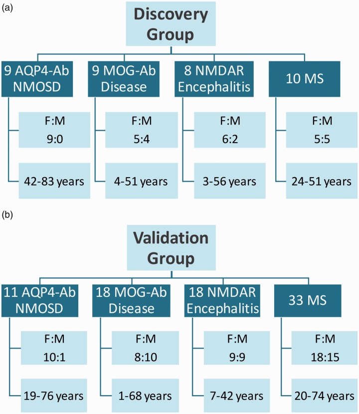
Methods: Serum and cerebrospinal fluid cytokine/chemokine levels were analysed using Procartaplex Multiplex Immunoassays. First, we analysed a panel of 32 cytokines/chemokines in a discovery group (nine aquaporin-4-antibody seropositive, nine myelin oligodendrocyte glycoprotein-antibody seropositive, eight encephalitis, 10 multiple sclerosis). Significantly dysregulated cytokines/chemokines were validated in a second cohort (11 aquaporin-4-antibody seropositive, 18 myelin oligodendrocyte glycoprotein-antibody seropositive, 18 encephalitis, 33 multiple sclerosis).

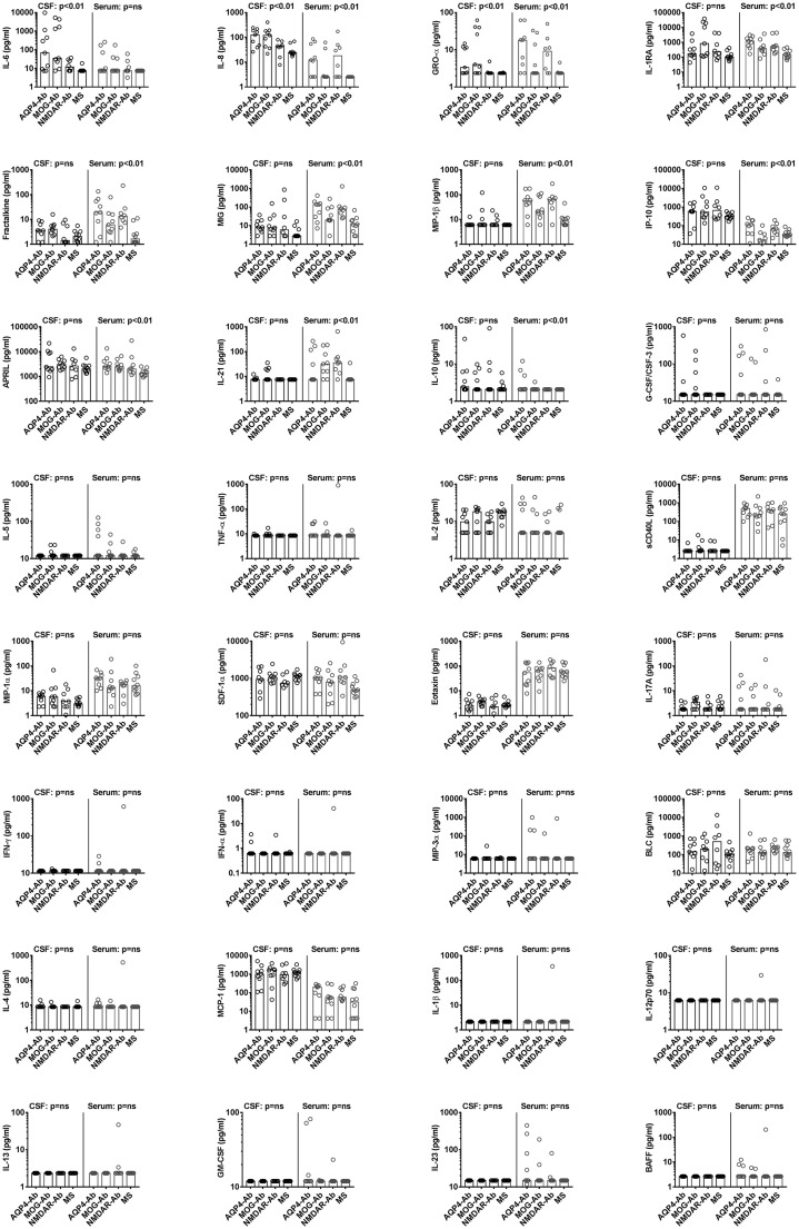
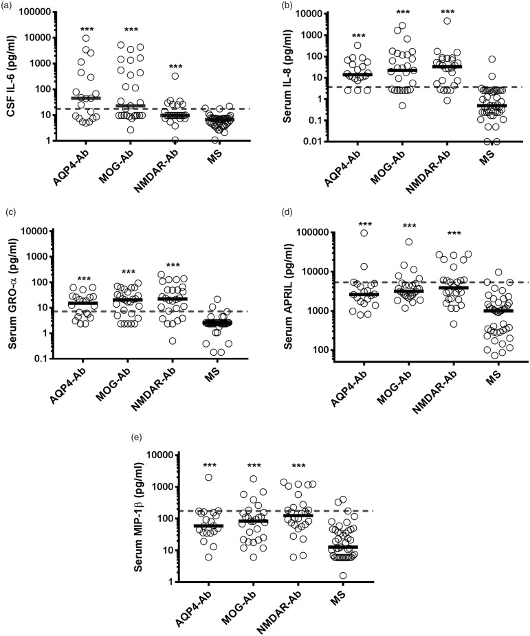
Results: We found 11 significantly altered cytokines/chemokines in cerebrospinal fluid and serum samples in the discovery group (a proliferation-inducing ligand, fractalkine=CX3CL1, growth-regulated oncogene-α, interleukin-1 receptor antagonist, interleukin-6, interleukin-8=CXCL8, interleukin-10, interleukin-21, interferon-ɣ-induced protein-10=CXCL10, monokine induced by interferon-ɣ=CXCL9, macrophage inflammatory protein-1ß=CCL4). Most of these cytokines/chemokines were up-regulated in autoantibodies against aquaporin-4 or autoantibodies against myelin-oligodendrocyte-glycoprotein positive patients compared to multiple sclerosis. We confirmed these results for cerebrospinal fluid interleukin-6 and serum interleukin-8, growth-regulated oncogene-α, a proliferation-inducing ligand and macrophage inflammatory protein-1β in the validation set. Receiver-operating characteristic analysis revealed increased levels of cerebrospinal fluid interleukin-6, serum interleukin-8 and growth-regulated oncogene-α in most patients with autoantibody-associated neurological diseases.

Conclusion: This study suggests that distinctive cerebrospinal fluid and serum cytokine/chemokine profiles are associated with autoantibody-mediated demyelination, but not with multiple sclerosis.

Keywords: Demyelinating diseases; aquaporin-4 antibody; chemokines; cytokines; multiple sclerosis; myelin-oligodendrocyte-glycoprotein antibody.


Figures

Figure 1.
Figure 1.
Demographic characteristics of participants from the (a) discovery and (b) validation group. Ab: antibody; AQP-4: aquaporin-4; F: female; M: male; MOG: myelin oligodendrocyte glycoprotein; MS: multiple sclerosis; NMDAR: N-methyl-D-aspartate receptor; NMOSD: neuromyelitis optica spectrum disorder.
Figure 2.
Figure 2.
Scatter dot plots of cytokines and chemokines in cerebrospinal fluid (CSF) and serum of the discovery set. Individual values for aquaporin-4-antibody (AQP4-Ab), myelin oligodendrocyte glycoprotein-antibody (MOG-Ab), anti-N-methyl-D-aspartate receptor-antibody (NMDAR-Ab) and multiple sclerosis (MS) patients are shown as circles and medians are shown as bars. Cytokines and chemokines are organised according to their p-values. Overall significance was assessed using the nonparametric Kruskal-Wallis test and p-values were adjusted for multiple comparisons using a false discovery rate (FDR) significance criterion of 10% based on the Benjamini-Hochberg correction. Between group comparisons were calculated using Dunn’s multiple comparison test. Significant changes in the CSF (AQP4-Ab and MOG-Ab versus MS) and serum (AQP4-Ab, MOG-Ab and NMDAR-Ab versus MS) are shown in the graphs.Ab: antibody; AQP-4: aquaporin-4; APRIL: a proliferation-inducing ligand; BAFF: B cell activating factor; BLC: B lymphocyte chemoattractant; G-CSF: granulocyte colony-stimulating factor; GM-CSF: granulocyte macrophage colony-stimulating factor; GRO: growth-regulated oncogene; IFN: interferon; IL: interleukin; IP: interferon-ɣ-induced protein; MCP: monocyte chemoattractant protein; MIG: monokine induced by interferon-ɣ; MIP: macrophage inflammatory protein; MOG: myelin oligodendrocyte glycoprotein; MS: multiple sclerosis; NMDAR: N-methyl-D-aspartate receptor; ns: not significant; SDF: stromal cell-derived factor; TNF: tumour necrosis factor.
Figure 3.
Figure 3.
Scatter dot plots of cytokines and chemokines in cerebrospinal fluid (CSF) and serum of the validation set. Individual values for aquaporin-4-antibody (AQP4-Ab), myelin oligodendrocyte glycoprotein-antibody (MOG-Ab), anti-N-methyl-D-aspartate receptor-antibody (NMDAR-Ab) and multiple sclerosis (MS) patients are shown as circles and medians are shown as bars. Cytokines and chemokines are organised according to their p-values. Overall significance was assessed using the nonparametric Kruskal Wallis test and p-values were adjusted for multiple comparisons using a false discovery rate (FDR) significance criterion of 10% based on the Benjamini-Hochberg correction. Between group comparisons were calculated using Dunn’s multiple comparison test. Significant changes in the CSF (AQP4-Ab and MOG-Ab versus MS) and serum (AQP4-Ab, MOG-Ab and NMDAR-Ab versus MS) are shown in the graphs.Ab: antibody; AQP-4: aquaporin-4; GRO: growth-regulated oncogene; IL: interleukin; IP: interferon-ɣ-induced protein; MCP: monocyte chemoattractant protein; MIG: monokine induced by interferon-ɣ; MIP: macrophage inflammatory protein; MOG: myelin oligodendrocyte glycoprotein; MS: multiple sclerosis; NMDAR: N-methyl-D-aspartate receptor; ns: not significant.
Figure 4.
Figure 4.
Dysregulated cytokines and chemokines confirmed in both cohorts. Pooled individual values for both cohorts are shown as circles and medians are shown as bars. Significance of group differences was analysed by the Kruskal Wallis test and significant differences (p-value <0.001) are shown by the asterisks.The grey dashed lines represent the cut-off value of CSF IL-6 (a) and of serum IL-8 (b), GRO-α (d), APRIL (d) and MIP-1β (d) to discriminate AQP4-Ab, MOG-Ab and NMDAR-Ab from MS patients with 95% specificity. Ab: antibody; APRIL: a proliferation-inducing ligand; AQP-4: aquaporin-4; CSF: cerebrospinal fluid; GRO: growth-regulated oncogene; IL: interleukin; MIP: macrophage inflammatory protein; MOG: myelin oligodendrocyte glycoprotein; MS: multiple sclerosis; NMDAR: N-methyl-D-aspartate receptor.

References

    1. Dalmau J, Graus F. Antibody-mediated encephalitis. N Engl J Med 2018; 378: 840–851. - PubMed
    1. Thompson AJ, Baranzini SE, Geurts J, et al. Multiple sclerosis. Lancet 2018; 391: 1622–1636. - PubMed
    1. Reindl M, Waters P. Myelin oligodendrocyte glycoprotein antibodies in neurological disease. Nat Rev Neurol 2019; 15: 89-102. DOI: 10.1038/s41582-018-0112-x. - DOI - PubMed
    1. Papadopoulos MC, Bennett JL, Verkman AS. Treatment of neuromyelitis optica: State-of-the-art and emerging therapies. Nat Rev Neurol 2014; 10: 493–506. - PMC - PubMed
    1. Wingerchuk DM, Banwell B, Bennett JL, et al. International consensus diagnostic criteria for neuromyelitis optica spectrum disorders. Neurology 2015; 85: 177–189. - PMC - PubMed