

Pentamidine Inhibits Titanium Particle-Induced Osteolysis In Vivo and Receptor Activator of Nuclear Factor-κB Ligand-Mediated Osteoclast Differentiation In Vitro
- PMID: 31205855
- PMCID: PMC6542890
- DOI: 10.1007/s13770-019-00186-y
Pentamidine Inhibits Titanium Particle-Induced Osteolysis In Vivo and Receptor Activator of Nuclear Factor-κB Ligand-Mediated Osteoclast Differentiation In Vitro
Abstract
Background: Wear debris-induced osteolysis leads to periprosthetic loosening and subsequent prosthetic failure. Since excessive osteoclast formation is closely implicated in periprosthetic osteolysis, identification of agents to suppress osteoclast formation and/or function is crucial for the treatment and prevention of wear particle-induced bone destruction. In this study, we examined the potential effect of pentamidine treatment on titanium (Ti) particle-induced osteolysis, and receptor activator of nuclear factor-κB ligand (RANKL)-induced osteoclastogenesis.
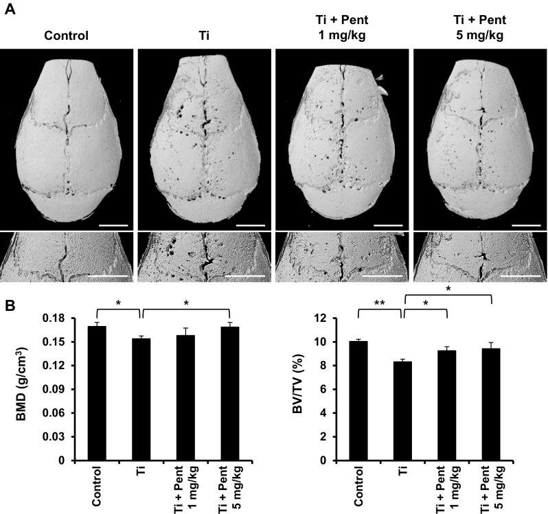
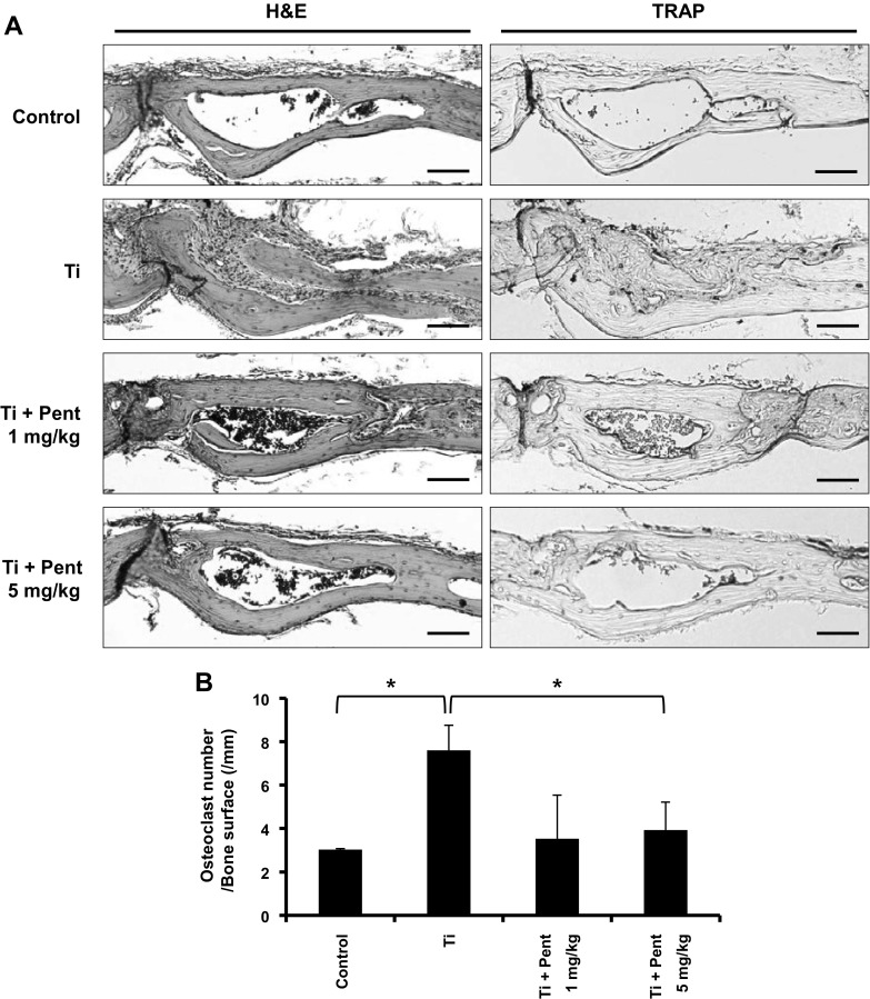
Methods: The effect of pentamidine treatment on bone destruction was examined in Ti particle-induced osteolysis mouse model. Ti particles were implanted onto mouse calvaria, and vehicle or pentamidine was administered for 10 days. Then, calvarial bone tissue was analyzed using micro-computed tomography and histology. We performed in vitro osteoclastogenesis assay using bone marrow-derived macrophages (BMMs) to determine the effect of pentamidine on osteoclast formation. BMMs were treated with 20 ng/mL RANKL and 10 ng/mL macrophage colony-stimulating factor in the presence or absence of pentamidine. Osteoclast differentiation was determined by tartrate-resistant acid phosphatase staining, real-time polymerase chain reaction, and immunofluorescence staining.
Results: Pentamidine administration decreased Ti particle-induced osteoclast formation significantly and prevented bone destruction compared to the Ti particle group in vivo. Pentamidine also suppressed RANKL-induced osteoclast differentiation and actin ring formation markedly, and inhibited the expression of nuclear factor of activated T cell c1 and osteoclast-specific genes in vitro. Additionally, pentamidine also attenuated RANKL-mediated phosphorylation of IκBα in BMMs.
Conclusion: These results indicate that pentamidine is effective in inhibiting osteoclast formation and significantly attenuates wear debris-induced bone loss in mice.
Keywords: Osteoclastogenesis; Osteolysis; Pentamidine; RANKL; Titanium.
Conflict of interest statement
Conflict of interestThe authors declare that they have no conflict of interest.
Figures


Similar articles
-
Inhibitory effects of triptolide on titanium particle-induced osteolysis and receptor activator of nuclear factor-κB ligand-mediated osteoclast differentiation.Int Orthop. 2015 Jan;39(1):173-82. doi: 10.1007/s00264-014-2596-3. Epub 2014 Nov 23. Int Orthop. 2015. PMID: 25416122
-
Rifampin suppresses osteoclastogenesis and titanium particle-induced osteolysis via modulating RANKL signaling pathways.Biochem Biophys Res Commun. 2017 Feb 26;484(1):64-70. doi: 10.1016/j.bbrc.2017.01.071. Epub 2017 Jan 18. Biochem Biophys Res Commun. 2017. PMID: 28108285
-
Theaflavin-3,3'-digallate represses osteoclastogenesis and prevents wear debris-induced osteolysis via suppression of ERK pathway.Acta Biomater. 2017 Jan 15;48:479-488. doi: 10.1016/j.actbio.2016.11.022. Epub 2016 Nov 9. Acta Biomater. 2017. PMID: 27838465
-
Mechanism of regulating macrophages/osteoclasts in attenuating wear particle-induced aseptic osteolysis.Front Immunol. 2023 Oct 4;14:1274679. doi: 10.3389/fimmu.2023.1274679. eCollection 2023. Front Immunol. 2023. PMID: 37860014 Free PMC article. Review.
-
Interleukin-27 prevents LPS-induced inflammatory osteolysis by inhibiting osteoclast formation and function.Am J Transl Res. 2019 Mar 15;11(3):1154-1169. eCollection 2019. Am J Transl Res. 2019. PMID: 30972153 Free PMC article. Review.
Cited by
-
Effects of the Local Bone Renin-Angiotensin System on Titanium-Particle-Induced Periprosthetic Osteolysis.Front Pharmacol. 2021 Jun 24;12:684375. doi: 10.3389/fphar.2021.684375. eCollection 2021. Front Pharmacol. 2021. PMID: 34248634 Free PMC article.
-
Periprosthetic Osteolysis: Mechanisms, Prevention and Treatment.J Clin Med. 2019 Dec 1;8(12):2091. doi: 10.3390/jcm8122091. J Clin Med. 2019. PMID: 31805704 Free PMC article. Review.
-
Zinc Sulfate Stimulates Osteogenic Phenotypes in Periosteum-Derived Cells and Co-Cultures of Periosteum-Derived Cells and THP-1 Cells.Life (Basel). 2021 Apr 30;11(5):410. doi: 10.3390/life11050410. Life (Basel). 2021. PMID: 33946199 Free PMC article.
-
Fermented Oyster Extract Prevents Ovariectomy-Induced Bone Loss and Suppresses Osteoclastogenesis.Nutrients. 2019 Jun 21;11(6):1392. doi: 10.3390/nu11061392. Nutrients. 2019. PMID: 31234292 Free PMC article.
-
The unfavorable role of titanium particles released from dental implants.Nanotheranostics. 2021 Mar 10;5(3):321-332. doi: 10.7150/ntno.56401. eCollection 2021. Nanotheranostics. 2021. PMID: 33732603 Free PMC article. Review.
References
Publication types
MeSH terms
Substances
LinkOut - more resources
Full Text Sources