. 2020 Jan;71(1):14-30.
doi: 10.1002/hep.30815. Epub 2019 Aug 9.

Analysis of Host Responses to Hepatitis B and Delta Viral Infections in a Micro-scalable Hepatic Co-culture System

Affiliations

Analysis of Host Responses to Hepatitis B and Delta Viral Infections in a Micro-scalable Hepatic Co-culture System

Benjamin Y Winer et al. Hepatology. 2020 Jan.

Abstract

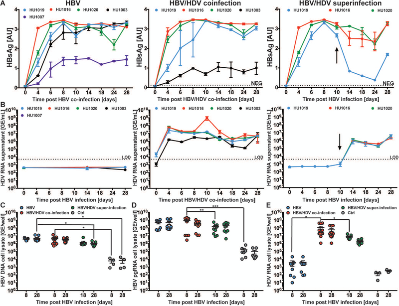
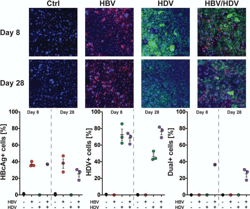
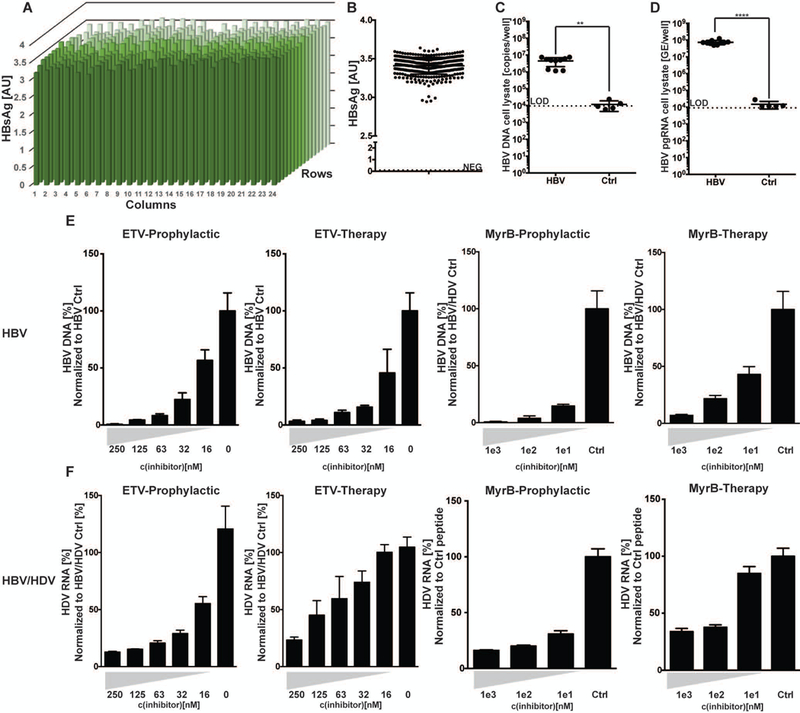
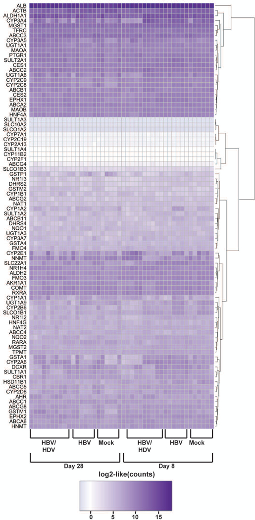
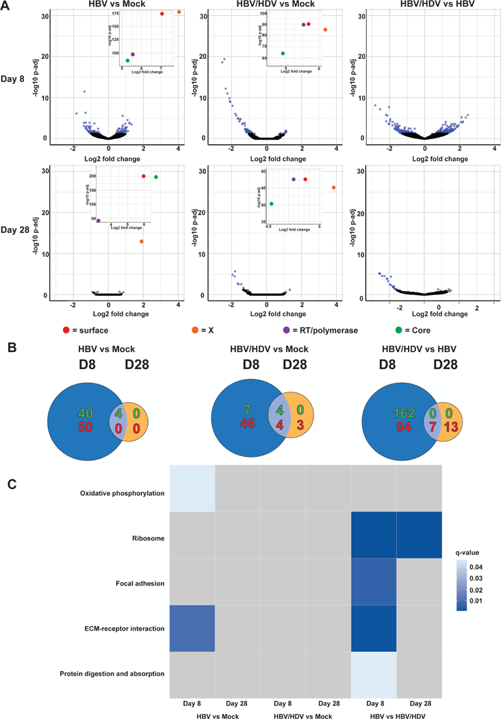
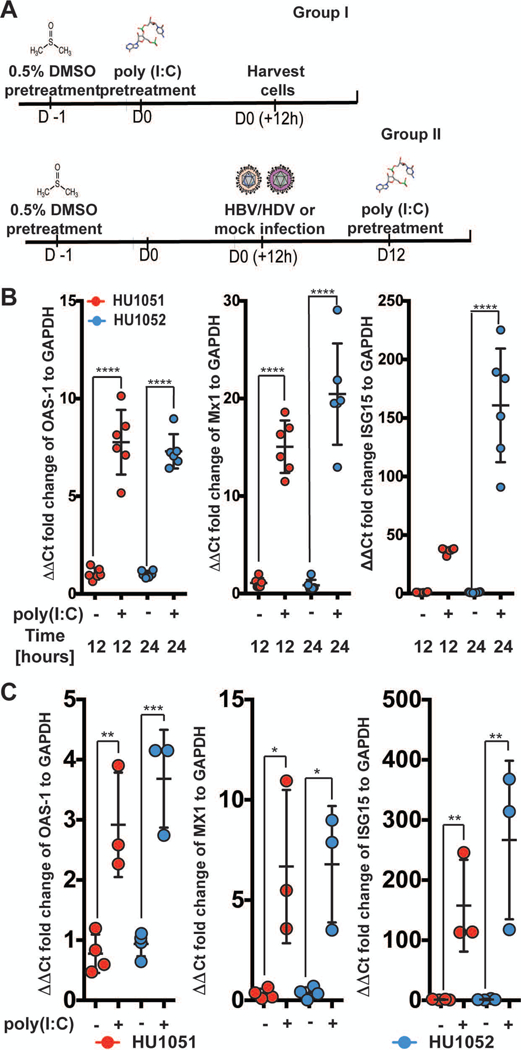
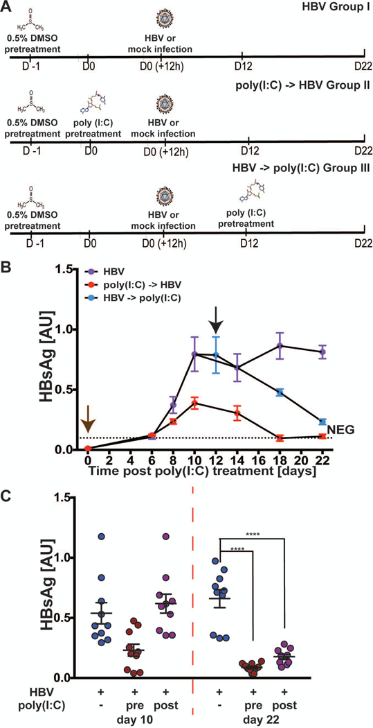
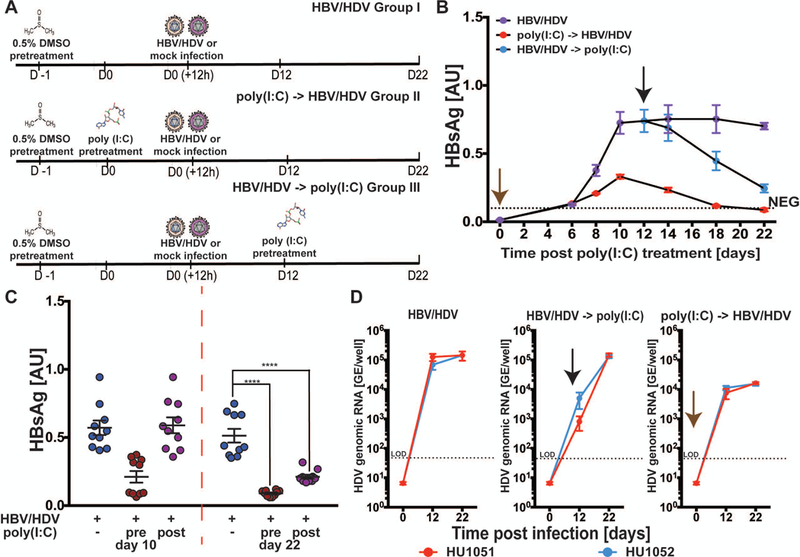
Hepatitis B virus (HBV) remains a major global health problem with 257 million chronically infected individuals worldwide, of whom approximately 20 million are co-infected with hepatitis delta virus (HDV). Progress toward a better understanding of the complex interplay between these two viruses and the development of novel therapies have been hampered by the scarcity of suitable cell culture models that mimic the natural environment of the liver. Here, we established HBV and HBV/HDV co-infections and super-infections in self-assembling co-cultured primary human hepatocytes (SACC-PHHs) for up to 28 days in a 384-well format and highlight the suitability of this platform for high-throughput drug testing. We performed RNA sequencing at days 8 and 28 on SACC-PHHs, either HBV mono-infected or HBV/HDV co-infected. Our transcriptomic analysis demonstrates that hepatocytes in SACC-PHHs maintain a mature hepatic phenotype over time, regardless of infection condition. We confirm that HBV is a stealth virus, as it does not induce a strong innate immune response; rather, oxidative phosphorylation and extracellular matrix-receptor interactions are dysregulated to create an environment that promotes persistence. Notably, HDV co-infection also did not lead to statistically significant transcriptional changes across multiple donors and replicates. The lack of innate immune activation is not due to SACC-PHHs being impaired in their ability to induce interferon stimulated genes (ISGs). Rather, polyinosinic:polycytidylic acid exposure activates ISGs, and this stimulation significantly inhibits HBV infection, yet only minimally affects the ability of HDV to infect and persist. Conclusion: These data demonstrate that the SACC-PHH system is a versatile platform for studying HBV/HDV co-infections and holds promise for performing chemical library screens and improving our understanding of the host response to such infections.


Conflict of interest statement

Conflicts of Interest.

The following authors declare competing financial interests. R.J., Am.P., C.C., A.S. are employees of the Hurel Corporation of which E.N. is also a stockholder. A.P. is a member of the Scientific Advisory Board of the Hurel Corporation. B.Y.W., J.M.G., Y.B., R.E.S., G.E.L., L.P., and R.L. do not have any conflicts of interest.

Figures

Fig. 1.
Fig. 1.. HBV mono-infection, HBV/HDV co-infection, and HBV/HDV super-infection kinetics in SACC-PHHs.
SACC-PHHs were infected with either HBV, HBV/HDV (co-infection), or were first persistently infected with HBV and then with HDV (super-infection). (A) Longitudinal HBsAg ELISA data. (B) HDV RNA RT-qPCR data from supernatants. HBV DNA (C), HBV pgRNA (D), or HDV RNA (E) in the cell lysates. All data are presented as mean ± SEM. Statistical significance was determined using ANOVA. *P < 0.05, **P < 0.01, ***P < 0.001, and ****P < 0.0001.
Fig. 2.
Fig. 2.. Quantification of infection in HBV, HDV, and co-infected SACC-PHHs by HDV vPLAYR/TSA and anti-HBcAg immunofluorescence staining.
SACC-PHHs were either infected with HBV (MOI=4,000), HDV (MOI=1,000), or both HBV/HDV (HBV MOI=4,000, HDV MOI=1,000). (A) At 8 (top) and 28 (bottom) days post infection, control and infected SACC-PHHs were fixed and stained for HDV genomic RNA (green) by a vPLAYR/TSA procedure and for HBcAg (red) by an anti-HBcAg antibody as well as with DAPI (blue) for nuclear DNA. Quantification of three different images for each experimental condition were performed (approximately ~800 cells total per condition) for (B) HBcAg positive cells; (C) HDV genomic RNA positive cells; and (D) dual-positive cells.
Fig. 3.
Fig. 3.. Assessment of clinically relevant drug treatments for HBV mono-infection and HBV/HDV co-infection of SACC-PHHs in a 384 well microwell format.
(A) SACC-PHHs in a 384 well format were infected with HBV and viremia assessed for each well by HBsAg ELISA. (B) Summary of HBsAg concentrations determined across plate shown in (A). Coefficient of variance across the 384 well plate equaled 3.2%. HBV DNA (C) and HBV pgRNA (D) from cell lysates of HBV infected vs control cultures as assessed by qPCR (HBV n=10; control n=5). For panel (E), SACC-PHHs were challenged with HBV. The x-axis equals drug concentration and the y-axis the amount of HBV DNA (for ETV) or HBsAg secretion (for MyrB) normalized to untreated, HBVcc-infected untreated control cells or HBVcc-infected cells treated with a control peptide, respectively. For panels (F) SACC-PHHs were co-infected with HBV/HDV and HDV RNA was quantified by RT-qPCR in supernatant (far left) and cell lysate (second from left). Co-infected SACC-PHHs were treated with the entry inhibitor MyrB prophylactically (second from right) or therapeutically (far right), with the x axis showing drug concentration and the y axis the amount of HDV RNA present in the supernatant normalized to that secreted by co-infected cells treated with a control peptide. All data are presented as mean ± SEM. Statistical significance was determined using ANOVA. *P < 0.05, **P < 0.01, ***P < 0.001, and ****P < 0.0001.
Fig. 4.
Fig. 4.. Regardless of experimental condition, minimal variation observed for 86 drug-metabolizing, liver-specific transcripts, in SACC-PHHs.
Regularized log2 transformed (rlog function in DESeq2) RNAseq counts from SACC-PHHs mono-infected with HBV, co-infected with HBV/HDV or mock-infected for 86 drug-metabolizing enzymes, predominantly regulated at the transcriptional level and expressed by the human liver, are shown.
Fig. 5.
Fig. 5.. Transcriptomic analysis of “early” and “late” stage of HBV mono-infected and HBV/HDV co-infected SACC-PHHs.
RNA was isolated at 8 and 28 dpi from SACC-PHHs HBV mono-, HBV/HDV co- or mock-infected. A cDNA library was then generated and sequenced. (A) Volcano plots of differentially expressed genes in mono-infected vs mock, co-infected vs mock, or co- vs mono-infected at 8 dpi (top row) and 28 dpi (bottom row). Each point represents a gene, with those having a padj ≤ 0.05 colored blue and those with a padj > 0.05 colored black. HBV genes are indicated in red, orange, purple or green. (B) Venn diagram of genes up- (shown in green) or down-regulated (shown in red) between mono-infected vs mock, co-infected vs mock, and co- vs mono-infected at 8 and 28 dpi. Genes were considered up- or down-regulated if the absolute value of log2(fold change) ≥ 0.5 and padj ≤ 0.05. (C) Significantly enriched pathways (q-value ≥ 0.07) determined using GAGE comparing mono-infected vs control, co-infected vs control, and co- vs mono-infected at 8 and 28 dpi.
Fig. 6:
Fig. 6:. HBV/HDV co-infection does not generally impair cell-intrinsic innate defenses.
(A) Schematic of experimental time course (B) Fold increase in the ISGs OAS-1 (left), MX1 (middle) and ISG15 (right) normalized to GAPDH, 12 and 24 hours post poly(I:C) transfection, quantified by RT-qPCR in the absence of infection. (C) Analysis of OAS-1 (left), MX1 (middle), and ISG15 (right) mRNA expression 12 hours post poly(I:C) exposure of SACC-PHHs co-infected 12 days prior. Different colors represent indicated hepatocyte donors. All data are presented as mean ± SEM. Statistical significance was determined using ANOVA. *P < 0.05, **P < 0.01, ***P < 0.001, and ****P < 0.0001. Abbreviations: GAPDH, glyceraldehyde 3‐phosphate dehydrogenase.
Fig. 7:
Fig. 7:. Induction of innate immunity by poly(I:C) treatment leads to suppression of HBV.
(A) Schematic of experimental time course for poly(I:C)-transfected SACC-PHHs infected with HBV. Longitudinal measurements of HBsAg by ELISA in supernatants (B-C) of SACC-PHHs +/− poly(I:C) as shown in panel A. Brown and black arrows indicate poly(I:C) transfection either pre- or post-establishment of persistent infection, respectively. Co-infected SACC-PHHs +/− poly(I:C) were lysed 12 and 22 dpi. A separate set of pre-infected SACC-PHHs for each donor were also lysed. Statistical significance was determined using ANOVA. ****P < 0.0001.
Fig. 8:
Fig. 8:. Induction of innate immunity has little effect on HDV infection.
(A) Schematic of experimental time course for poly(I:C)-transfected SACC-PHHs co-infected with HBV/HDV. Longitudinal measurements of HBsAg by ELISA in supernatants of co-infected (B-C) SACC-PHHs +/− poly(I:C) as shown in panel A. Brown and black arrows indicate poly(I:C) transfection either pre- or post-establishment of persistent infection, respectively. Co-infected SACC-PHHs +/− poly(I:C) were lysed 12 and 22 dpi. A separate set of pre-infected SACC-PHHs for each donor were also lysed. (D) Quantification of intracellular HDV genomic RNA by RT-qPCR at 12 and 22 dpi in cultures mock treated (left), or exposed to poly(I:C) post- (middle) or pre- (right) infection. Statistical significance was determined using ANOVA. ****P < 0.0001.

References

    1. Seeger C, Mason WS. HBV replication, pathobiology and therapy: Unanswered questions. J Hepatol 2016;64:S1–S3. - PubMed
    1. Yan H, Zhong G, Xu G, He W, Jing Z, Gao Z, Huang Y, et al. Sodium taurocholate cotransporting polypeptide is a functional receptor for human hepatitis B and D virus. Elife 2012;1:e00049. - PMC - PubMed
    1. Lempp FA, Ni Y, Urban S. Hepatitis delta virus: insights into a peculiar pathogen and novel treatment options. Nat Rev Gastroenterol Hepatol 2016;13:580–589. - PubMed
    1. Winer BY, Ploss A. Determinants of hepatitis B and delta virus host tropism. Curr Opin Virol 2015;13:109–116. - PMC - PubMed
    1. Hetzel U, Szirovicza L, Smura T, Prahauser B, Vapalahti O, Kipar A, Hepojoki J. Identification of a Novel Deltavirus in Boa Constrictors. MBio 2019;10. - PMC - PubMed

Publication types

MeSH terms