. 2019 Jun 17;17(6):e3000328.
doi: 10.1371/journal.pbio.3000328. eCollection 2019 Jun.

Star nanoparticles delivering HIV-1 peptide minimal immunogens elicit near-native envelope antibody responses in nonhuman primates

Affiliations

Star nanoparticles delivering HIV-1 peptide minimal immunogens elicit near-native envelope antibody responses in nonhuman primates

Joseph R Francica et al. PLoS Biol. .

Abstract

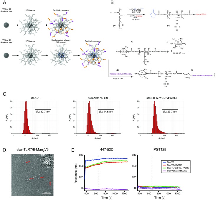
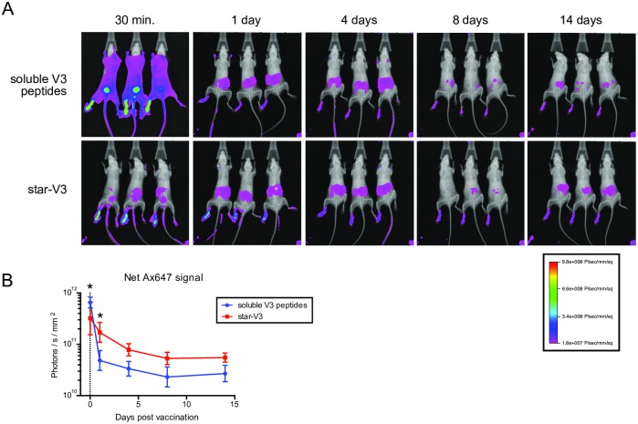
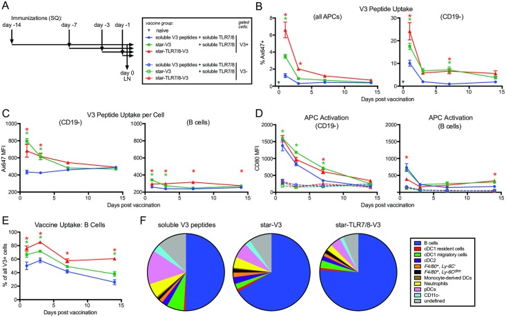
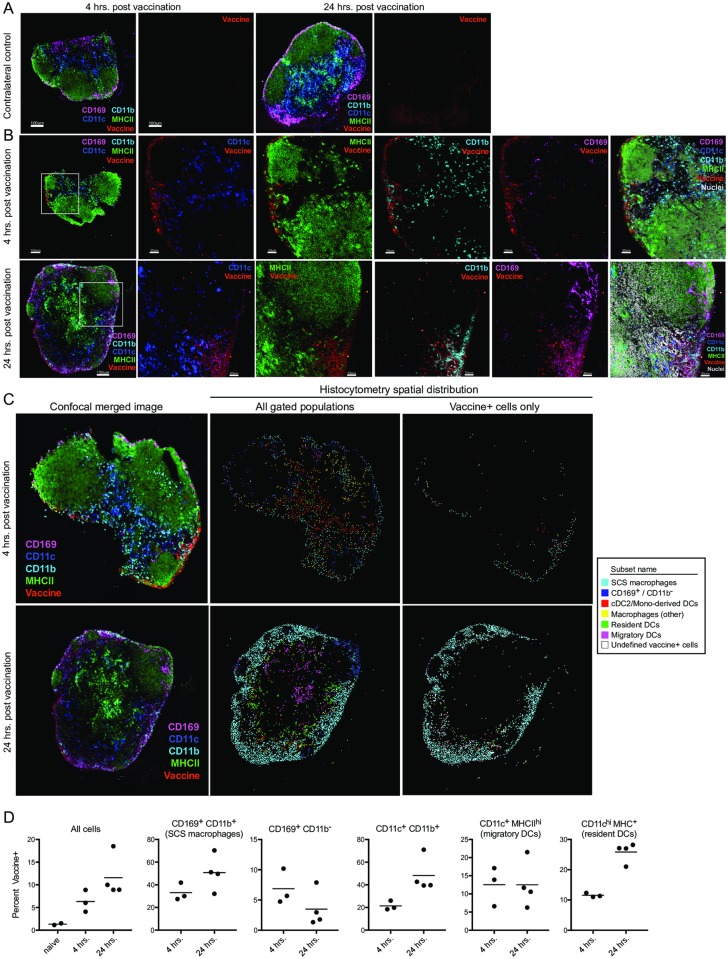
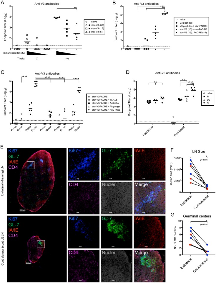
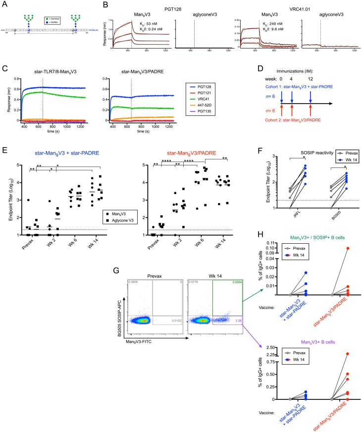
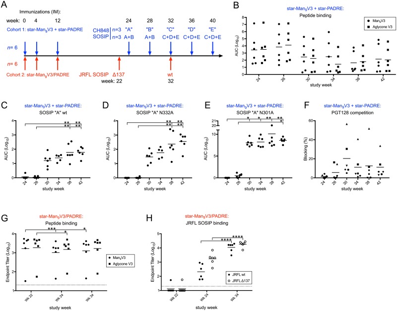
Peptide immunogens provide an approach to focus antibody responses to specific neutralizing sites on the HIV envelope protein (Env) trimer or on other pathogens. However, the physical characteristics of peptide immunogens can limit their pharmacokinetic and immunological properties. Here, we have designed synthetic "star" nanoparticles based on biocompatible N-[(2-hydroxypropyl)methacrylamide] (HPMA)-based polymer arms extending from a poly(amidoamine) (PAMAM) dendrimer core. In mice, these star nanoparticles trafficked to lymph nodes (LNs) by 4 hours following vaccination, where they were taken up by subcapsular macrophages and then resident dendritic cells (DCs). Immunogenicity optimization studies revealed a correlation of immunogen density with antibody titers. Furthermore, the co-delivery of Env variable loop 3 (V3) and T-helper peptides induced titers that were 2 logs higher than if the peptides were given in separate nanoparticles. Finally, we performed a nonhuman primate (NHP) study using a V3 glycopeptide minimal immunogen that was structurally optimized to be recognized by Env V3/glycan broadly neutralizing antibodies (bnAbs). When administered with a potent Toll-like receptor (TLR) 7/8 agonist adjuvant, these nanoparticles elicited high antibody binding titers to the V3 site. Similar to human V3/glycan bnAbs, certain monoclonal antibodies (mAbs) elicited by this vaccine were glycan dependent or targeted the GDIR peptide motif. To improve affinity to native Env trimer affinity, nonhuman primates (NHPs) were boosted with various SOSIP Env proteins; however, significant neutralization was not observed. Taken together, this study provides a new vaccine platform for administration of glycopeptide immunogens for focusing immune responses to specific bnAb epitopes.


Conflict of interest statement

The authors have declared that no competing interests exist.

Figures

Fig 1
Fig 1. Star nanoparticle synthesis and characterization.
(A) Cartoon depiction of star nanoparticles, composed of a dendrimer core and HPMA-based arms, to which peptide immunogens (yellow and purple) are attached. Small molecule adjuvants may also be attached, shown as blue polygons in the lower row scheme. (B) Star nanoparticles are synthesized by polymerization of HPMA monomers (1) into 10-kDa chains (2), then conjugating these to G5 PAMAM dendrimers by acylation (3) to yield unloaded nanoparticles (4); finally, peptide immunogens (5) are conjugated using to the HPMA grafts by CuI catalyzed cycloaddition to yield the loaded vaccine nanoparticle (6). (C) Dynamic light scattering analysis of star nanoparticles displaying monodispersity, with hydrodynamic radius (Rh) shown. (D) Star nanoparticles bearing a TLR7/8 agonist and glycosylated V3 peptides (Man9V3, described in Fig 6) as visualized by electron microscopy, magnification 100,000×; red arrows indicate individual nanoparticles. (E) The antigenicity of star nanoparticles bearing HIV V3 and PADRE peptides and a TLR7/8 agonist was analyzed by biolayer interferometry. Antibodies 447-52D (specific for V3 loop apex) and PGT128 (specific for glycan and V3 loop base) were immobilized on sensors; binding of star nanoparticles in solution was then measured. Nanoparticles displaying a V3 epitope comprising the unglycosylated loop base (purple trace) serve as a negative control. Data associated with this figure can be found in S1 Data. ACVA, 4,4′-azobis(4-cyanovaleric acid); CuI, copper(I); G5, fifth-generation; HPMA, N-[(2-hydroxypropyl)methacrylamide]; Man9V3, Env variable loop 3 oligomannose-9 glycopeptide; PADRE, pan DR epitope; PAMAM, poly(amidoamine); TLR7/8, Toll-like receptor 7/8; V3, Env variable loop 3.
Fig 2
Fig 2. Star nanoparticles restrict the biodistribution and prolong the localization of minimal peptide immunogens.
Mice were immunized subcutaneously in the left footpad with star nanoparticles bearing Ax647-labeled V3 peptides; control mice were immunized with soluble Ax647-labeled V3 peptides. (A) Mice were imaged at the indicated time points following vaccination. Composite overlays of X-ray and fluorescent images are shown. (B) Vaccine retention at the site of injection was measured by quantifying fluorescence in the left footpad at the time points shown above. Data points indicate group geometric means and 95% confidence intervals; vertical line indicates immunization; asterisk (*), statistical difference by ANOVA, comparing between groups at each time point. Data associated with this figure can be found in S1 Data. Ax647, Alexa Fluor 647; V3, Env variable loop 3.
Fig 3
Fig 3. APC uptake of star nanoparticles in the draining LN.
Mice were immunized subcutaneously with star nanoparticles bearing Ax647-labeled V3 peptides formulated with a TLR7/8 adjuvant via direct conjugation (red symbols), or admixed with soluble TLR7/8 (green symbols); control mice were immunized with soluble V3 peptides and a soluble TLR7/8 agonist (blue symbols). T-cell help was provided via unlabeled nanoparticles bearing PADRE peptides to all groups. (A) Immunization scheme: vaccines were administered 14, 7, 3, or 1 day(s) before day 0; on day 0, popliteal LNs were harvested and analyzed by flow cytometry. (B) V3 peptide uptake was measured by gating on Ax647+ cells. Left graph shows positive cells from all APCs; right graph shows non–B cell APCs only. (C) V3 peptide uptake per cell was measured by Ax647 MFI in the V3+ gate. Left graph shows non–B cell APCs only; right graph shows B cells only. (D) APC activation, as measured by CD80 expression. Left graph shows non–B cell APCs only; right graph shows B cells only. (E) Vaccine uptake by B cells, graphed as a percentage of the total number of Ax647+ cells. Asterisk (*), statistical difference by ANOVA, compared with the soluble V3 peptides group for star-V3 group (green) or star-TLR7/8-V3 group (red). (F) Vaccine uptake by APC phenotype, 24 hours after vaccination. Data associated with this figure can be found in S1 Data. APC, antigen-presenting cell; Ax647, Alexa Fluor 647; CD, cluster of differentiation; cDC, conventional dendritic cell; LN, lymph node; MFI, median fluorescence intensity; PADRE, pan DR epitope; pDC, plasmacytoid dendritic cell; SC, subcutaneous; TLR7/8, Toll-like receptor 7/8; V3, Env variable loop 3.
Fig 4
Fig 4. Star nanoparticle spatial distribution on APCs in draining LNs.
Mice were vaccinated in the footpad, with star nanoparticles bearing Ax647-labeled V3 peptides and unlabeled nanoparticles bearing PADRE peptides and a soluble TLR7/8 agonist. (A) Confocal microscopy of contralateral control LNs at 4 and 24 hours or (B) vaccinated draining ipsilateral LNs at 4 hours (top row) or 24 hours (bottom row). Colocalization of the vaccine with various APC markers is shown. (C,D) Histocytometry analysis of vaccine+ APC populations. (B) Confocal images from (B, left panels) were gated into the indicated populations based on marker co-expression, then replotted by their x- and y- coordinates. Center panels depict all gated subsets; right panels show only vaccine+ cells. (D) Vaccine+ events are graphed as a percentage of each gated population, from draining LNs of multiple mice. Data associated with this figure can be found in S1 Data. APC, antigen-presenting cell; Ax647, Alexa Fluor 647; CD, cluster of differentiation; cDC, conventional dendritic cell; DC, dendritic cell; LN, lymph node; MHCII, major histocompatibility complex class II; PADRE, pan DR epitope; SCS, subcapsular sinus; TLR7/8, Toll-like receptor 7/8; V3, Env variable loop 3.
Fig 5
Fig 5. Optimization of nanoparticle immunogenicity.
(A) Mice were immunized subcutaneously with star nanoparticles bearing 5, 15, or 30 V3 peptides per particle, with or without star nanoparticles bearing PADRE peptides for T help. The V3 peptide dose (5 μg) was constant across all groups; all vaccines were adjuvanted by admixing with a soluble TLR7/8 agonist. (B) Mice were immunized with soluble V3 peptides, with or without star nanoparticles bearing PADRE peptides, with star nanoparticles bearing V3 or PADRE peptides on separate particles, or with star nanoparticles bearing both V3 and PADRE peptides on the same particle; the density of V3 peptides (15/nanoparticle) was constant across all groups; the V3 peptide dose (5 μg) was constant across all nanoparticle groups; all vaccines were adjuvanted by admixing with soluble a TLR7/8 agonist. (C) Comparison of different adjuvants for use with star nanoparticles. Nanoparticles bearing V3 and PADRE peptides were left unadjuvanted, or were admixed with a TLR7/8 agonist, the emulsion adjuvant AddaVax, or the alum formulations Alhydrogel or Adju-Phos. (D) Comparison of different vaccination routes. Star nanoparticles bearing TLR7/8 agonists, V3, and PADRE peptides were administered intramuscularly (IM), subcutaneously (SC), or intravenously (IV). In all studies, serum antibody responses were measured by ELISA after two homologous immunizations, except where indicated; bars indicate group geometric means; horizontal lines indicate ELISA assay limit of detection. *p < 0.05; **p < 0.01; ***p < 0.001; ****p < 0.0001; n.s., not significant by the Kruskal-Wallis test. (E-G) Germinal center formation in popliteal LNs 10 days after footpad immunization, with star nanoparticles bearing Ax647-labeled V3 peptides and unlabeled nanoparticles bearing PADRE peptides and a soluble TLR7/8 agonist. (E) Germinal centers are visualized by confocal microscopy with Ki67 and GL-7 staining in the vaccinated draining (ipsilateral) or unvaccinated (contralateral) LNs; scale bars indicate 200 μm and 30 μm, respectively. (F) General inflammation was quantified by measuring the size of ipsilateral and contralateral LNs. (G) Germinal centers were enumerated in the ipsilateral and contralateral LNs. Red and blue symbols indicate samples from two independent experiments; p-values are derived from the Wilcoxon matched-pairs signed-rank test. Data associated with this figure can be found in S1 Data. Ax647, Alexa Fluor 647; CD, cluster of differentiation; GC, germinal center; IM, intramuscularly; IV, intravenously; LN, lymph node; n.s., not significant; PADRE, pan DR epitope; SC, subcutaneously; TLR7/8, Toll-like receptor 7/8; V3, Env variable loop 3.
Fig 6
Fig 6. Immunogenicity of star nanoparticles in NHP.
(A) The synthetic minimal peptide immunogen, Man9V3, was designed to mimic the epitopes bound by the bnAbs PGT128 and VRC41. (B) Binding affinity was measured by BLI, in which the biotinylated glycosylated (Man9) or unglycosylated (aglycone) peptide immunogens were immobilized and then associated with PGT128 (left graph) or VRC41.01 (right graph). Vertical lines indicate the transition from association to dissociation phases. (C) The antigenicity of the star nanoparticles was confirmed by BLI, in which various V3-directed mAbs were immobilized and then associated with star nanoparticles bearing Man9V3 alone (left graph) or with PADRE (right graph). (D) NHPs were homologously immunized in two cohorts at 0, 4, and 12 weeks. The first cohort was given a mixture of star nanoparticles bearing either Man9V3 or PADRE peptides. The second cohort was given nanoparticles bearing both Man9V3 and PADRE peptides. (E) Serum binding titers measured after each immunization to ELISA plates coated with Man9V3 or aglycone (non-glycosylated) V3 peptides. Cohort 1, left graph; cohort 2, right graph. Bars indicate group geometric means; horizontal lines indicate ELISA assay limit of detection. *p < 0.05, **p < 0.01, ****p < 0.0001 by the Kruskal-Wallis test. (F) Serum binding titers from cohort 1 measured after the third immunization to ELISA plates coated with JRFL SOSIP trimers. *p < 0.05 by the Wilcoxon test. (G,H) B cell immunophenotyping to identify vaccine-specific responses (Man9V3 probe) and cross-reactive SOSIP responses (BG505 SOSIP probe). (G) Representative flow cytometry plots showing prevaccination and peak vaccine responses. (H) B cell immunophenotyping by cohort for vaccine-specific responses (bottom graph) and cross-reactive SOSIP responses (top graph). Data associated with this figure can be found in S1 Data. APC, antigen-presenting cell; BLI, biolayer interferometry; bnAb, broadly neutralizing antibody; GlcNAc, N-acetylglucosamine; IgG+, immunoglobulin G; mAb, monoclonal antibody; Man9V3, Env variable loop 3 oligomannose-9 glycopeptide; NHP, nonhuman primate; PADRE, pan DR epitope; TLR7/8, Toll-like receptor 7/8; V3, Env variable loop 3.
Fig 7
Fig 7. Immune responses following SOSIP trimer boosting of nanoparticle-primed NHP.
(A) Immunization scheme for SOSIP boosts. (B) Serum binding titers measured from cohort 1 after each immunization to ELISA plates coated with Man9V3 or aglycone (non-glycosylated) V3 peptides. ELISA reactivity of cohort 1 sera to (C) SOSIP “A” wt, (D) SOSIP “A” N332A, and (E) SOSIP “A” N301A. (F) Competition from cohort 1 sera to PGT128 binding after each immunization. (G) Serum binding titers measured from cohort 2 after each immunization to ELISA plates coated with Man9V3 or aglycone (non-glycosylated) V3 peptides. (H) Serum binding titers measured from cohort 2 after each immunization to ELISA plates coated with wt or ΔN137 JRFL SOSIP. Data associated with this figure can be found in S1 Data. AUC, area under the curve; Man9V3, Env variable loop 3 oligomannose-9 glycopeptide; NHP, nonhuman primate; PADRE, pan DR epitope; V3, Env variable loop 3; wt, wild-type.

Similar articles

Cited by

References

    1. Kwong PD, Mascola JR, Nabel GJ. Broadly neutralizing antibodies and the search for an HIV-1 vaccine: the end of the beginning. Nature reviews Immunology. 2013;13(9):693–701. 10.1038/nri3516 . - DOI - PubMed
    1. Rerks-Ngarm S, Pitisuttithum P, Nitayaphan S, Kaewkungwal J, Chiu J, Paris R, et al. Vaccination with ALVAC and AIDSVAX to prevent HIV-1 infection in Thailand. The New England journal of medicine. 2009;361(23):2209–20. 10.1056/NEJMoa0908492 . - DOI - PubMed
    1. Pitisuttithum P, Gilbert P, Gurwith M, Heyward W, Martin M, van Griensven F, et al. Randomized, double-blind, placebo-controlled efficacy trial of a bivalent recombinant glycoprotein 120 HIV-1 vaccine among injection drug users in Bangkok, Thailand. The Journal of infectious diseases. 2006;194(12):1661–71. 10.1086/508748 . - DOI - PubMed
    1. Flynn NM, Forthal DN, Harro CD, Judson FN, Mayer KH, Para MF, et al. Placebo-controlled phase 3 trial of a recombinant glycoprotein 120 vaccine to prevent HIV-1 infection. The Journal of infectious diseases. 2005;191(5):654–65. 10.1086/428404 . - DOI - PubMed
    1. Hraber P, Seaman MS, Bailer RT, Mascola JR, Montefiori DC, Korber BT. Prevalence of broadly neutralizing antibody responses during chronic HIV-1 infection. Aids. 2014;28(2):163–9. 10.1097/QAD.0000000000000106 . - DOI - PMC - PubMed

Publication types