. 2020 Jan;18(1):222-238.
doi: 10.1111/pbi.13190. Epub 2019 Jun 26.

GbCYP86A1-1 from Gossypium barbadense positively regulates defence against Verticillium dahliae by cell wall modification and activation of immune pathways

Affiliations

GbCYP86A1-1 from Gossypium barbadense positively regulates defence against Verticillium dahliae by cell wall modification and activation of immune pathways

Guilin Wang et al. Plant Biotechnol J. 2020 Jan.

Abstract

Suberin acts as stress-induced antipathogen barrier in the root cell wall. CYP86A1 encodes cytochrome P450 fatty acid ω-hydroxylase, which has been reported to be a key enzyme for suberin biosynthesis; however, its role in resistance to fungi and the mechanisms related to immune responses remain unknown. Here, we identified a disease resistance-related gene, GbCYP86A1-1, from Gossypium barbadense cv. Hai7124. There were three homologs of GbCYP86A1 in cotton, which are specifically expressed in roots and induced by Verticillium dahliae. Among them, GbCYP86A1-1 contributed the most significantly to resistance. Silencing of GbCYP86A1-1 in Hai7124 resulted in severely compromised resistance to V. dahliae, while heterologous overexpression of GbCYP86A1-1 in Arabidopsis improved tolerance. Tissue sections showed that the roots of GbCYP86A1-1 transgenic Arabidopsis had more suberin accumulation and significantly higher C16-C18 fatty acid content than control. Transcriptome analysis revealed that overexpression of GbCYP86A1-1 not only affected lipid biosynthesis in roots, but also activated the disease-resistant immune pathway; genes encoding the receptor-like kinases (RLKs), receptor-like proteins (RLPs), hormone-related transcription factors, and pathogenesis-related protein genes (PRs) were more highly expressed in the GbCYP86A1-1 transgenic line than control. Furthermore, we found that when comparing V. dahliae -inoculated and noninoculated plants, few differential genes related to disease immunity were detected in the GbCYP86A1-1 transgenic line; however, a large number of resistance genes were activated in the control. This study highlights the role of GbCYP86A1-1 in the defence against fungi and its underlying molecular immune mechanisms in this process.

Keywords: GbCYP86A1-1; Gossypium barbadense; Verticillium dahliae; cell wall modification; cytochrome P450 fatty acid ω-hydroxylase; immune pathway.


Conflict of interest statement

The authors declared that they had no competing interests.

Figures

Figure 1
Figure 1
Expression patterns of CYP86 genes in different tissues and upon Verticillium dahliae challenge in cotton. (a) Transcriptional profiling of CYP86 genes in different tissues and organs in G. hirsutum acc. TM‐1. Root, stem, leaf, petal, stamen, ovule at ‐3, 0 and 3 dpa, fibre at 5, 10, 20 and 25 dpa were used for the comparative transcriptome analysis. The expression data were converted to log2 (FPKM) to calculate the expression levels of the CYP86 genes. Coloured squares indicated expression levels from ‐3 (blue) to 3 (red). The RNA‐seq data used here could be downloaded from http://www.ncbi.nlm.nih.gov/bioproject/ PRJNA248163/. (b) Expression patterns of CYP86 genes in response to V. dahliae infection were analysed by qRTPCR in the resistant cultivar G. barbadense cv. Hai7124 and the susceptible cultivar G. hirsutum cv. Junmian 1, respectively. Error bars represent the standard deviation of three biological replicates. The statistical analyses were performed by comparing expression levels at different time points after V. dahliae infection to 0 h without inoculation using Student's t‐test (*< 0.05, **< 0.01), respectively.
Figure 2
Figure 2
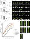
Silencing of GbCYP86A1‐1, GbCYP86A1‐2, GbCYP86A1‐3 and H3091 in the resistant cultivar G. barbadense cv. Hai7124 leads to increased susceptibility to Verticillium dahliae infection. Gene‐specific DNA fragment for specifically silencing one of the three GbCYP86A1s and conserved region of these three genes (named as H3091) were selected as the target, respectively.(a) Verification of GbCYP86A1‐1, GbCYP86A1‐2 and GbCYP86A1‐3 silencing by qRTPCR in different VIGS plants. Asterisks indicate statistically significant differences, as determined by Student's t‐test (**< 0.01). (b) Disease symptoms of GbCYP86A1‐1, GbCYP86A1‐2, GbCYP86A1‐3 and H3091 silenced cotton plants at 20, 25 and 30 days after V. dahliae inoculation. (c)Disease progression curves in GbCYP86A1‐1, GbCYP86A1‐2, GbCYP86A1‐3 and H3091 silenced cotton plants after V. dahliae inoculation. Each biological repeat contains at least 30 seedlings. Error bars represent the standard deviation of three biological replicates. (d) Vascular discoloration in GbCYP86A1‐1, GbCYP86A1‐2, GbCYP86A1‐3 and H3091 silenced cotton plants compared with the controls (Hai7124 and TRV:00) after inoculation with V991 and Hai7124 without inoculation(H2O). Photographs were taken by stereoscope (Olympus MVX10, Tokyo, Japan) at 15 dpi.
Figure 3
Figure 3
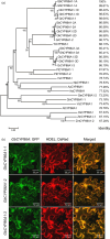
Characterization of GbCYP86A1 genes. (a) Phylogenetic trees of CYP86A1s from G. raimondii (Gr), G. hirsutum (Gh), G. barbadense (Gb), Theobroma cacao (Tc), Populus trichocarpa (Pt), Vitis vinifera (Vv), Nicotiana attenuata (Na), Artemisia annua (Aa), Helianthus annuus (Ha), Glycine max (Gm), Vigna angularis (Va), Arabidopsis thaliana (At), Oryza sativa (Os) and Zea mays (Zm) plants. The neighbour‐joining tree was constructed using the MEGA5.1 program ( http://www.megasoftware.net/). Identity value was relative to GbCYP86A1‐1 and was calculated using software DNAMAN ( http://www.lynnon.com/). (b) Localization of GbCYP86A1s in tobacco epidermal cells by GFP fusion. HDEL: DsRed (red) is an endoplasmic reticulum (ER) marker. The results show that GbCYP86A1s colocalize with the ER marker. Bars = 50 μm.
Figure 4
Figure 4
GbCYP86A1s overexpression confers resistance to Verticillium dahliae infection in Arabidopsis. (a) Identification of Arabidopsis transgenic lines overexpressing GbCYP86A1s. The presence of GbCYP86A1s genes was verified by PCR using genomic DNA (lower panel), and GbCYP86A1 transcript levels in each line were quantified by qRTPCR using AtUbq5 (At3g62250) as the internal control (upper panel). WT, Arabidopsis Columbia‐0 (Col‐0); OE1 to OE6 represents the different GbCYP86A1 overexpression lines. Error bars represent the standard deviation of three biological replicates.(b) GbCYP86A1s overexpression enhanced resistance to V. dahliae infection in Arabidopsis. 4‐week‐old Arabidopsis plants were inoculated with V. dahliae and replanted in soil, with at least 30 plants for each line, and photographed two weeks after inoculation. The mock plants were WT without V. dahliae infection. (c) Statistical analysis of disease grade and disease index in transgenic Arabidopsis plants and the WT control. The disease grade was classified as five levels as described in Materials and methods. The data were generated from three replicates, with each contains 30 Arabidopsis plants. (d) qPCR analysis of fungal biomass in different transgenic and the WT Arabidopsis plants. DNAs of roots, stems and leaves were extracted from plants 10 days post‐inoculation by V. dahliae. Error bars represent the standard deviation of three biological replicates. Statistical analyses were performed using Student's t‐test (*< 0.05, **< 0.01). (e) Fungal recovery experiments. Stem sections of Arabidopsis transgenic lines and the WT control 7 days post‐inoculation were cut and placed on potato dextrose agar plates and incubated at 25°C. Photographs were taken at 3d after culture.
Figure 5
Figure 5
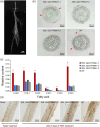
GbCYP86A1‐1 overexpression promotes lipid production in cell walls and hinders root invasion by Verticillium dahliae in Arabidopsis. (a) Zone of Arabidopsis root used for cross section analysis. The cross section was marked by red dashed line. (b) Bright field microscopic picture of representative cross sections of Arabidopsis root stained with the lipophilic dye Sudan 7B. The red‐stained parts represent suberin deposited in the periderm of the roots in secondary developmental stage cell walls. Red arrow marks the periderm cell. Bars = 50 μm. (c) Relative content of fatty acid in the WT and transgenic line roots. Error bars represent the standard deviation of three biological replicates. The asterisks indicate statistically significant differences between the transgenic and WT plants (*P < 0.05, **< 0.01, Student's t‐test). (d) Visualization of V. dahliae accumulation in Arabidopsis root. Bright field microscopic picture showed representative longitudinal section of the Arabidopsis root and was taken 3 days post‐inoculation, Bars = 100 μm. The red arrow marks the invading black mycelium.
Figure 6
Figure 6
RNA‐seq reveals that GbCYP86A1‐1 overexpression affects lipid‐related pathways and immunity‐related pathways in root of Arabidopsis.(a) GO enrichment analysis of the differentially expressed genes in roots of GbCYP86A1‐1 transgenic line and the WT control. The RNAs isolated from roots of three individual plants were used for RNA‐seq. P value of 0.05 adjusted by false discovery rate (FDR). Rich factor: Percentage of enriched genes comparing with background in corresponding GO term. (b) The number of differentially expressed genes between GbCYP86A1‐1 transgenic Arabidopsis plants and WT after challenged with Verticillium dahliae for 3 days. GO terms were separately shown by enrichment analysis. P value of 0.05 adjusted by false discovery rate (FDR).
Figure 7
Figure 7
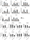
Pathogenesis‐related (PR) gene expression assays.(a) The relative expression of PR genes in resistant cultivar Hai7124 and susceptible cultivar Junmian 1 roots after inoculation with Verticillium dahliae strain V991. The statistical analyses were performed by comparing expression levels at different time points after V. dahliae infection to 0 h without inoculation. (b) The effects of GbCYP86A1‐1 silencing on PR genes expression in Hai7124. Error bars show the standard deviation of three biological replicates. Statistical analyses were performed using Student's t‐test (*< 0.05, **< 0.01).
Figure 8
Figure 8
Model of GbCYP86A1‐1 function in Verticillium dahliae –host interaction. The cell wall of plant root epidermis is the first barrier against pathogen attack, and expression of GbCYP86A1‐1 will accelerate suberization in cell walls and induce structural resistance. Fatty acids can be polymerized into the suberin monomers in the endoplasmic reticulum (ER), which then transported to the cell wall. LTPs and ABCs are involved in transport of suberin monomers across the plasma membrane (PM). Suberin accumulating in the cell walls plays a role in defence against the invasion of V. dahliae. Additionally, the changed lipids or the cell wall modification may activate intracellular immune responses. Pectate lyase is involved in the hydrolysis of pectin in the cell wall and the release of OGs, which may serve as signalling molecules and activate the immune system. Receptor‐like protein kinases (RLKs) and receptor‐like proteins (RLPs) located in the cell wall or plasma membrane are involved in the recognition and transmission of signals. Production of defence‐related phytohormones, such as ethylene (ET), salicylic acid (SA), jasmonate (JA), and activation of transcription factors (ERF, WRKY) lead to up‐regulation of pathogenesis‐related (PRs) genes and prevent the pathogen propagation.

Similar articles

Cited by

References

    1. Álvarez, K. and Vasquez, G. (2017) Damage‐associated molecular patterns and their role as initiators of inflammatory and auto‐immune signals in systemic lupus erythematosus. Int. Rev. Immunol. 36, 259–270. - PubMed
    1. Anders, S. and Huber, W. (2010) Differential expression analysis for sequence count data. Genome Biol. 11, R106. - PMC - PubMed
    1. Andersen, T.G. , Barberon, M. and Geldner, N. (2011) Suberization ‐ the second life of an endodermal cell. Curr. Opin. Plant Biol. 28, 9–15. - PubMed
    1. Bacete, L. , Mélida, H. , Miedes, E. and Molina, A. (2018) Plant cell wall‐mediated immunity: Cell wall changes trigger disease resistance responses. Plant J. 93, 614–636. - PubMed
    1. Beisson, F. , Li‐Beisson, Y. and Pollard, M. (2012) Solving the puzzles of cutin and suberin polymer biosynthesis. Curr. Opin. Plant Biol. 15, 329–337. - PubMed

Publication types

LinkOut - more resources