. 2019 Sep 1:136:1176-1187.
doi: 10.1016/j.ijbiomac.2019.05.149. Epub 2019 Jun 15.

Pyrimidine biosynthesis in pathogens - Structures and analysis of dihydroorotases from Yersinia pestis and Vibrio cholerae

Affiliations

Pyrimidine biosynthesis in pathogens - Structures and analysis of dihydroorotases from Yersinia pestis and Vibrio cholerae

Joanna Lipowska et al. Int J Biol Macromol. .

Abstract

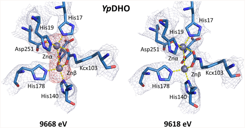
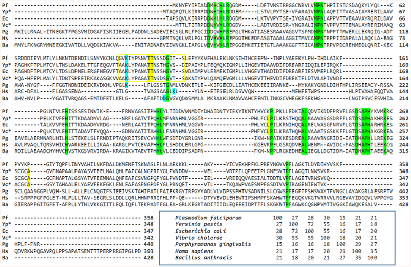
The de novo pyrimidine biosynthesis pathway is essential for the proliferation of many pathogens. One of the pathway enzymes, dihydroorotase (DHO), catalyzes the reversible interconversion of N-carbamoyl-l-aspartate to 4,5-dihydroorotate. The substantial difference between bacterial and mammalian DHOs makes it a promising drug target for disrupting bacterial growth and thus an important candidate to evaluate as a response to antimicrobial resistance on a molecular level. Here, we present two novel three-dimensional structures of DHOs from Yersinia pestis (YpDHO), the plague-causing pathogen, and Vibrio cholerae (VcDHO), the causative agent of cholera. The evaluations of these two structures led to an analysis of all available DHO structures and their classification into known DHO types. Comparison of all the DHO active sites containing ligands that are listed in DrugBank was facilitated by a new interactive, structure-comparison and presentation platform. In addition, we examined the genetic context of characterized DHOs, which revealed characteristic patterns for different types of DHOs. We also generated a homology model for DHO from Plasmodium falciparum.

Keywords: Crystal structure; Dihydroorotase; Drug target; Plasmodium falciparum; Vibrio cholera; Yersinia pestis.


Figures

Fig. 1.
Fig. 1.
Enzymes involved in the de novo pyrimidine biosynthesis pathway (left) and the reaction catalyzed by DHO (right). In bacteria and other microbes, each step of the reaction is catalyzed by a distinct enzyme. In mammals, CPSase, ATCase, and DHO together form the CAD multienzyme.
Fig. 2.
Fig. 2.
The homodimeric assemblies of YpDHO (A) and VcDHO (D), and the structures of their respective monomers (B, E) and active sites (C, F). Both YpDHO and VcDHO monomers adopt the TIM barrel structure–eight α-helices colored in red, eight parallel β-strands colored in yellow–with surrounding loops colored in green and an additional small adjacent domain colored in magenta. The metal-binding site of both enzymes is highly conserved, and it is composed of four histidine residues, one aspartate residue, and one carboxylated lysine residue. Zinc ions are presented as grey spheres, water molecules are presented as red spheres, and coordination bonds are shown as yellow dashed lines.
Fig. 3.
Fig. 3.
Comparison between electron density maps in active site regions of YpDHO calculated for datasets collected at energies above and below the zinc absorption edge (9668 eV and 9618 eV, respectively). The 2mFo-DFc maps are colored in grey and contoured at 1σ; anomalous electron density maps are colored in red and contoured at 4σ. The anomalous map calculated for the dataset above the zinc absorption edge shows strong peaks correlated with the metal ions’ positions in the binding site. Below the zinc absorption edge, these peaks completely disappear. For interactive visualizations of the electron density maps in all chains of YpDHO, the reader is referred to https://molstack.bioreproducibility.org/project/view/SlDqq9NMH7F1CPuVuAuW/.
Fig. 4.
Fig. 4.
(A) Flexible loop (defined as residues 106–116) movement upon malate binding in the active site of YpDHO. Zinc ions are shown as grey spheres, oxygen atoms in red, nitrogen atoms in blue, carbon atoms of YpDHO residues in cyan, carbon atoms of malate in light-blue, the flexible loop of chain A in magenta, side chains of Thr110 and Thr111 in red.(B) Malate-binding residues. Hydrogen bonds and coordination bonds are shown as green dashed lines with distances labelled. The malate ligand, the active site, and the respective electron density maps, including omit maps, can be inspected using an interactive figure created with Molstack at http://molstack.bioreproducibility.org/project/view/QMDKqLGs2eeM4OyXCc2P/.
Fig. 5.
Fig. 5.
Comparison of the overall structure, active sites, and main characteristic features for bacterial type I (BaDHO, PDB ID: 3MPG), bacterial type II (EcDHO, PDB ID: 1J79), mammalian CAD (HsDHO, PDB ID: 4C6C), and PfDHO (the structure by homology modeling).
Fig. 6.
Fig. 6.
Multiple sequence alignment for representatives of different DHO types, such as Bacillus anthracis (bacterial type I), Escherichia coli (bacterial type II), Porphyromonas gingivalis (bacterial type III) and Homo sapiens (CAD), together with sequences of YpDHO, VcDHO and PfDHO (Pf; NCBI Reference Sequence: XP_001348871.1). Sequences for DHO from Y. pestis and V. cholerae are starred. Residues conserved in all sequences are highlighted in green. Residues involved in zinc and substrate binding only in bacterial type II DHO are highlighted in yellow. Highly conserved residues in the flexible loop region in bacterial type II DHO are bracketed in yellow. The variable zinc-binding residue is highlighted in blue. A sequence identity matrix reflects the comparisons between any two of the seven sequences shown.

References

    1. O’Neill J, Review on antimicrobial resistance, Antimicrobial Resistance: Tackling a Crisis for the Health and Wealth of Nations, 2014.
    1. Perry RD, Fetherston JD, Yersinia pestis—etiologic agent of plague, Clin. Microbiol. Rev 10 (1) (1997) 35–66. - PMC - PubMed
    1. Welch TJ, et al., Multiple antimicrobial resistance in plague: an emerging public health risk, PLoS One 2 (3) (2007). - PMC - PubMed
    1. Sjolund-Karlsson M, et al., Drug-resistance mechanisms in Vibrio cholerae O1 outbreak strain, Haiti, 2010, Emerg. Infect. Dis 17 (11) (2011) 2151–2154. - PMC - PubMed
    1. Pang B, et al., The transmission and antibiotic resistance variation in a multiple drug resistance clade of Vibrio cholerae circulating in multiple countries in Asia, PLoS One 11 (3) (2016). - PMC - PubMed

LinkOut - more resources