Review
. 2019 Jun 14;20(12):2911.
doi: 10.3390/ijms20122911.

Nervous-Like Circuits in the Ribosome Facts, Hypotheses and Perspectives

Affiliations
Review

Nervous-Like Circuits in the Ribosome Facts, Hypotheses and Perspectives

Youri Timsit et al. Int J Mol Sci. .

Abstract

In the past few decades, studies on translation have converged towards the metaphor of a "ribosome nanomachine"; they also revealed intriguing ribosome properties challenging this view. Many studies have shown that to perform an accurate protein synthesis in a fluctuating cellular environment, ribosomes sense, transfer information and even make decisions. This complex "behaviour" that goes far beyond the skills of a simple mechanical machine has suggested that the ribosomal protein networks could play a role equivalent to nervous circuits at a molecular scale to enable information transfer and processing during translation. We analyse here the significance of this analogy and establish a preliminary link between two fields: ribosome structure-function studies and the analysis of information processing systems. This cross-disciplinary analysis opens new perspectives about the mechanisms of information transfer and processing in ribosomes and may provide new conceptual frameworks for the understanding of the behaviours of unicellular organisms.

Keywords: complexity; evolution; information processing; nervous circuit; network; neuron; protein interface; ribosome; translation.


Conflict of interest statement

The authors declare no conflict of interest.

Figures
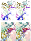
Figure 1
Figure 1
Sensing the decoding centre. Stereoviews of key r-protein and rRNA elements contacting the codon-anticodon interaction between the tRNA-A and the mRNA within the decoding centre. (A) overall view; (B) detail of the interactions; the rRNA is represented by a transparent surface coloured in the function of the RNA domains (from blue to red in the 5′-3′ direction) (pdb_id: 4y4p).
Figure 2
Figure 2
Sensing the P- and E-tRNA sites. Stereoviews of the tiny r-protein tRNA interactions in the (A) P- and (B) E-tRNA sites (pdb_id: 4y4p).
Figure 3
Figure 3
Sensing the peptidyl transferase centre (PTC). Stereoview of the PTC showing the pseudosymmetric interaction of uL2 (top) and uL3 (bottom) with the two uracile bases U 2585 and U 2518 (E. coli numbering) that undergo conformational changes upon tRNA binding. The backbones of the PTC rRNA helices are represented by red cartoons (pdb_id: 4y4p).
Figure 4
Figure 4
Sensing the interior of the tunnel. Side stereoview of the peptide tunnel showing the interactions between the r-protein uL4 (green) and uL22 (brown) with the stalled nascent peptide VemP (blue). rRNA is represented by a white transparent surface (pdb_id: 5nwy).
Figure 5
Figure 5
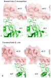
Sensing the rotational state of the two subunits. Stereoviews of the interaction in the inter-subunit bridge formed by uL5 and uS13. (A) the rotated state observed in a high-resolution structure of T. thermophilus ribosome containing the 3 tRNAs (pdb_id: 4y4p). (B) The un-rotated state observed in a high-resolution structure of the E. coli ribosome without bound tRNA (pdb_id: 4ybb).
Figure 6
Figure 6
The possible communication pathways along the r-proteins located around the PTC. (A) The view of the r-proteins are represented by transparent surfaces coloured according to the amino acids; yellow: aromatic amino acids; blue: basic amino acids. (B) The same view without the surface focused on the universal uL3–uL13 interaction. The tiny interface uL3–uL13 is represented by transparent surfaces. The position of the mutant W255C that causes translation defects in yeast is indicated by the red arrow.
Figure 7
Figure 7
The schematic representation of possible allosteric mechanisms along and between r-proteins. (A) The hypothetical mechanism for a cooperative switch formed by the stacking of an rRNA base in the r-protein array of cation-π interactions. Basic (blue), aromatic residues (yellow) or rRNA base (orange) are proposed to transfer charges or electrostatic perturbation (vertical arrow). The signal can be propagated if the rRNA base is stacked in the array and the switch is ON. The signal cannot be propagated if the base is stacked in the rRNA helix and the switch is OFF. (B) Summary of the possible combination of several allosteric mechanisms (dielectric and classic). Dielectric allostery propagates an electrostatic perturbation through array of basic (blue) and aromatic (yellow) residues (for example, coming from the binding of tRNA). This may transiently change the charge of a distant neutral residue (N) into a negative one (-) thus inducing a conformational change triggered by a local charge repulsion (or attraction).
Figure 8
Figure 8
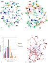
The comparison of bacterial and eukaryotic r-protein networks. Three-dimensional simplified representations of the bacterial (A) eukaryotic (B) r-protein networks. The centres of mass of the r-proteins (coloured spheres) are connected by red lines. The proteins are coloured in the function of their number of interacting partners. White: 0; blue: 1; cyan: 2; green: 3; yellow: 4; orange: 5; brown: 6; red brick: 7; red: 8. (C) 3D simplified representation of the universal network; the three tRNAs and the mRNA are represented by transparent surfaces. (D) Histogram reporting the degree distribution (inter-protein contacts) in bacterial (blue) and eukaryotic (orange) ribosome r-protein networks.
Figure 9
Figure 9
The representation of the bacterial r-protein network. (A) A schematic representation of the r-protein nodes (coloured circles), tRNAs (grey squares), mRNA (pale blue line) and the functional centres (coloured ellipsoids) interconnected by black lines that join either the globular domains (circles) or the r-protein extensions (codified by symbols represented in the box). The colours are used to differentiate the functional module or sub-networks. (B) Stereoview of the r-proteins and tRNAs in the bacterial ribosome from the A-site tRNA. The modular organisation of r-proteins is shown by colour codes used in (A).
Figure 9
Figure 9
The representation of the bacterial r-protein network. (A) A schematic representation of the r-protein nodes (coloured circles), tRNAs (grey squares), mRNA (pale blue line) and the functional centres (coloured ellipsoids) interconnected by black lines that join either the globular domains (circles) or the r-protein extensions (codified by symbols represented in the box). The colours are used to differentiate the functional module or sub-networks. (B) Stereoview of the r-proteins and tRNAs in the bacterial ribosome from the A-site tRNA. The modular organisation of r-proteins is shown by colour codes used in (A).
Figure 10
Figure 10
The possible communication pathways depicted by the blue, yellow and red arrays, between the functional sites within the r-protein network.

Similar articles

Cited by

References

    1. Bray D. Protein molecules as computational elements in living cells. Nature. 1995;376:307–310. doi: 10.1038/376307a0. - DOI - PubMed
    1. Poirot O., Timsit Y. Neuron-Like networks between ribosomal proteins within the ribosome. Sci. Rep. 2016;6:26485. doi: 10.1038/srep26485. - DOI - PMC - PubMed
    1. Bullock H., Horridge A. Structure and Function in the Nervous Systems of Invertebrates. W.H. Freeman; San Francisco, CA, USA: 1965.
    1. Moroz L.L. On the independent origins of complex brains and neurons. Brain Behav. Evol. 2009;74:177–190. doi: 10.1159/000258665. - DOI - PMC - PubMed
    1. Schmeing T.M., Ramakrishnan V. What recent ribosome structures have revealed about the mechanism of translation. Nature. 2009;461:1234–1242. doi: 10.1038/nature08403. - DOI - PubMed

LinkOut - more resources