Review
. 2019 Jun 14;10(6):453.
doi: 10.3390/genes10060453.

The cGMP Pathway and Inherited Photoreceptor Degeneration: Targets, Compounds, and Biomarkers

Affiliations
Review

The cGMP Pathway and Inherited Photoreceptor Degeneration: Targets, Compounds, and Biomarkers

Arianna Tolone et al. Genes (Basel). .

Abstract

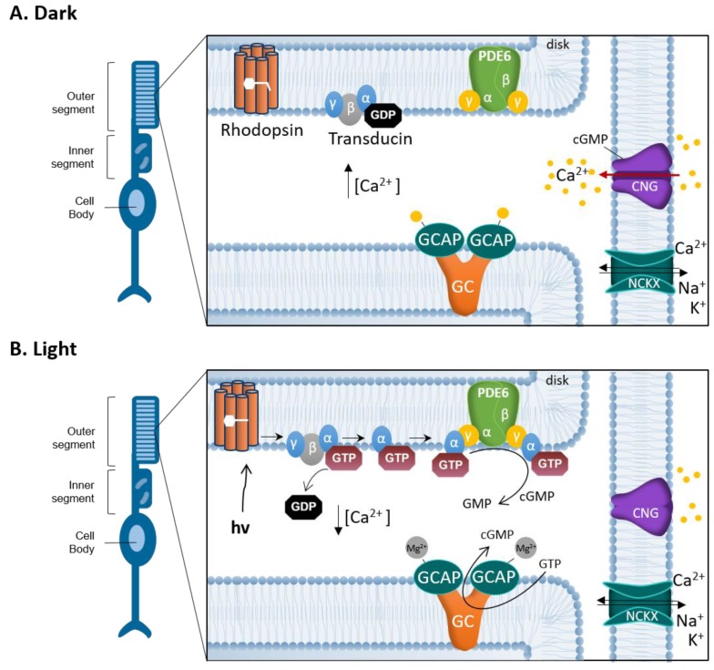
Photoreceptor physiology and pathophysiology is intricately linked to guanosine-3',5'-cyclic monophosphate (cGMP)-signaling. Here, we discuss the importance of cGMP-signaling for the pathogenesis of hereditary retinal degeneration. Excessive accumulation of cGMP in photoreceptors is a common denominator in cell death caused by a variety of different gene mutations. The cGMP-dependent cell death pathway may be targeted for the treatment of inherited photoreceptor degeneration, using specifically designed and formulated inhibitory cGMP analogues. Moreover, cGMP-signaling and its down-stream targets may be exploited for the development of novel biomarkers that could facilitate monitoring of disease progression and reveal the response to treatment in future clinical trials. We then briefly present the importance of appropriate formulations for delivery to the retina, both for drug and biomarker applications. Finally, the review touches on important aspects of future clinical translation, highlighting the need for interdisciplinary cooperation of researchers from a diverse range of fields.

Keywords: apoptosis; cyclic GMP; drug delivery systems; necrosis; retina; translational medicine.


Conflict of interest statement

The authors declare no conflicts of interest.

Figures

Figure 1
Figure 1
Schematic drawings of a healthy retina and a inherited retinal degeneration (IRD) retina. (A) Illustration of the various layers of an intact, healthy retina, from the retinal pigment epithelium (RPE) to the ganglion cell layer. Rod photoreceptors in the outer retina are shown in black, while cones are indicated by red, green, and blue. (B) Degenerated IRD retina. The outer nuclear layer is almost completely lost and the outer plexiform layer has essentially disappeared. Curiously, when the retina has lost all functionality, a small number of cone photoreceptors may still be present, possibly for many years beyond the loss of rod photoreceptors. BC = bipolar cell; GC = ganglion cell; MC = Müller glial cell. Note that the retinal structure has been simplified for clarity and that not all retinal cell types are shown.
Figure 2
Figure 2
Phototransduction and the photoreceptor cGMP-Ca2+ feedback loop. Schematic representation of the interplay between cGMP and Ca2+ in the photoreceptor outer segment (OS). (A) In darkness, cGMP binds to the cyclic nucleotide-gated channel (CNGC). The opening of CNGC allows for an influx of Na+ and Ca2+ into the photoreceptor OS. At the same time K+ and Ca2+ ions are constantly extruded via Na+/Ca2+/K+ exchanger (NCKX) creating a continuous influx and outflow of ions called the dark current. Ca2+ binds GC activating proteins (GCAPs), which inhibit the synthesis of cGMP by limiting guanylyl cyclase (GC) activity. (B) In light, photon (hν) absorption induces conformational changes in the rhodopsin protein. Rhodopsin stimulates the GTP-binding protein transducin to detach from heteromeric G-protein complex, by replacing bound GDP with GTP. The activated transducin α subunit binds to the PDE6 complex, abolishing the inhibitory effect exerted by its γ subunits. Activated phosphodiesterase-6 (PDE6) hydrolyses cGMP to GMP, which in turn limits the CNGC opening and leads to a reduction of Ca2+ influx. The closure of the CNGC and a hyperpolarization of the OS, due to continued activity of NCKX, promote the generation of an electro-chemical signal that is transmitted to second order neurons. When OS Ca2+ concentration is reduced, Mg2+ replaces the Ca2+ bound to GCAP, reactivating GCAP and stimulating GC to synthesize cGMP, opening the CNGC again.
Figure 3
Figure 3
Schematic structure of Rp-cGMPS analogues. Arrows illustrate typical structural positions in Rp-cGMPS used for the introduction of substituents to generate Rp-cGMPS analogues with improved inhibitory potency for cGMP-dependent protein kinase I or II. (A) Position 1, N2: Addition of β-phenyl-1,N2-etheno-modifications (PET) with varying additional substituents (R1) to Rp-cGMPS [65,66]. (B) Position 8: Addition of halogens (e.g., bromine (Br)) or sulfur-connected aromatic ring systems with varying additional substituents (R2) [65,67].
Figure 4
Figure 4
Incorporation of NAD+ as a biomarker for retinal cell death. At post-natal day 11 (P11), when compared to wild-type (WT) retina (A), photoreceptors in the rd1 mouse model (B) display marked incorporation of biotinylated NAD+. This is highly correlated with the TUNEL assay for cell death, which at the same age detects only few cells in the WT situation (C), while large numbers are detected in the rd1 outer nuclear layer (ONL; D). INL = inner nuclear layer, GCL = ganglion cell layer.

Similar articles

Cited by

References

    1. Hoon M., Okawa H., Della Santina L., Wong R.O. Functional architecture of the retina: Development and disease. Prog. Retin. Eye Res. 2014;42:44–84. doi: 10.1016/j.preteyeres.2014.06.003. - DOI - PMC - PubMed
    1. Verbakel S.K., van Huet R.A.C., Boon C.J.F., den Hollander A.I., Collin R.W.J., Klaver C.C.W., Hoyng C.B., Roepman R., Klevering B.J. Non-syndromic retinitis pigmentosa. Prog. Retin. Eye Res. 2018;66:157–186. doi: 10.1016/j.preteyeres.2018.03.005. - DOI - PubMed
    1. Chizzolini M., Galan A., Milan E., Sebastiani A., Costagliola C., Parmeggiani F. Good epidemiologic practice in retinitis pigmentosa: From phenotyping to biobanking. Curr. Genom. 2011;12:260–266. doi: 10.2174/138920211795860071. - DOI - PMC - PubMed
    1. Kennan A., Aherne A., Humphries P. Light in retinitis pigmentosa. Trends Genet. 2005;21:103–110. doi: 10.1016/j.tig.2004.12.001. - DOI - PubMed
    1. Hamel C.P. Cone rod dystrophies. Orphanet. J. Rare. Dis. 2007;2:7. doi: 10.1186/1750-1172-2-7. - DOI - PMC - PubMed

Publication types

MeSH terms

Substances

LinkOut - more resources