. 2019 Jun 14;20(12):2915.
doi: 10.3390/ijms20122915.

Cigarette Smoke Induces the Risk of Metabolic Bone Diseases: Transforming Growth Factor Beta Signaling Impairment via Dysfunctional Primary Cilia Affects Migration, Proliferation, and Differentiation of Human Mesenchymal Stem Cells

Affiliations

Cigarette Smoke Induces the Risk of Metabolic Bone Diseases: Transforming Growth Factor Beta Signaling Impairment via Dysfunctional Primary Cilia Affects Migration, Proliferation, and Differentiation of Human Mesenchymal Stem Cells

Romina H Aspera-Werz et al. Int J Mol Sci. .

Abstract

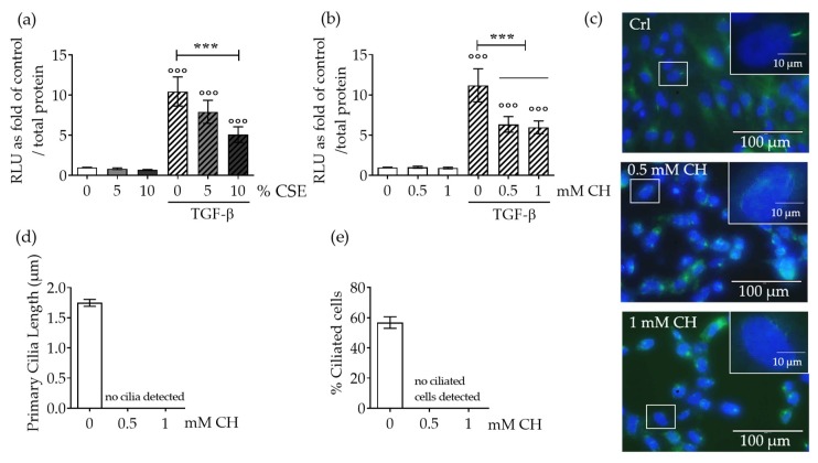
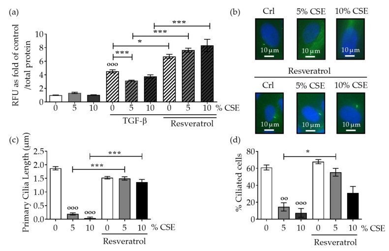
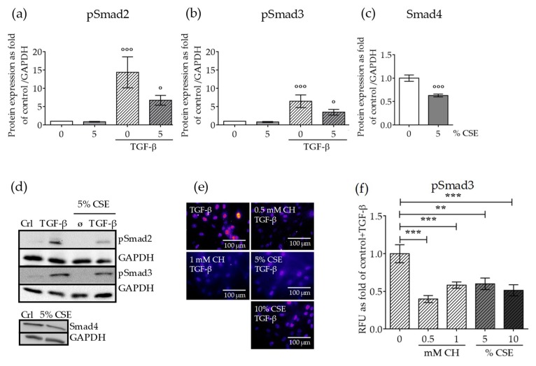
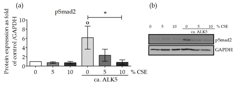
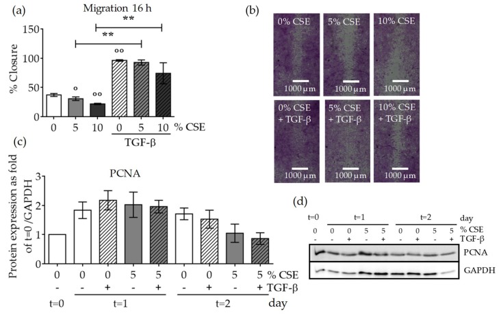
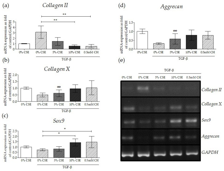
It is well established that smoking has detrimental effects on bone integrity and is a preventable risk factor for metabolic bone disorders. Following orthopedic surgeries, smokers frequently show delayed fracture healing associated with many complications, which results in prolonged hospital stays. One crucial factor responsible for fracture repair is the recruitment and differentiation of mesenchymal stem cells (MSCs) at early stages, a mechanism mediated by transforming growth factor β (TGF-β). Although it is known that smokers frequently have decreased TGF-β levels, little is known about the actual signaling occurring in these patients. We investigated the effect of cigarette smoke on TGF-β signaling in MSCs to evaluate which step in the pathway is affected by cigarette smoke extract (CSE). Single-cell-derived human mesenchymal stem cell line (SCP-1 cells) were treated with CSE concentrations associated with smoking up to 20 cigarettes a day. TGF-β signaling was analyzed using an adenovirus-based reporter assay system. Primary cilia structure and downstream TGF-β signaling modulators (Smad2, Smad3, and Smad4) were analyzed by Western blot and immunofluorescence staining. CSE exposure significantly reduced TGF-β signaling. Intriguingly, we observed that protein levels of phospho-Smad2/3 (active forms) as well as nuclear translocation of the phospho-Smad3/4 complex decreased after CSE exposure, phenomena that affected signal propagation. CSE exposure reduced the activation of TGF-β modulators under constitutive activation of TGF-β receptor type I (ALK5), evidencing that CSE affects signaling downstream of the ALK5 receptor but not the binding of the cytokine to the receptor itself. CSE-mediated TGF-β signaling impaired MSC migration, proliferation, and differentiation and ultimately affected endochondral ossification. Thus, we conclude that CSE-mediated disruption of TGF-β signaling in MSCs is partially responsible for delayed fracture healing in smokers.

Keywords: MSCs; Smad signaling; TGF-β signaling; bone metabolic diseases; cigarette smoke; fracture; osteoporosis; primary cilia; smokers.


Conflict of interest statement

The authors declare no conflict of interest.

Figures

Figure A1
Figure A1
Adenovirus infection efficiency was approximately 90%. Micrographs of SCP-1 cells infected with Ad5-green fluorescent protein (GFP) after 24 h are shown. Micrographs represent (a) bright field, (b) GFP fluorescence, and (c) the merged picture.
Figure 1
Figure 1
CSE exposure decreased TGF-β signaling by disrupting MSC primary cilia. Single-cell-derived human mesenchymal stem cell line (SCP-1 cells (N = 4, n  =  8)) infected with an adenoviral Ad5-CAGA9-MLP-Luciferase reporter constructs (Smad2/3 reporter) were exposed overnight, either with or without Cigarette smoke extract (CSE) (a; 5–10%) or chloral hydrate (CH) (b; 0.5–1 mM). Next, cultures were incubated with recombinant human transforming growth factor beta one (rhTGF-β1) 10 ng/mL for 48 h, and luciferase activity was measured in cell lysates. The results were normalized to total protein content and expressed as relative luminesce units (RLU). Results represent mean ± standard error of the mean (SEM). Statistical significance was determined by the Kruskal–Wallis H test, followed by Dunn’s post-test. Significance was established as *** p < 0.001 compared to TGF-β-treated cells and °°° p < 0.001 compared to untreated cells. (c) Representative immunostaining images of SCP-1 cells stained for acetylated α-tubulin (green), and nuclei (blue), after CH exposure. (d) Primary cilia length quantification of SCP-1 cells treated with and without CH. (e) Percentage of ciliated SCP-1 cells following CH treatment.
Figure 2
Figure 2
Resveratrol preserves the primary cilia structure from CSE and reestablishes TGF-β signaling. SCP-1 cells (N  =  3, n  =  6) infected with Ad5-CAGA9-MLP-Luciferase reporter constructs (Smad2/3 reporter) were co-incubated overnight, either with or without CSE (5–10%) and resveratrol (1 µM). Next, cultures were incubated with rhTGF-β1 (10 ng/mL) for 48 h, and (a) luciferase activity was measured in cell lysates. The results were normalized to total protein content and expressed as relative luminesce units (RLU). Results represent mean ± standard error of the mean (SEM). Statistical significance was determined by the Kruskal–Wallis H test, followed by Dunn’s post-test. Significance was established as * p < 0.05, *** p < 0.001 compared to TGF-β-treated cells and °°° p < 0.001 compared to untreated cells. (b) Representative immunostaining images of SCP-1 cells stained for acetylated α-tubulin (green), and nuclei (blue), after incubation with CSE and resveratrol. (c) Primary cilia length quantification of SCP-1 cells treated with and without CSE and resveratrol. (d) Percentage of ciliated SCP-1 cells following resveratrol treatment.
Figure 3
Figure 3
CSE exposure affected protein expression levels of canonical TGF-β signaling mediators and their nuclear translocation. SCP-1 cells (N  =  3, n  =  3) were exposed to CSE (5%) twice a week. After 14 days, the cells were treated with rhTGF-β1 (10 ng/mL) for 1 h. Protein expression of phospho-Smad2 (a), phospho-Smad3 (b), and Smad4 (c) was measured in cell lysates by Western blot and normalized to Glyceraldehyde 3-phosphate dehydrogenase (GAPDH). (d) A representative Western blot of the measured proteins is shown. SCP-1 cells (N  =  3, n = 3) were treated overnight with or without CSE (5–10%) or CH (0.5–1 mM). Next, cultures were incubated with rhTGF-β1 (10 ng/mL) for 24 h and then stained for Smad3. (e) Representative immunostaining images of nuclear localization of Smad3. The immunofluorescence signal was pseudocolored for better visualization using the fire tool in ImageJ. (f) Quantification of Smad3 nuclear translocation was performed with ImageJ. The results are expressed as mean ± SEM. Statistical significance was determined by the Kruskal–Wallis H, test followed by Dunn’s post-test. Significance was established as ** p < 0.01 or *** p < 0.001 compared to TGF-β and ° p < 0.05 or °°° p < 0.001 compared to untreated cells.
Figure 4
Figure 4
CSE reduced the protein expression of phospho-Smad2, despite a constitutively active TGF-β receptor type I (ca. ALK5). SCP-1 cells (N  =  3, n  =  3) infected with Ad5-ca. ALK5 were treated with CSE (5–10%). After 24 h, the total protein expression of phospho-Smad2 (a) was measured in cell lysates by Western blot and normalized to GAPDH. (b) A representative Western blot is shown. The results are expressed as mean ± SEM. Statistical significance was determined by the Kruskal–Wallis H test, followed by Dunn’s post-test. Significance was established as * p < 0.05 compared to circa ALK5-infected cells and ° p < 0.05 compared to untreated cells.
Figure 5
Figure 5
CSE exposure decreased MSC migration and proliferation. In order to investigate the effect of CSE on SCP-1 cell migration, a scratch assay was performed. SCP-1 cells (N ≥ 3, n ≥ 3) were co-incubated with CSE (5–10%) and rhTGF-β1 (10 ng/mL). Wound closure was determined from microscopic pictures (equation: (100 − wound area at 16 h/wound area at 0 h) × 100) with ImageJ software. (a) SCP-1 cell migration after 16 h. (b) Representative migration pictures. Cells were visualized with sulforhodamine B (SRB) staining. (c) Normalized proliferating cell nuclear antigen (PCNA) protein expression in SCP-1 cells at day 0 (t = 0) and after 24 h (t = 1) or 48 h (t = 2) stimulation with rhTGF-β1 (10 ng/mL) and with or without CSE (5% v/v). (d) A representative PCNA Western blot. The results are expressed as mean ± SEM. Statistical significance was determined by the Kruskal–Wallis H test, followed by Dunn’s post-test. Significance was established as ** p < 0.01 compared to TGF-β1-treated cells and ° p < 0.05 or °° p < 0.01 compared to untreated cells.
Figure 6
Figure 6
Disruption of TGF-β signaling with CSE-mediated primary cilia disruption affected MSC chondrogenic differentiation. In order to evaluate the gene expression of chondrocyte markers under impaired TGF-β signaling, SCP-1 cells (N = 3, n = 2) were differentiated with CSE (5–10%) and rhTGF-β1 (10 ng/mL) for 14 days. Gene expression analysis was performed with semi-quantitative RT-PCR from 10 ng cDNA. The graph represents gene expression, normalized to the GAPDH (housekeeping gene), of (a) Collagen II, (b) Collagen X, (c) Sox9, and (d) Aggrecan. The results are expressed as mean ± SEM. Statistical significance was determined by the Kruskal–Wallis H test, followed by Dunn’s post-test. Significance was established as ** p < 0.01 or * p < 0.05 compared to TGF-β1-treated cells or °° p < 0.01 compared to untreated cells. (e) A representative semi-quantitative reverse-transcription polymerase chain reaction (RT-PCR) gel picture.

References

    1. Abate M., Vanni D., Pantalone A., Salini V. Cigarette smoking and musculoskeletal disorders. Muscles Ligaments Tendons J. 2013;3:63–69. doi: 10.32098/mltj.02.2013.03. - DOI - PMC - PubMed
    1. Burns D.M. Tobacco-related diseases. Semin. Oncol. Nurs. 2003;19:244–249. doi: 10.1053/j.soncn.2003.08.001. - DOI - PubMed
    1. Kanis J.A., Johnell O., Oden A., Johansson H., De Laet C., Eisman J.A., Fujiwara S., Kroger H., McCloskey E.V., Mellstrom D., et al. Smoking and fracture risk: A meta-analysis. Osteoporos. Int. 2005;16:155–162. doi: 10.1007/s00198-004-1640-3. - DOI - PubMed
    1. Yoon V., Maalouf N.M., Sakhaee K. The effects of smoking on bone metabolism. Osteoporos. Int. 2012;23:2081–2092. doi: 10.1007/s00198-012-1940-y. - DOI - PubMed
    1. Ward K.D., Klesges R.C. A meta-analysis of the effects of cigarette smoking on bone mineral density. Calcif. Tissue Int. 2001;68:259–270. doi: 10.1007/BF02390832. - DOI - PMC - PubMed

MeSH terms

Substances