

Environment and Behavior: Neurochemical Effects of Different Diets in the Calf Brain
- PMID: 31207977
- PMCID: PMC6617313
- DOI: 10.3390/ani9060358
Environment and Behavior: Neurochemical Effects of Different Diets in the Calf Brain
Abstract
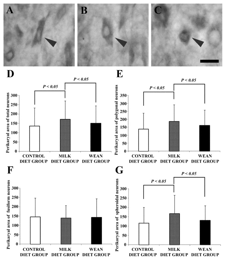
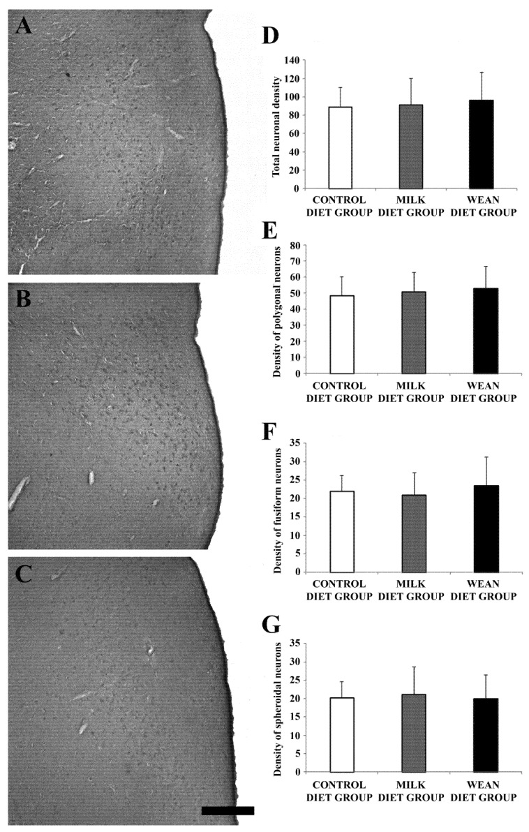
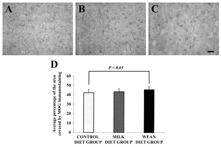
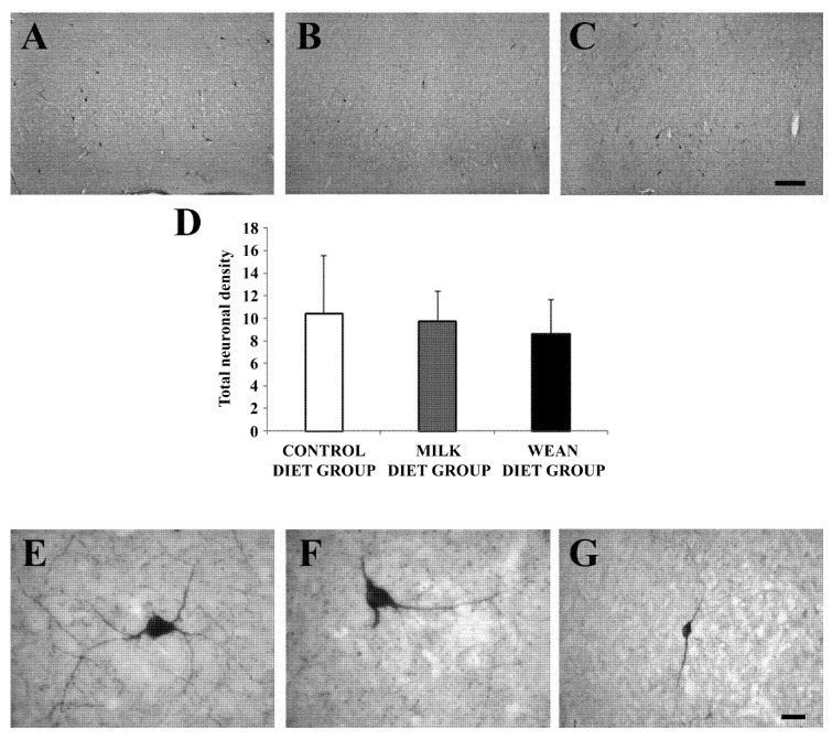
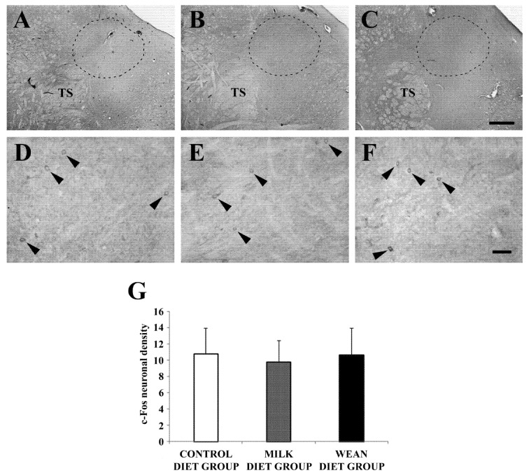
Calves reared for the production of white veal are subjected to stressful events due to the type of liquid diet they receive. Stress responses are mediated by three main stress-responsive cerebral regions: the prefrontal cortex, the paraventricular nucleus of the hypothalamus, and the nucleus of the solitary tract of the brainstem. In the present study, we have investigated the effects of different diets on these brain regions of ruminants using immunohistochemical methods. In this study, 15 calves were used and kept in group housing systems of five calves each. They were fed with three different diets: a control diet, a milk diet, and a weaned diet. Brain sections were immunostained to evaluate the distribution of neuronal nitric oxide synthase and myelin oligodendrocyte glycoprotein immunoreactivity in the prefrontal cortex; the expression of oxytocin in the paraventricular nucleus; and the presence of c-Fos in the A2 group of the nucleus of the solitary tract. The main results obtained indicate that in weaned diet group the oxytocin activity is lower than in control diet and milk diet groups. In addition, weaning appears to stimulate myelination in the prefrontal cortex. In summary, this study supports the importance of maintaining a nutritional lifestyle similar to that occurring in natural conditions.
Keywords: calf; diets; nucleus of the solitary tract; paraventricular nucleus; prefrontal cortex.
Conflict of interest statement
The authors declare no conflict of interest.
Figures
Similar articles
-
The effect of increasing the nutrient and amino acid concentration of milk diets on dairy heifer individual feed intake, growth, development, and lactation performance.J Dairy Sci. 2013 Oct;96(10):6539-49. doi: 10.3168/jds.2012-6489. Epub 2013 Aug 16. J Dairy Sci. 2013. PMID: 23958020 Clinical Trial.
-
Medial prefrontal cortex depressor response: role of the solitary tract nucleus in the rat.Neuroscience. 1999;89(4):1331-46. doi: 10.1016/s0306-4522(98)00389-3. Neuroscience. 1999. PMID: 10362318
-
Starch-protein interaction in the rumen of weaned dairy calves.J Dairy Sci. 2021 May;104(5):5445-5456. doi: 10.3168/jds.2020-19990. Epub 2021 Mar 6. J Dairy Sci. 2021. PMID: 33685686
-
Invited review: Transitioning from milk to solid feed in dairy heifers.J Dairy Sci. 2016 Feb;99(2):885-902. doi: 10.3168/jds.2015-9975. Epub 2015 Dec 17. J Dairy Sci. 2016. PMID: 26709160 Review.
-
A 100-Year Review: Calf nutrition and management.J Dairy Sci. 2017 Dec;100(12):10151-10172. doi: 10.3168/jds.2017-13062. J Dairy Sci. 2017. PMID: 29153160 Review.
Cited by
-
Using Non-Invasive Monitoring Technologies to Capture Behavioural, Physiological and Health Responses of Dairy Calves to Different Nutritional Regimes during the First Ten Weeks of Life.Animals (Basel). 2019 Oct 2;9(10):760. doi: 10.3390/ani9100760. Animals (Basel). 2019. PMID: 31581685 Free PMC article.
References
-
- Webb L.E., Bokkers E.A.M., Engel B., Berends H., Gerrits W.J.J., van Reenen C.G. Behaviour and welfare of veal calves fed different amounts of solid feed supplemented to a milk replacer ration adjusted for similar growth. Appl. Anim. Behav. Sci. 2012;136:108–116. doi: 10.1016/j.applanim.2011.12.004. - DOI
-
- Herman J.P., Figueiredo H., Mueller N.K., Ulrich-Lai Y., Ostrander M.M., Choi D.C., Cullinan W.E. Central mechanisms of stress integration: Hierarchical circuitry controlling hypothalamo-pituitary-adrenocortical responsiveness. Front. Neuroendocrinol. 2003;24:151–180. doi: 10.1016/j.yfrne.2003.07.001. - DOI - PubMed
-
- Fuster J.M. The Prefrontal Cortex. 4th ed. Academic Press; London, UK: 2008. pp. 221–270.
-
- Campos A.C., Piorino E.M., Ferreira F.R., Guimarães F.S. Increased nitric oxide-mediated neurotransmission in the medial prefrontal cortex is associated with the long lasting anxiogenic-like effect of predator exposure. Behav. Brain Res. 2013;256:391–397. doi: 10.1016/j.bbr.2013.08.006. - DOI - PubMed
LinkOut - more resources
Full Text Sources
