Case Reports
. 2019 Jun 16;16(12):2129.
doi: 10.3390/ijerph16122129.

Aluminium in Brain Tissue in Epilepsy: A Case Report from Camelford

Affiliations
Case Reports

Aluminium in Brain Tissue in Epilepsy: A Case Report from Camelford

Matthew Mold et al. Int J Environ Res Public Health. .

Abstract

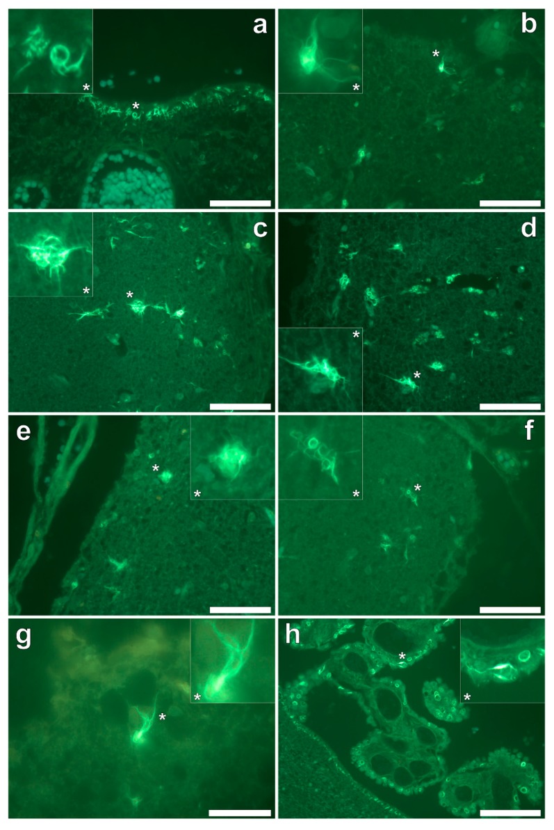
(1) Introduction: Human exposure to aluminium is a burgeoning problem. In 1988, the population of the Cornish town of Camelford was exposed to exceedingly high levels of aluminium in their potable water supply. Herein we provide evidence that aluminium played a role in the death of a Camelford resident following development of late-onset epilepsy. (2) Case summary: We have measured the aluminium content of brain tissue in this individual and demonstrated significant accumulations of aluminium in the hippocampus (4.35 (2.80) µg/g dry wt.) and the occipital lobe (2.22 (2.23) µg/g dry wt., mean, SD, n = 5), the latter being associated with abnormal calcifications. Aluminium-specific fluorescence microscopy confirmed the presence of aluminium in both of these tissues and made the consistent observation of aluminium-loaded glial cells in close proximity to aluminium-rich cell/neuronal debris. These observations support an inflammatory component in this case of late-onset epilepsy. Congo red failed to identify any amyloid deposits in any tissue while thioflavin S showed extensive extracellular and intracellular tau pathologies. (3) Discussion: We present the first data showing aluminium in brain tissue in epilepsy and suggest, in light of complementary evidence from scientific literature, the first evidence that aluminium played a role in the advent of this case of late-onset adult epilepsy.

Keywords: Camelford in Cornwall; aluminium in brain tissue; aluminium-specific fluorescence; epilepsy; occipital calcifications; tau pathologies.


Conflict of interest statement

The authors declare no conflict of interest.

Figures

Figure 1
Figure 1
Aluminium-loaded cells in the frontal cortex, morphologically compatible with glia, identified by punctate orange fluorescence, are in close proximity to aluminium-rich extracellular debris (a) Lumogallion staining of the parietal lobe (b) is negative for the presence of aluminium with cortical lipofuscin deposits highlighted via yellow fluorescence. Magnified inserts are denoted by asterisks, including a bright-field overlay in the lower panels. Magnification × 400, scale bars: 50 μm.
Figure 2
Figure 2
Intracellular aluminium in microglia in close proximity to extracellular deposits of aluminium-rich debris (a) and in astrocyte-like cells (b) in the occipital cortex. Magnified inserts are depicted (asterisks) with the bright field overlay shown in lower panels. Magnification × 400, scale bars: 50 μm.
Figure 3
Figure 3
Extracellular aluminium highlighted via an intense orange fluorescence emission in the white matter of the temporal lobe (a). Intracellular aluminium was also noted in the temporal cortex (b) as highlighted via punctate orange fluorescence within inflammatory cells in the vessel wall. Magnified inserts and their bright field overlays are denoted by asterisks. Magnification × 400, scale bars: 50 μm.
Figure 4
Figure 4
Intracellular aluminium in glial-like cells and within surrounding cellular debris in the parahippocampal gyrus (a) and in glial cells exhibiting astrocyte-like processes in the hippocampus (b). Magnified inserts are depicted (asterisks) with the bright field channel overlaid. Magnification × 400, scale bars: 50 μm.
Figure 5
Figure 5
Congo red stained serial tissue sections of the occipital lobe, revealing multiple spherulites. Spherulites were noted via a Maltese-Cross diffraction pattern (white circles) under fully polarised light (a,c) and an ovoid-like morphology (grey circles) under bright field illumination (b,d). Spherulites were exclusively distributed across the grey-white matter interface of the occipital lobe. Magnification × 200, scale bars: 100 μm.
Figure 6
Figure 6
Thioflavin S (ThS) staining of the frontal (a,b), parietal (c,d), occipital (e,f) and temporal lobes (g) and hippocampus (h) revealed cortical neurofibrillary tangles (NFTs) as highlighted via a green fluorescence emission. ThS-reactive NFTs were noted in pia mater, as demonstrated in the frontal cortex (a) and hippocampus (h). Ring-like NFTs were also noted in the choroid plexus (h). Magnified inserts are denoted with asterisks. Magnifications: (af): × 40, (g): × 1000, (h): × 200, scale bars: (af): 50 μm, (g): 20 μm, (h): 100 μm.

Similar articles

Cited by

References

    1. Exley C., Esiri M. Severe cerebral congophilic angiopathy coincident with increased brain aluminium in a resident of Camelford, Cornwall, UK. J. Neurol. Neurosurg. Psychiat. 2006;77:877–879. doi: 10.1136/jnnp.2005.086553. - DOI - PMC - PubMed
    1. Mirza A., King A., Troakes C., Exley C. The identification of aluminium in human brain tissue using lumogallion and fluorescence microscopy. J. Alzh. Dis. 2016;54:1333–1338. doi: 10.3233/JAD-160648. - DOI - PMC - PubMed
    1. King A., Troakes C., Aizpurua M., Mirza A., Hodges A., Al-Sarraj S., Exley C. Unusual neuropathological features and increased brain aluminium in a resident of Camelford, UK. Neuropathol. Appl. Neurobiol. 2017;43:537–541. doi: 10.1111/nan.12417. - DOI - PubMed
    1. Mold M., Cottle J., King A., Exley C. Intracellular aluminium in inflammatory and glial cells in cerebral amyloid angiopathy: A case report. Int. J. Environ. Res. Public Health. 2019;16:1459. doi: 10.3390/ijerph16081459. - DOI - PMC - PubMed
    1. Mirza A., King A., Troakes C., Exley C. Aluminium in brain tissue in familial Alzheimer’s disease. J. Trace Elem. Med. Biol. 2017;40:30–36. doi: 10.1016/j.jtemb.2016.12.001. - DOI - PubMed

Publication types