

Cerebrovascular plasticity: Processes that lead to changes in the architecture of brain microvessels
- PMID: 31208241
- PMCID: PMC6681538
- DOI: 10.1177/0271678X19855875
Cerebrovascular plasticity: Processes that lead to changes in the architecture of brain microvessels
Abstract
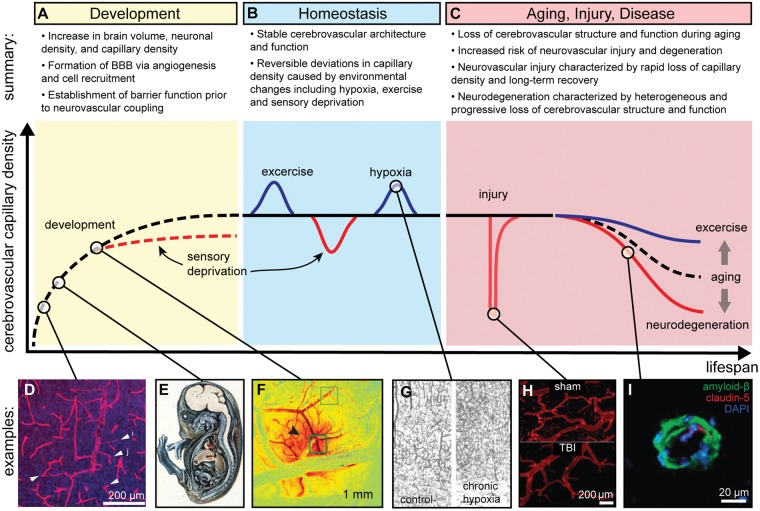
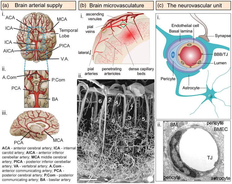
The metabolic demands of the brain are met by oxygen and glucose, supplied by a complex hierarchical network of microvessels (arterioles, capillaries, and venules). Transient changes in neural activity are accommodated by local dilation of arterioles or capillaries to increase cerebral blood flow and hence nutrient availability. Transport and communication between the circulation and the brain is regulated by the brain microvascular endothelial cells that form the blood-brain barrier. Under homeostatic conditions, there is very little turnover in brain microvascular endothelial cells, and the cerebrovascular architecture is largely static. However, changes in the brain microenvironment, due to environmental factors, disease, or trauma, can result in additive or subtractive changes in cerebrovascular architecture. Additions occur by angiogenesis or vasculogenesis, whereas subtractions occur by vascular pruning, injury, or endothelial cell death. Here we review the various processes that lead to changes in the cerebrovascular architecture, including sustained changes in the brain microenvironment, development and aging, and injury, disease, and repair.
Keywords: Cerebrovascular plasticity; blood–brain barrier; brain microvascular endothelial cells; cerebrovascular architecture; neurovascular coupling.
Figures
Similar articles
-
Neurovascular dysfunction in dementia - human cellular models and molecular mechanisms.Clin Sci (Lond). 2018 Feb 14;132(3):399-418. doi: 10.1042/CS20160720. Print 2018 Feb 14. Clin Sci (Lond). 2018. PMID: 29444850 Review.
-
Seizure-induced microvascular injury is associated with impaired neurovascular coupling and blood-brain barrier dysfunction.Epilepsia. 2019 Feb;60(2):322-336. doi: 10.1111/epi.14631. Epub 2019 Jan 4. Epilepsia. 2019. PMID: 30609012
-
Revisiting the neurovascular unit.Nat Neurosci. 2021 Sep;24(9):1198-1209. doi: 10.1038/s41593-021-00904-7. Epub 2021 Aug 5. Nat Neurosci. 2021. PMID: 34354283 Free PMC article. Review.
-
Neuronal regulation of the blood-brain barrier and neurovascular coupling.Nat Rev Neurosci. 2020 Aug;21(8):416-432. doi: 10.1038/s41583-020-0322-2. Epub 2020 Jul 7. Nat Rev Neurosci. 2020. PMID: 32636528 Free PMC article. Review.
-
The Cerebrovascular Side of Plasticity: Microvascular Architecture across Health and Neurodegenerative and Vascular Diseases.Brain Sci. 2024 Sep 28;14(10):983. doi: 10.3390/brainsci14100983. Brain Sci. 2024. PMID: 39451997 Free PMC article. Review.
Cited by
-
Aging Impairs Cerebrovascular Reactivity at Preserved Resting Cerebral Arteriolar Tone and Vascular Density in the Laboratory Rat.Front Aging Neurosci. 2019 Nov 8;11:301. doi: 10.3389/fnagi.2019.00301. eCollection 2019. Front Aging Neurosci. 2019. PMID: 31780917 Free PMC article.
-
Modeling hyperosmotic blood-brain barrier opening within human tissue-engineered in vitro brain microvessels.J Cereb Blood Flow Metab. 2020 Jul;40(7):1517-1532. doi: 10.1177/0271678X19867980. Epub 2019 Aug 8. J Cereb Blood Flow Metab. 2020. PMID: 31394959 Free PMC article.
-
Functional and Therapeutic Potential of Cynara scolymus in Health Benefits.Nutrients. 2024 Mar 17;16(6):872. doi: 10.3390/nu16060872. Nutrients. 2024. PMID: 38542782 Free PMC article. Review.
-
Dynamics of Endothelial Cell Diversity and Plasticity in Health and Disease.Cells. 2024 Jul 29;13(15):1276. doi: 10.3390/cells13151276. Cells. 2024. PMID: 39120307 Free PMC article. Review.
-
Electroacupuncture ameliorates cerebrovascular impairment in Alzheimer's disease mice via melatonin signaling.CNS Neurosci Ther. 2023 Mar;29(3):917-931. doi: 10.1111/cns.14027. Epub 2022 Nov 15. CNS Neurosci Ther. 2023. PMID: 36382345 Free PMC article.
References
-
- Aiello LC, Wheeler P. The expensive-tissue hypothesis – The brain and the digestive-system in human and primate evolution. Curr Anthropol 1995; 36(2): 199–221.
-
- Zlokovic BV. Neurovascular mechanisms of Alzheimer’s neurodegeneration. Trends Neurosci 2005; 28(4): 202–208. - PubMed
Publication types
MeSH terms
Grants and funding
LinkOut - more resources
Full Text Sources
Miscellaneous
