. 2020 Apr;16(4):600-614.
doi: 10.1080/15548627.2019.1632620. Epub 2019 Jun 28.

GABARAPs and LC3s have opposite roles in regulating ULK1 for autophagy induction

Affiliations

GABARAPs and LC3s have opposite roles in regulating ULK1 for autophagy induction

Douglas S Grunwald et al. Autophagy. 2020 Apr.

Abstract

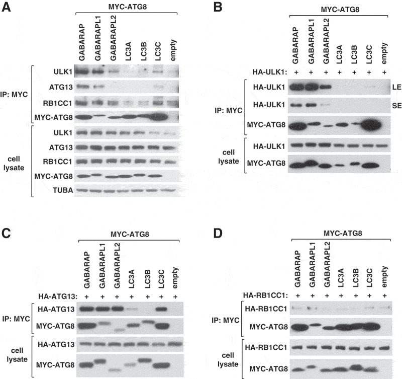
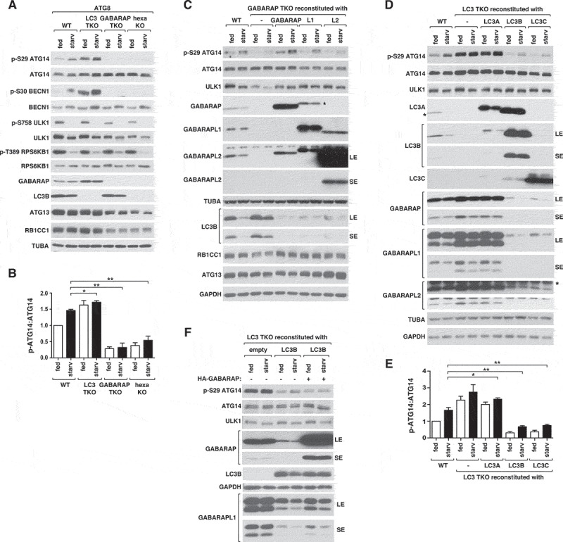
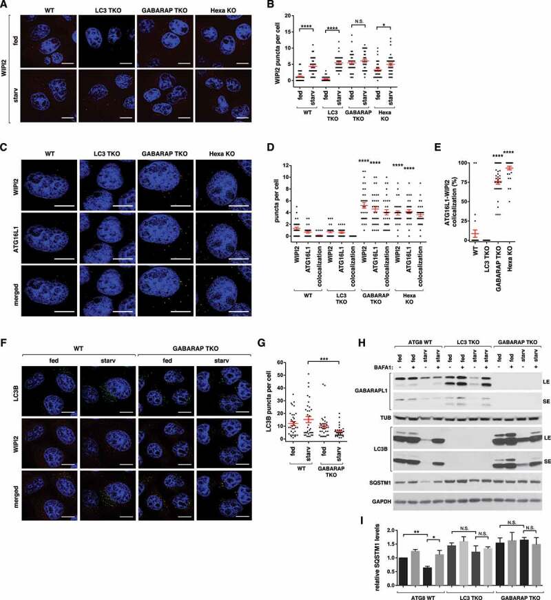
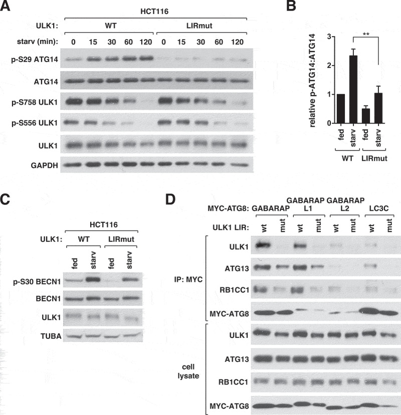
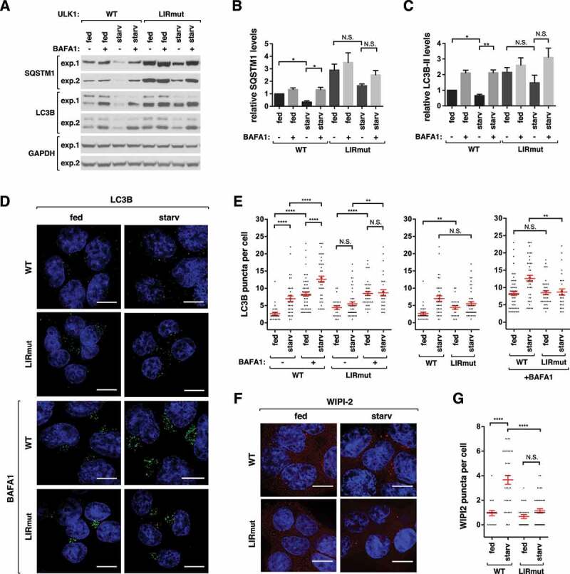
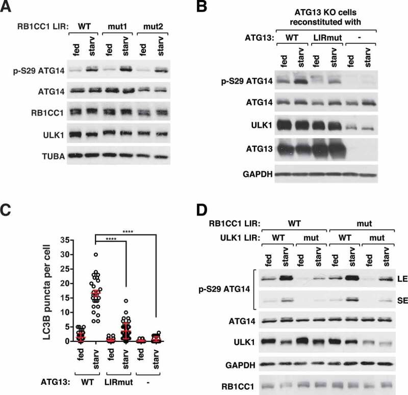
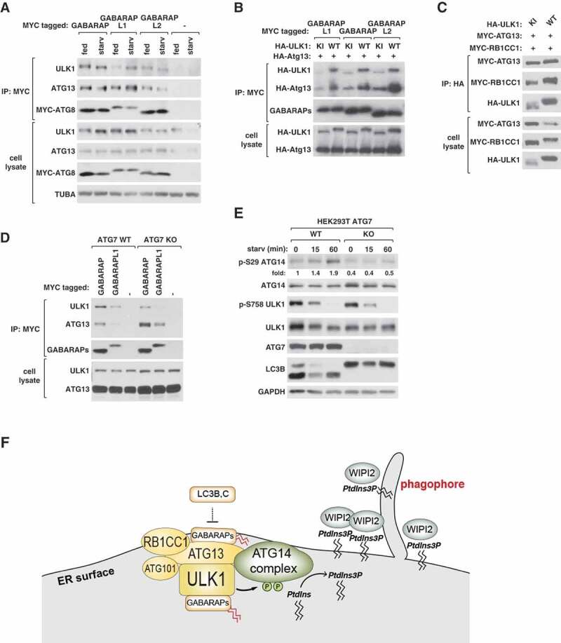
ULK1 (unc-51 like autophagy activating kinase 1) is the key mediator of MTORC1 signaling to macroautophagy/autophagy. ULK1 functions as a protein complex by interacting with ATG13, RB1CC1/FIP200, and ATG101. How the ULK1 complex is regulated to trigger autophagy induction remains unclear. In this study, we have determined roles of Atg8-family proteins (ATG8s) in regulating ULK1 activity and autophagy. Using human cells depleted of each subfamily of ATG8, we found that the GABARAP subfamily positively regulates ULK1 activity and phagophore and autophagosome formation in response to starvation. In contrast, the LC3 subfamily negatively regulates ULK1 activity and phagophore formation. By reconstituting ATG8-depleted cells with individual ATG8 members, we identified GABARAP and GABARAPL1 as positive and LC3B and LC3C as negative regulators of ULK1 activity. To address the role of ATG8 binding to ULK1, we mutated the LIR of endogenous ULK1 to disrupt the ATG8-ULK1 interaction by genome editing. The mutation drastically reduced the activity of ULK1, autophagic degradation of SQSTM1, and phagophore formation in response to starvation. The mutation also suppressed the formation and turnover of autophagosomes in response to starvation. Similar to the mutation of the ULK1 LIR, disruption of the ATG13-ATG8 interaction suppressed ULK1 activity and autophagosome formation. In contrast, RB1CC1 did not show any specific binding to ATG8s, and mutation of its LIR did not affect ULK1 activity. Together, this study demonstrates differential binding and opposite regulation of the ULK1 complex by GABARAPs and LC3s, and an important role of the ULK1- and ATG13-ATG8 interactions in autophagy induction.Abbreviations: ATG5: autophagy related 5; ATG7: autophagy related 7; ATG8: autophagy related 8; ATG13: autophagy related 13; ATG14: autophagy related 14; ATG16L1: autophagy related 16 like 1; ATG101: autophagy related 101; BAFA1: bafilomycin A1; BECN1: beclin 1; Cas9: CRISPR associated protein 9; CRISPR: clustered regularly interspaced short palindromic repeats; EBSS: earle's balanced salt solution; DAPI: 4'-6-diamidino-2-phenylindole; GABARAP: GABA type A receptor-associated protein; GABARAPL1: GABA type A receptor-associated protein like 1; GABARAPL2: GABA type A receptor-associated protein like 2; GAPDH: glyceraldehyde-3-phosphate dehydrogenase; GFP: green fluorescence protein; gRNA: guide RNA; KI: kinase inactive mutant; KO: knockout; LC3A: microtubule associated protein 1 light chain 3 alpha; LC3B: microtubule associated protein 1 light chain 3 beta; LC3C: microtubule associated protein 1 light chain 3 gamma; LIR: LC3-interacting region; MTORC1: mechanistic target of rapamycin kinase complex 1; PBS: phosphate buffered saline; PCR: polymerase chain reaction; PE: phosphatidylethanolamine; PtdIns3P: phosphatidylinositol-3-phosphate; qPCR: quantitative PCR; RB1CC1/FIP200: RB1 inducible coiled-coil 1; RPS6KB1: ribosomal protein S6 kinase B1; SEM: standard error of the mean; SQSTM1/p62: sequestosome 1; TALEN: transcription activator-like effector nuclease; TUBA: tubulin alpha; ULK1: unc-51 like autophagy activating kinase 1; WB: western blotting; WIPI2: WD repeat domain phosphoinositide interacting 2; WT: wild type.

Keywords: ATG8; GABARAP; LC3; LIR; ULK1.


Figures

Figure 1.
Figure 1.
GABARAPs and LC3s interact with endogenous ULK1 and ATG13 with differential binding affinities. (A) GABARAP, GABARAPL1, GABARAPL2 and LC3C, but not LC3A or LC3B, interact with endogenously expressed ULK1 complex proteins. MYC-tagged ATG8 proteins were transiently expressed in HEK293T cells. Endogenous ULK1, ATG13, and RB1CC1 co-immunoprecipitated with MYC-ATG8 were analyzed by western blotting (WB). (B–D) ATG8 proteins interact with ULK1 and ATG13 with varying affinities. MYC-tagged ATG8 proteins were co-expressed with HA-tagged ATG13, ULK1, or RB1CC1 in HEK293T cells. The amounts of HA-tagged proteins co-immunoprecipitated with MYC-ATG8 were analyzed by WB. LE, long exposure; SE, short exposure.
Figure 2.
Figure 2.
GABARAPs and LC3s have opposite effects on ULK1 activity. (A) GABARAPs are necessary for basal and starvation-induced activation of ULK1, whereas LC3s have negative effects on ULK1 activity. Wild-type (WT) or ATG8-depleted HeLa cells were cultured in full medium (fed) or EBSS (starv) for 1 h. ULK1 activity was assessed by WB using polyclonal antibodies specific to ATG14 phosphorylation at Ser29 and BECN1 phosphorylation at Ser30. (B) Quantitative analysis of ATG14 Ser29 phosphorylation in (A). Bar values are mean ± SEM (*p < 0.05; **p < 0.01; Student t-test, n = 3). (C) GABARAP and GABARAPL1 have positive roles for ULK1 activity. Individual GABARAPs without any tag at either N- or C-terminus were stably expressed in GABARAP TKO HeLa cells and treated as described in (A). GABARAPL1 marked by * indicates cross-reactivity with anti-GABARAP antibody. (D) LC3B and LC3C have negative effects on ULK1 activity. Individual members of LC3s without any tag were stably expressed in LC3 TKO HeLa cells and treated as described in (A). The bands marked by * are LC3B that cross-reacted with anti-LC3A antibody and a protein that is non-specifically recognized by anti-GABARAPL2. (E) Quantitative analysis of ATG14 Ser29 phosphorylation in (D). Bar values are mean ± SEM (*p < 0.05; **p < 0.01 relative to starved WT cells; Student t-test, n = 3). (F) The negative effect of LC3B on ULK1 activity does not require downregulation of GABARAP expression. LC3 TKO HeLa cells stably reconstituted with an empty vector or untagged LC3B were transiently transfected to express untagged GABARAP. The fed and starvation conditions were as described in (A).
Figure 3.
Figure 3.
GABARAPs are required for proper control of autophagy initiation and flux. (A) Wild-type (WT) or ATG8-depleted HeLa cells were cultured in full medium or EBSS for 1 h. Endogenous WIPI2 (red) was detected by immunostaining using anti-WIPI2 antibody (EMD-Millipore, MABC91). Nuclei were stained with DAPI (blue). Scale bar: 10 μm. (B) Quantitative analysis of (A). Red bars are mean ± SEM (*p < 0.05; ****p < 0.0001; N.S., not significant; Student t-test, n > 30). (C) Endogenous WIPI2 (red) and ATG16L1 (green) were detected in HeLa cells described in (A) by immunostaining cells cultured in full medium. Scale bar: 10 μm. (D) Quantitative analysis of (C). Red bars are mean ± SEM. The statistical significance (****p < 0.0001; Student t-test, n > 30) is relative to WIPI2 and ATG16L1 puncta in WT cells. (E) Percent of WIPI2 puncta that colocalize with ATG16L1 puncta per cell. The values were obtained from the quantitative analysis of (D). (F) WT or GABARAP TKO HeLa cells were cultured in full medium or EBSS for 3 h. Endogenous LC3B (green) was detected by immunostaining using anti-LC3B antibody (MBL, PM036). Nuclei were stained with DAPI (blue). Scale bar: 10 μm. (G) Quantitative analysis of (F). Red bars are mean ± SEM (***p < 0.001; Student t-test, n > 30) (H) GABARAPs are important for autophagic flux of LC3B and autophagic degradation of SQSTM1. WT, LC3 TKO, or GABARAP TKO HeLa cells were cultured in full medium or EBSS for 3 h in the presence or absence of 100 nM bafilomycin A1 (BAFA1). (I) Quantitative analysis of SQSTM1 levels in (H) (*p < 0.05; **p < 0.01; N.S., not significant; Student t-test).
Figure 4.
Figure 4.
ATG8 binding to ULK1 is important for ULK1 activity. (A) ATG8 binding to ULK1 is important for the phosphorylation of ATG14 Ser29 by ULK1 in response to starvation. HCT116 cells whose genome was modified to express LIR-mutated ULK1 (LIRmut) and unmodified HCT116 cells (WT) were cultured in EBSS for the indicated periods of time. (B) Quantitative analysis of ATG14 Ser29 phosphorylation at 60 min of starvation in (A). Bar values are mean ± SEM (**p < 0.01; Student t-test, n = 3). (C) ATG8 binding to ULK1 is important for the phosphorylation of BECN1 Ser30 by ULK1 in response to starvation. WT or ULK1 LIR mutant HCT116 cells were cultured in full medium or EBSS for 1 h. (D) ATG8 binding to ULK1 stabilizes the interactions of ATG13 and RB1CC1 with ATG8. MYC-ATG8s were transiently expressed in WT or ULK1 LIR mutant HEK293T cells and immunoprecipitated using anti-MYC antibody.
Figure 5.
Figure 5.
ATG8 binding to ULK1 is important for autophagy. (A) ATG8 binding to ULK1 is important for autophagic degradation of SQSTM1 and autophagic flux of LC3B. WT or ULK1 LIR mutant HCT116 cells were cultured in full media or EBSS for 2 h in the presence or absence of 100 nM BAFA1. Blots from 2 independent experiments are shown. (B-C) Quantitative analysis of SQSTM1 and LC3B-II from (A). The protein levels were normalized based on the levels of GAPDH. Bar values are mean ± SEM (*p < 0.05; **p < 0.01; Student t-test, n = 3). (D) ATG8 binding to ULK1 is important for starvation induced LC3B puncta formation. WT or ULK1 LIR mutant HCT116 cells were cultured in full medium or EBSS for 2 h in the presence or absence of 100 nM BAFA1. LC3B (green) was detected by immunostaining of endogenous LC3B using anti-LC3B antibody (MBL, PM036). Nuclei were stained with DAPI (blue). Scale bar: 10 μm. (E) Quantitative analysis of (D). Red bars are mean ± SEM (**p < 0.01; ****p < 0.0001; Student t-test, n > 25). (F) ATG8 binding to ULK1 is important for starvation induced WIPI2 puncta formation. WT or ULK1 LIR mutant HCT116 cells were cultured in full medium or EBSS for 1 h. WIPI2 (red) was detected by immunostaining endogenous WIPI2 using anti-WIPI2 antibody (EMD-Millipore, MABC91). Scale bar: 10 μm. (G) Quantitative analysis of (F). Red bars are mean ± SEM (****p < 0.0001; Student t-test, n > 20).
Figure 6.
Figure 6.
ATG8 binding to ATG13, but not RB1CC1, is important for ULK1 activity. (A) RB1CC1 LIR mutation does not affect ULK1 activity. WT or RB1CC1 LIR mutant HCT116 cells were cultured in full medium or EBSS for 1 h. Two LIR mutant clones (mut1 and mut2) were analyzed. (B) ATG8 binding to ATG13 is important for ULK1 activity. ATG13-depleted HCT116 cells stably reconstituted with ATG13 WT, ATG13 LIR mutant (I447A/D448A) or empty vector were cultured in full medium or EBSS for 1 h. (C) ATG8 binding to ATG13 is important for starvation-induced formation of LC3B puncta. Red bars are mean ± SEM (****p < 0.0001; Student t-test, n > 25). (D) Disruption of both the ULK1-ATG8 interaction and the RB1CC1-ATG8 interaction does not completely suppress ULK1 activity. The ULK1 LIR mutation was introduced in RB1CC1 LIR mutant cells by CRISPR-Cas9-assisted genome editing. Cells were cultured in full medium or EBSS for 1 h.
Figure 7.
Figure 7.
GABARAP bindings to ULK1 and ATG13 are regulated by starvation and PE conjugation. (A) Amino acid starvation enhances the interaction of GABARAPL1 with ULK1 and ATG13. MYC-GABARAPs were transiently expressed in HEK293T cells. Cells were cultured in full medium or EBSS for 1 h. The amounts of endogenous ULK1 and ATG13 interacting with MYC-GABARAPs were analyzed by co-immunoprecipitation and WB. (B) The interactions of GABARAPs with ULK1 and ATG13 depend on the kinase activity of ULK1. MYC-tagged GABARAPs were transiently expressed in HEK293T cells together with HA-tagged ATG13 and HA-tagged WT or kinase inactive (KI) ULK1. The amounts of HA-ULK1 and HA-ATG13 co-immunoprecipitated with MYC-GABARAPs were analyzed by WB. (C) ULK1 kinase activity is not required for the interaction between ULK1, ATG13, and RB1CC1. HEK293T cells were transiently transfected to express the indicated proteins. (D) ATG7 depletion suppresses the interaction of GABARAPs with ULK1 and enhances the interaction with ATG13. MYC-GABARAPs were transiently expressed in ATG7 WT and ATG7-depleted (KO) HEK293T cells. Cells were cultured in full medium or EBSS for 1 h before co-immunoprecipitation was conducted as described in (A). (E) ATG7 is important for starvation-induced activation of ULK1. ATG7 WT or KO HEK293T cells were treated as described in (A). Numbers below the p-ATG14 blot represent fold changes of the band intensities relative to the intensity of the first lane band. (F) Diagram showing the interactions and relations between the autophagy initiation machinery and ATG8s.

Similar articles

Cited by

References

    1. Jung CH, Jun CB, Ro SH, et al. ULK-Atg13-FIP200 complexes mediate mTOR signaling to the autophagy machinery. Mol Biol Cell. 2009. April;20(7):1992–2003. - PMC - PubMed
    1. Mercer CA, Kaliappan A, Dennis PB.. A novel, human Atg13 binding protein, Atg101, interacts with ULK1 and is essential for macroautophagy. Autophagy. 2009. July;5(5):649–662. - PubMed
    1. Hosokawa N, Hara T, Kaizuka T, et al. Nutrient-dependent mTORC1 association with the ULK1-Atg13-FIP200 complex required for autophagy. Mol Biol Cell. 2009. April;20(7):1981–1991. - PMC - PubMed
    1. Ganley IG, Lam Du H, Wang J, et al. ULK1.ATG13.FIP200 complex mediates mTOR signaling and is essential for autophagy. J Biol Chem. 2009. May 1;284(18):12297–12305. - PMC - PubMed
    1. Kim J, Kundu M, Viollet B, et al. AMPK and mTOR regulate autophagy through direct phosphorylation of Ulk1. Nat Cell Biol. 2011. February;13(2):132–141. - PMC - PubMed

Publication types

MeSH terms

Substances