. 2019 Jun 17;20(1):223.
doi: 10.1186/s12882-019-1394-y.

Protective effect of anisodamine in rats with glycerol-induced acute kidney injury

Affiliations

Protective effect of anisodamine in rats with glycerol-induced acute kidney injury

Yun-Feng Li et al. BMC Nephrol. .

Abstract

Background: Anisodamine is used for the treatment of reperfusion injury in various organs. In this study, we investigated the effectiveness and mechanisms of action of anisodamine in promoting recovery from glycerol-induced acute kidney injury (AKI).

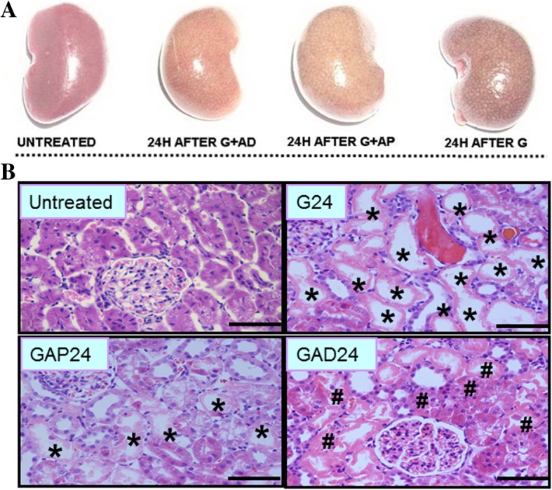
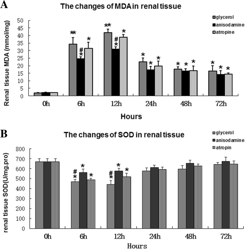
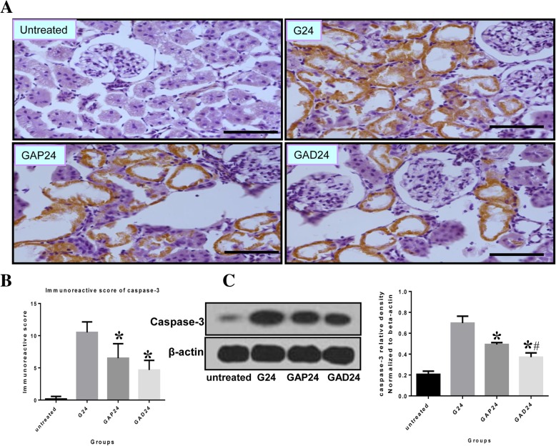
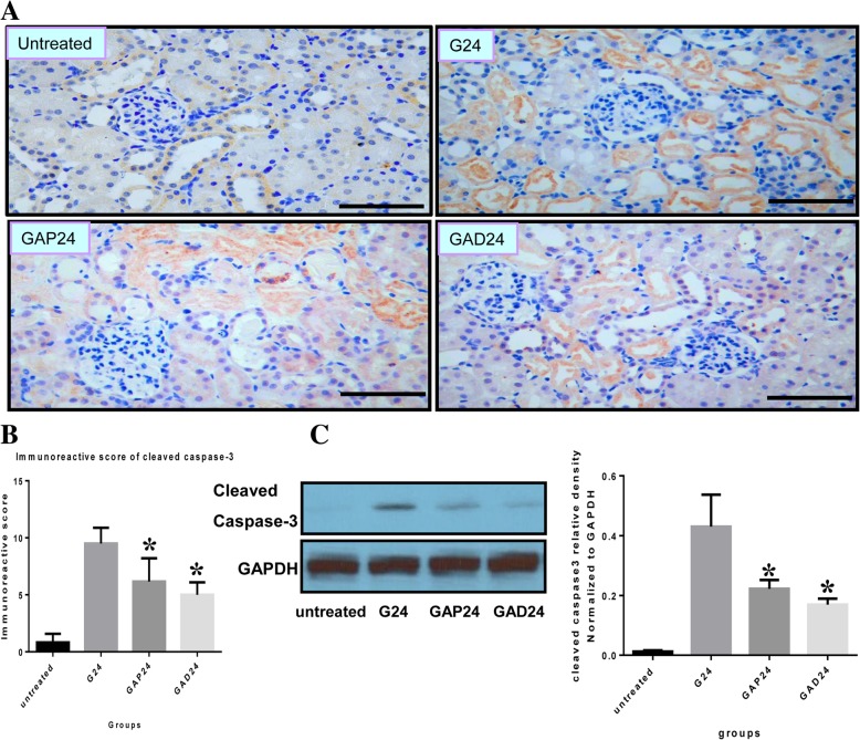
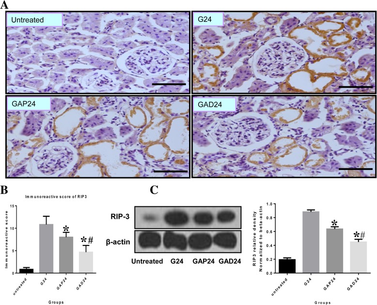
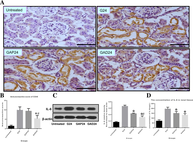
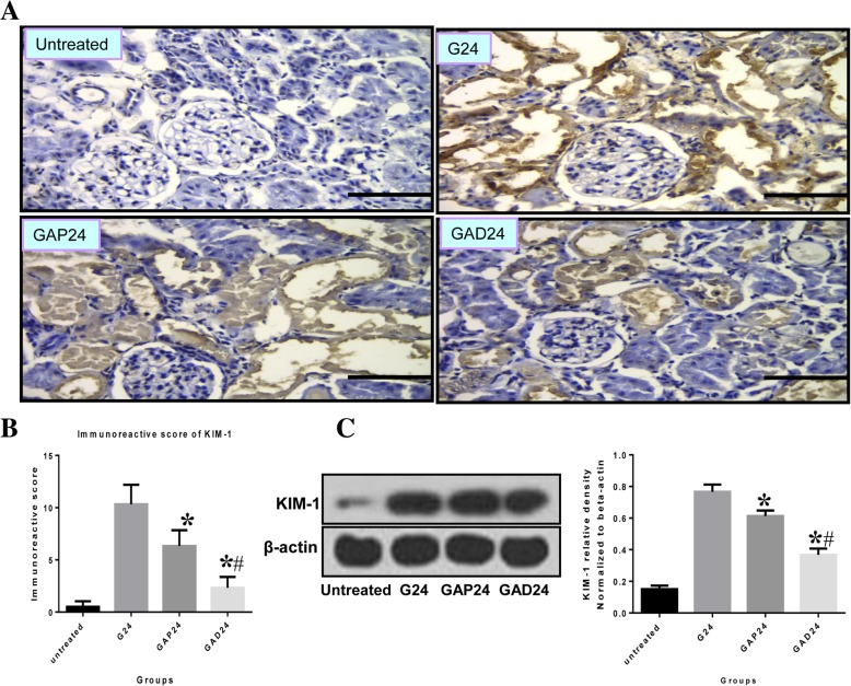
Methods: We compared the protective effects of atropine and anisodamine in the rat model of glycerol-induced AKI. We examined signaling pathways involved in oxidative stress, inflammation and apoptosis, as well as expression of kidney injury molecule-1 (KIM-1). Renal injury was assessed by measuring serum creatinine and urea, and by histologic analysis. Rhabdomyolysis was evaluated by measuring creatine kinase levels, and oxidative stress was assessed by measuring malondialdehyde (MDA) and superoxide dismutase (SOD) levels in kidney tissues. Inflammation was assessed by quantifying interleukin 6 (IL-6) and CD45 expression. Apoptosis and necrosis were evaluated by measuring caspase-3 (including cleaved caspase 3) and RIP3 levels, respectively.

Results: Glycerol administration resulted in a higher mean histologic damage score, as well as increases in serum creatinine, urea, creatine kinase, reactive oxygen species (ROS), MDA, IL-6, caspase-3 and KIM-1 levels. Furthermore, glycerol reduced kidney tissue SOD activity. All of these markers were significantly improved by anisodamine and atropine. However, the mean histologic damage score and levels of urea, serum creatinine, creatine kinase, ROS and IL-6 were lower in the anisodamine treatment group compared with the atropine treatment group.

Conclusion: Pretreatment with anisodamine ameliorates renal dysfunction in the rat model of glycerol-induced rhabdomyolytic kidney injury by reducing oxidative stress, the inflammatory response and cell death.

Keywords: Acute kidney injury; Anisodamine; Atropine; Rhabdomyolysis.


Conflict of interest statement

The authors declare that they have no competing interests.

Figures

Fig. 1
Fig. 1
Representative morphological changes in kidney shown with H&E staining at different time points after glycerol treatment. *:desquamation and necrosis of tubular epithelial cells in the proximal and distal tubules; #: regenerative epithelial cells in the proximal and distal tubules
Fig. 2
Fig. 2
Effect of anisodamine and atropine on macroscopic and morphological changes. a Representative macroscopic changes in the kidney at 24 h with anisodamine and atropine administration. b Representative morphological changes in the kidney assessed by H&E staining at 24 h with anisodamine and atropine. Scale bar:50 μm. G, glycerol; (GAP), glycerol + atropine; (GAD): glycerol + anisodamine
Fig. 3
Fig. 3
Effect of advanced administration of anisodamine/atropine on kidneys in rats subjected to glycerol-induced AKI. a Serum Scr in groups of male SD rats (n = 6) given intramuscular injections of 50% glycerol (10 mL/kg) with anisodamine (1 mg/kg) and atropine (0.05 mg/kg) by intraperitoneal injection 20 min before glycerol treatment. The control group did not receive any treatment. Data are expressed as the mean ± SEM (n = 6). #, statistically significantly different from respective 0 h controls; *, statistically significantly different from rats receiving glycerol treatment alone at the corresponding time point (P < 0.05). b Serum BUN in groups of male SD rats (n = 6) given intramuscular injections of 50% glycerol (10 mL/kg), with anisodamine (1 mg/kg) and atropine (0.05 mg/kg) by intraperitoneal injection 20 min before glycerol treatment. The control group did not receive any treatment. Data are expressed as the mean ± SEM (n = 6). #, statistically significantly different from respective 0 h controls; *, statistically significantly different from rats receiving glycerol treatment alone at the corresponding time point (P < 0.05). c Serum CK in groups of male SD rats (n = 6) injected intramuscularly with 50% glycerol (10 mL/kg), with anisodamine (1 mg/kg) and atropine (0.05 mg/kg) by intraperitoneal injection 20 min before glycerol treatment. The control group did not receive any treatment. Data are expressed as the mean ± S.E. (n = 6). #, statistically significantly different from respective 0 h controls (P < 0.05). *, statistically significantly different from rats receiving glycerol treatment alone at the corresponding time point (P < 0.05)
Fig. 4
Fig. 4
Effect of advanced administration of adnisodamine/atropine on renal ROS in rats subjected to glycerol-induced AKI. a tissue MDA in groups of male SD rats (n = 7) that were given intramuscular injections 50% glycerol (10 ml/kg), with anisodamine (1 mg/kg) and atropine (0.05 mg/kg) with intraperitoneal injection before 20 min glycerol treatment. One more group did not receive any treatment. Data are expressed as mean ± SEM (n = 6). #, statistically significant from respective 0 h controls. *, statistically significant from rats receiving glycerol treatment alone at the corresponding time point (p < 0.05). b tissue SOD in groups of male SD rats (n = 6) that were given intramuscular injections 50% glycerol (10 ml/kg), with anisodamine (1 mg/kg) and atropine (0.05 mg/kg) with intraperitoneal injection before 20 min glycerol treatment. One more group did not receive any treatment. Data are expressed as mean ± SEM (n = 6), statistically significant from respective 0 h controls. *, statistically significant from rats receiving glycerol treatment alone at the corresponding time point (p < 0.05). *P < 0.05;P < 0.01 versus 6, 12,24,48,72 h; #P < 0.05; versus the value of anisodamine group at the same time point
Fig. 5
Fig. 5
Effect of administration of adnisodamine/atropine on caspase-3 in rats subjected to glycerol-induced AKI. a Caspase-3 immunostaining in cross sections of rat kidney at 24 h in untreated, glycerol-treated, and adnisodamine/atropine intervention after glycerol treatment groups over the time course. Caspase-3 positive staining was observed on the proximal tubular epithelial cells and damaged tubules. U, untreated; G, glycerol treatment alone group; GAP, glycerol atropine treatment group. GAD, glycerol adnisodamine treatment group. Bars = 50 μm. Data are the mean ± SEM from three separate experiments. b. The statistic evaluation of immunostaining of caspase-3 (*P < 0.05 versus G24 group). c Total kidney tissue extracts were analyzed for caspase-3 protein levels by western blot. Data are expressed as mean ± S.E. (n = 3), statistically significant from respective 0 h controls. *, statistically significant from rats receiving glycerol treatment alone at the corresponding time point (p < 0.05)
Fig. 6
Fig. 6
Effect of administration of adnisodamine/atropine on cleaved caspase-3 in rats subjected to glycerol-induced AKI. a Cleaved caspase-3 immunostaining in cross sections of rat kidney at 24 h in untreated, glycerol-treated, and adnisodamine/atropine intervention after glycerol treatment groups over the time course. Cleaved caspase-3 positive staining was observed on the proximal tubular epithelial cells and damaged tubules. U, untreated; G, glycerol treatment alone group; GAP, glycerol atropine treatment group. GAD, glycerol adnisodamine treatment group. Bars = 50 μm. Data are the mean ± SEM from three separate experiments. b. The statistic evaluation of immunostaining of Cleaved caspase-3 (*P < 0.05 versus G24 group). c. Total kidney tissue extracts were analyzed for cleaved caspase-3 protein levels by western blot. Data are expressed as mean ± S.E. (n = 3), statistically significant from respective 0 h controls. *, statistically significant from rats receiving glycerol treatment alone at the corresponding time point (p < 0.05)
Fig. 7
Fig. 7
Effect of administration of adnisodamine/atropine on in rats RIP-3 subjected to glycerol-induced AKI. a RIP-3 immunostaining in cross sections of rat kidney at 24 h in untreated, glycerol-treated, and adnisodamine/atropine intervention after glycerol treatment groups over the time course. RIP-3 positive staining was observed on the proximal tubular epithelial cells and damaged tubules (arrows). U, untreated; G, glycerol treatment alone group; GAP, glycerol atropine treatment group. GAD, glycerol adnisodamine treatment group. Bars = 50 μm. Data are the mean ± SEM from three separate experiments. b The statistic evaluation of immunostaining of RIP3 (*P < 0.05 versus G24 group). c Total kidney tissue extracts were analyzed for RIP-3 protein levels by western blot. Data are expressed as mean ± S.E. (n = 3), statistically significant from respective 0 h controls. *, statistically significant from rats receiving glycerol treatment alone at the corresponding time point (p < 0.05)
Fig. 8
Fig. 8
Effect of administration of adnisodamine/atropine on in rats IL-6 subjected to glycerol-induced AKI. a CD45 immunostaining in cross sections of rat kidney at 24 h in untreated, glycerol-treated, and adnisodamine/atropine intervention after glycerol treatment groups over the time course. CD45 positive staining was observed on the proximal tubular epithelial cells and damaged tubules (arrows). U, untreated; G, glycerol treatment alone group; GAP, glycerol atropine treatment group. GAD, glycerol adnisodamine treatment group. Bars = 50 μm. Data are the mean ± SEM from three separate experiments (b) The statistic evaluation of immunostaining of CD45 (*P < 0.05 versus G24 group) (c) Total kidney tissue extracts were analyzed for IL-6 protein levels by western blot. d Total kidney tissue extracts were assayed for IL-6 activity by ELISA (*P < 0.05 versus G24 group) .Data are expressed as mean ± S.E. (n = 3)
Fig. 9
Fig. 9
Effect of administration of adnisodamine/atropine on KIM-1 in rats subjected to glycerol-induced AKI. a KIM-1 immunostaining in cross sections of rat kidney at 24 h in untreated, glycerol-treated, and adnisodamine/atropine intervention after glycerol treatment groups over the time course. KIM-1 positive staining was observed on the proximal tubular epithelial cells and damaged tubules (arrows). U, untreated; G, glycerol treatment alone group; GAP, glycerol atropine treatment group. GAD, glycerol adnisodamine treatment group. Bars = 50 μm. Data are the mean ± SEM from three separate experiments. b The statistic evaluation of immunostaining of KIM-1 (*P < 0.05 versus G24 group). c Total kidney tissue extracts were analyzed for KIM-1 protein levels by western blot. Data are expressed as mean ± S.E. (n = 3)

References

    1. Holt SG, Moore KP. Pathogenesis and treatment of renal dysfunction in rhabdomyolysis. Intensive Care Med. 2001;27:803–811. doi: 10.1007/s001340100878. - DOI - PubMed
    1. Melli G, Chaudhry V, Cornblath DR. Rhabdomyolysis:an evaluation of 475 hospitalized patients. Medicine (Baltimore) 2005;84:377–385. doi: 10.1097/01.md.0000188565.48918.41. - DOI - PubMed
    1. Mousleh R, Al Laham S, Al-Manadili A. The preventive role of pioglitazone in glycerol-induced acute kidney injury in rats during two different treatment periods. Iran J Med Sci. 2018;43(2):184–194. - PMC - PubMed
    1. Gburek J, Birn H, Verroust PJ, Goj B, Jacobsen C, Moestrup SK, Willnow TE, Christensen EI. Renal uptake of myoglobin is mediated bythe endocytic receptors megalin and cubilin. Am J Physiol Renal Physiol. 2003;285:F451–F458. doi: 10.1152/ajprenal.00062.2003. - DOI - PubMed
    1. Reeder BJ, Wilson MT. Hemoglobin and myoglobin assoeiated oxidative stress: from moleular mehanisms to disease states. Curr Med Chem. 2005;12:2741–2751. doi: 10.2174/092986705774463021. - DOI - PubMed

Publication types

MeSH terms