

Disorders of mitochondrial dynamics in peripheral neuropathy: Clues from hereditary neuropathy and diabetes
- PMID: 31208522
- PMCID: PMC11533248
- DOI: 10.1016/bs.irn.2019.05.002
Disorders of mitochondrial dynamics in peripheral neuropathy: Clues from hereditary neuropathy and diabetes
Abstract
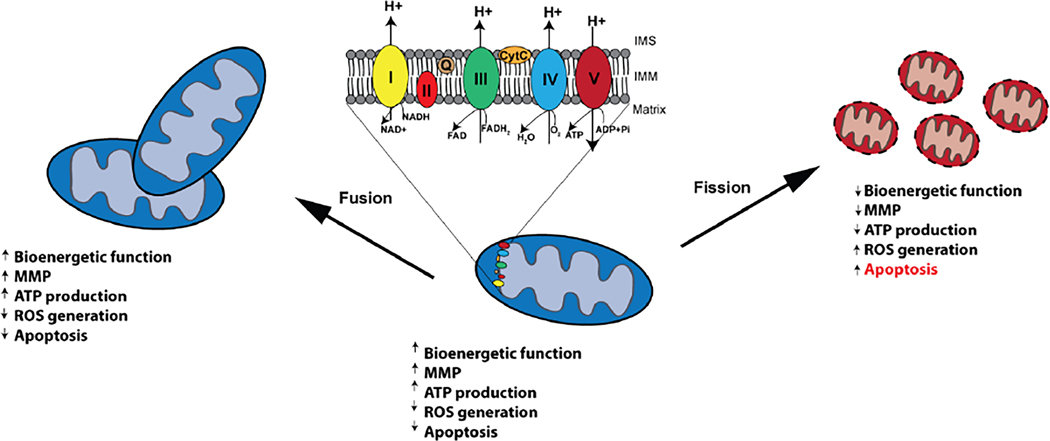
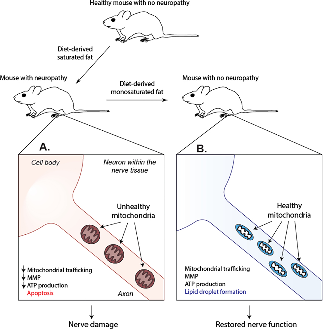
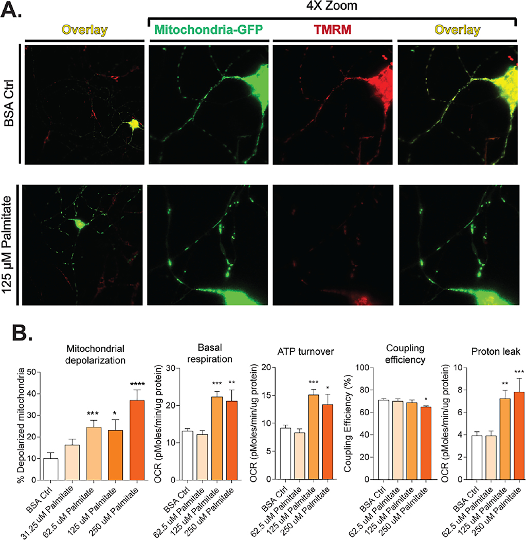
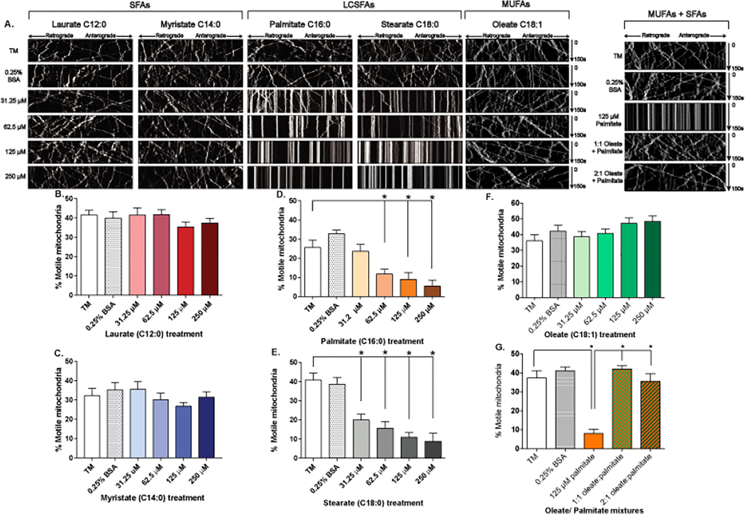
Peripheral neuropathy is a common and debilitating complication of diabetes and prediabetes. Recent clinical studies have identified an association between the development of neuropathy and dyslipidemia in prediabetic and diabetic patients. Despite the prevalence of this complication, studies identifying molecular mechanisms that underlie neuropathy progression in prediabetes or diabetes are limited. However, dysfunctional mitochondrial pathways in hereditary neuropathy provide feasible molecular targets for assessing mitochondrial dysfunction in neuropathy associated with prediabetes or diabetes. Recent studies suggest that elevated levels of dietary saturated fatty acids (SFAs) associated with dyslipidemia impair mitochondrial dynamics in sensory neurons by inducing mitochondrial depolarization, compromising mitochondrial bioenergetics, and impairing axonal mitochondrial transport. This causes lower neuronal ATP and apoptosis. Conversely, monounsaturated fatty acids (MUFAs) restore nerve and sensory mitochondrial function. Understanding the mitochondrial pathways that contribute to neuropathy progression in prediabetes and diabetes may provide therapeutic targets for the treatment of this debilitating complication.
Keywords: Bioenergetics; Charcot-Marie-Tooth disease; Diabetes; Fission; Fusion; Hereditary neuropathy; Mitochondria; Mitochondrial associated membranes; Mitochondrial trafficking; Prediabetes.
© 2019 Elsevier Inc. All rights reserved.
Figures
Similar articles
-
A Role for Fatty Acids in Peripheral Neuropathy Associated with Type 2 Diabetes and Prediabetes.Antioxid Redox Signal. 2022 Sep;37(7-9):560-577. doi: 10.1089/ars.2021.0155. Epub 2022 Apr 26. Antioxid Redox Signal. 2022. PMID: 35152728 Free PMC article. Review.
-
The Divergent Roles of Dietary Saturated and Monounsaturated Fatty Acids on Nerve Function in Murine Models of Obesity.J Neurosci. 2019 May 8;39(19):3770-3781. doi: 10.1523/JNEUROSCI.3173-18.2019. Epub 2019 Mar 18. J Neurosci. 2019. PMID: 30886017 Free PMC article.
-
High Dietary Fat Consumption Impairs Axonal Mitochondrial Function In Vivo.J Neurosci. 2021 May 12;41(19):4321-4334. doi: 10.1523/JNEUROSCI.1852-20.2021. Epub 2021 Mar 30. J Neurosci. 2021. PMID: 33785643 Free PMC article.
-
Dyslipidemia impairs mitochondrial trafficking and function in sensory neurons.FASEB J. 2018 Jan;32(1):195-207. doi: 10.1096/fj.201700206R. Epub 2017 Sep 13. FASEB J. 2018. PMID: 28904018 Free PMC article.
-
The role of aberrant mitochondrial bioenergetics in diabetic neuropathy.Neurobiol Dis. 2013 Mar;51:56-65. doi: 10.1016/j.nbd.2012.03.016. Epub 2012 Mar 9. Neurobiol Dis. 2013. PMID: 22446165 Review.
Cited by
-
Amyloid Proteins and Peripheral Neuropathy.Cells. 2020 Jun 26;9(6):1553. doi: 10.3390/cells9061553. Cells. 2020. PMID: 32604774 Free PMC article. Review.
-
Clinical Pathobiochemistry of Vitamin B12 Deficiency: Improving Our Understanding by Exploring Novel Mechanisms with a Focus on Diabetic Neuropathy.Nutrients. 2023 Jun 1;15(11):2597. doi: 10.3390/nu15112597. Nutrients. 2023. PMID: 37299560 Free PMC article. Review.
-
A Role for Fatty Acids in Peripheral Neuropathy Associated with Type 2 Diabetes and Prediabetes.Antioxid Redox Signal. 2022 Sep;37(7-9):560-577. doi: 10.1089/ars.2021.0155. Epub 2022 Apr 26. Antioxid Redox Signal. 2022. PMID: 35152728 Free PMC article. Review.
-
Multimodal Comparison of Diabetic Neuropathy in Aged Streptozotocin-Treated Sprague-Dawley and Zucker Diabetic Fatty Rats.Biomedicines. 2022 Dec 22;11(1):20. doi: 10.3390/biomedicines11010020. Biomedicines. 2022. PMID: 36672528 Free PMC article.
-
Identification and validation of NAD+ metabolism-related biomarkers in patients with diabetic peripheral neuropathy.Front Endocrinol (Lausanne). 2024 Feb 23;15:1309917. doi: 10.3389/fendo.2024.1309917. eCollection 2024. Front Endocrinol (Lausanne). 2024. PMID: 38464965 Free PMC article.
References
Publication types
MeSH terms
Grants and funding
LinkOut - more resources
Full Text Sources
Medical
