. 2019 Aug 12;39(17):e00080-19.
doi: 10.1128/MCB.00080-19. Print 2019 Sep 1.

MafB Is Important for Pancreatic β-Cell Maintenance under a MafA-Deficient Condition

Affiliations

MafB Is Important for Pancreatic β-Cell Maintenance under a MafA-Deficient Condition

Gulibaikelamu Xiafukaiti et al. Mol Cell Biol. .

Abstract

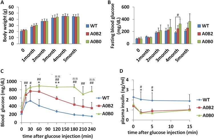
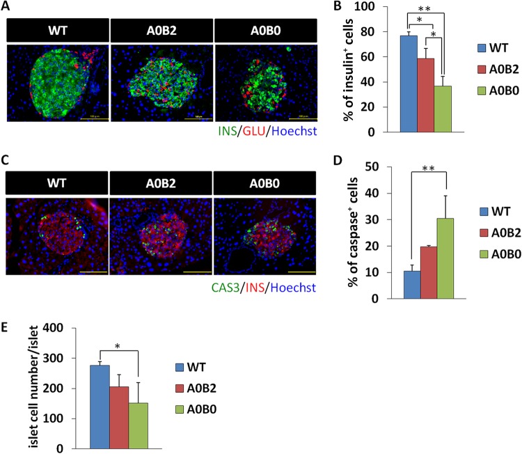
The pancreatic-islet-enriched transcription factors MafA and MafB have unique expression patterns in β cells in rodents. MafA is specifically expressed in β cells and is a key regulatory factor for maintaining adult β-cell function, whereas MafB plays an essential role in β-cell development during embryogenesis, and its expression in β cells gradually decreases and is restricted to α cells after birth in rodents. However, it was previously observed that MafB started to be reexpressed in insulin-positive (insulin+) β cells in MafA-deficient adult mice. To elucidate how MafB functions in the adult β cell under MafA-deficient conditions, we generated MafA and MafB double-knockout (A0B0) mice in which MafB was specifically deleted from β cells. As a result, the A0B0 mice became more vulnerable to diabetes under a high-fat diet (HFD) treatment, with impaired islet formation and a decreased number of insulin+ β cells because of increased β-cell apoptosis, indicating MafB can take part in the maintenance of adult β cells under certain pathological conditions.

Keywords: MafA; MafB; diabetes mellitus; pancreatic β cells.


Figures

FIG 1
FIG 1
The effect of MafB on insulin-producing β cells does not last into adulthood. (A) Insulin (green) and glucagon (red) immunoreactivities in MafBflox/flox::Cre and MafB CKO mice at E18.5, P7, and 6 months of age (6M). (B) Fasting blood glucose (FBG) levels of mice at P7. Six or 7 mice from each genotype were used. F/F, MafBflox/flox. (C) Insulin contents of mice at P7. Four mice from each genotype were used. (D) Fasting blood glucose levels of mice at 6 months of age. Five mice from each genotype were used. (E) Glucose tolerance tests (ipGTT) after intraperitoneal loading with 2 g glucose/kg were performed on 6-month-old mice of the indicated genotypes following a 16-h fasting period. (F) In vivo GSIS tests after intraperitoneal loading with 3 g glucose/kg were performed on 6-month-old mice of the indicated genotypes following a 16-h fasting period. The data are from 5 male mice of each genotype. *, P < 0.05. The error bars indicate SD.
FIG 2
FIG 2
Deletion of MafB in β cells showing slightly impaired glucose tolerance in the A0B0 group compared to the WT and A0B2 groups. (A and B) Body weight and fasting blood glucose (FBG) levels of mice from different genotypes over 20 weeks. The data are from 3 to 6 male mice of each genotype. (C) Glucose tolerance tests (ipGTT) after intraperitoneal loading with 2 g glucose/kg were performed on 3- month-old (right) and 9-month-old (left) female mice of the indicated genotypes following a 16-h fasting period. (D) In vivo GSIS testing after intraperitoneal loading with 3 g glucose/kg was performed on 9-month-old female mice of the indicated genotypes following a 16-h fasting period. The data are from 3 or 4 female mice of each genotype. *, A0B2 and WT, P < 0.05; **, A0B02 and WT, P < 0.01; #, A0B0 and WT, P < 0.05; ##, A0B0 and WT, P < 0.01; ※, A0B2 and A0B0, P < 0.05; ※※, A0B2 and A0B0, P < 0.01. The error bars indicate SD.
FIG 3
FIG 3
Impaired islet structure is more significant in the A0B0 pancreas than in the WT and A0B2 pancreas. (A) Insulin (green) and glucagon (red) immunoreactivities in mouse pancreatic islets from 9-month-old female mice of each genotype. (B) Insulin-positive cell number/total islet cell number ratio in pancreatic islets of each mice group. (C) Insulin contents of mice from different genotypes. The data are from 3 to 9 males of each genotype at 9 months of age. (D) Ins1 and Ins2 gene expression in islets from each genotype. The amount of each transcript was normalized to the amount of the Hprt transcript. The expression levels of the Ins1 and Ins2 genes in the WT were set as 1. The data are from 3 or 4 female mice of each genotype at 9 months. (E) Glucagon-positive cell number/total islet cell number ratio in pancreatic islets of each genotype. (F) Glucagon contents of mice from different genotypes. The data are from 3 to 9 males of each genotype at 9 months. (G) Gcg gene expression of islets from each genotype. The amount of glucagon transcript was normalized by the amount of the Hprt transcript. (H) Glucagon-positive cell number/insulin-positive cell number ratio in pancreatic islets of each genotype. The data are from 3 or 4 female mice of each genotype at 9 months. *, P < 0.05; **, P < 0.01; NS, not significant. The error bars indicate SD.
FIG 4
FIG 4
Blood glucose tolerance is severely impaired in A0B0 mice under HFD treatment. (A and B) Body weights and fasting blood glucose levels from mice with different genotypes during the 5-month HFD feeding period. The mice were fed an HFD from 8 weeks of age. The data are from 3 or 4 male mice of each genotype. (C) Glucose tolerance tests (ipGTT) after intraperitoneal loading with 2 g glucose/kg were performed on male mice of the indicated genotypes following a 16-h fast after 5 months of HFD treatment. (D) An in vivo glucose-stimulated insulin secretion (GSIS) test after intraperitoneal loading with 3 g glucose/kg was performed on 5-month-HFD-treated male mice of the indicated genotypes following a 16-h fasting period. The data are from 3 or 4 male mice of each genotype. *, A0B2 and WT, P < 0.05; **, A0B02 and WT, P < 0.01; #, A0B0 and WT, P < 0.05; ##, A0B0 and WT, P < 0.01; ※, A0B2 and A0B0, P < 0.05; ※※, A0B2 and A0B0, P < 0.01. The error bars indicate SD.
FIG 5
FIG 5
A0B0 mice with an HFD had decreased total islet numbers, and the islets failed to properly expand. (A) Hematoxylin-and-eosin staining of mice from different genotypes after 5 months of HFD feeding. (B) Morphometric analysis of islet diameters in pancreases from each mouse group after 5 months of HFD feeding. (C) Total islet numbers in pancreases from each mouse group. The data are from 3 or 4 mice of each genotype. *, P < 0.05; **, P < 0.01; NS, not significant. The error bars indicate SD.
FIG 6
FIG 6
Insulin production is severely impaired in islets of A0B0 mice compared to WT and A0B2 mice. (A) Insulin (green) and glucagon (red) immunoreactivities in mouse pancreatic islets from each genotype after HFD treatment. (B) Insulin-positive cell number/total islet cell number ratio in pancreatic islets of each mouse group. (C) Cleaved caspase 3 (green) and insulin (red) immunoreactivities in mouse pancreatic islets from each genotype after HFD treatment. Scale bars, 100 μm. (D) Cleaved caspase 3-positive cell number/total islet cell number ratio in pancreatic islets of each mouse group. (E) Total islet cell number per pancreatic islet of each mouse group. The data are from 3 or 4 mice of each genotype. *, P < 0.05; **, P < 0.01. The error bars indicate SD.

Similar articles

Cited by

References

    1. Alberti KG, Zimmet PZ. 1998. Definition, diagnosis and classification of diabetes mellitus and its complications. Part1. Diagnosis and classification of diabetes mellitus; provisional report of a WHO consultation. Diabet Med 15:539–553. doi:10.1002/(SICI)1096-9136(199807)15:7<539::AID-DIA668>3.0.CO;2-S. - DOI - PubMed
    1. Rorsman P, Renström E. 2003. Insulin granule dynamics in pancreatic beta cells. Diabetologia 46:1029–1045. doi:10.1007/s00125-003-1153-1. - DOI - PubMed
    1. Hang Y, Stein R. 2011. MafA and MafB activity in pancreatic β-cells. Trends Endocrinol Metab 22:364–373. doi:10.1016/j.tem.2011.05.003. - DOI - PMC - PubMed
    1. Artner I, Hang Y, Mazur M, Yamamoto T, Guo M, Lindner J, Magnuson MA, Stein R. 2010. MafA and MafB regulate beta-cells in a unique temporal manner. Diabetes 59:2530–2539. doi:10.2337/db10-0190. - DOI - PMC - PubMed
    1. Nishimura W, Kondo T, Salameh T, Ei Khattabi I, Dodge R, Bonner-Weir S, Sharma A. 2006. A switch from MafB to MafA expression accompanies differentiation to pancreatic beta-cells. Dev Biol 293:526–539. doi:10.1016/j.ydbio.2006.02.028. - DOI - PMC - PubMed

Publication types

MeSH terms

Substances