Review
. 2019 Jul 1;218(7):2086-2095.
doi: 10.1083/jcb.201811160. Epub 2019 Jun 17.

Clathrin-containing adhesion complexes

Affiliations
Review

Clathrin-containing adhesion complexes

John G Lock et al. J Cell Biol. .

Abstract

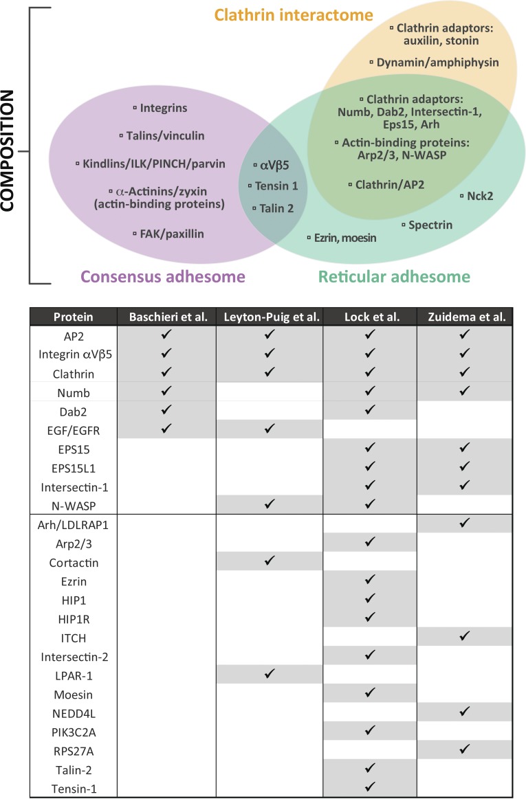
An understanding of the mechanisms whereby cell adhesion complexes (ACs) relay signals bidirectionally across the plasma membrane is necessary to interpret the role of adhesion in regulating migration, differentiation, and growth. A range of AC types has been defined, but to date all have similar compositions and are dependent on a connection to the actin cytoskeleton. Recently, a new class of AC has been reported that normally lacks association with both the cytoskeleton and integrin-associated adhesome components, but is rich in components of the clathrin-mediated endocytosis machinery. The characterization of this new type of adhesion structure, which is emphasized by mitotic cells and cells in long-term culture, identifies a hitherto underappreciated link between the adhesion machinery and clathrin structures at the plasma membrane. While this discovery has implications for how ACs are assembled and disassembled, it raises many other issues. Consequently, to increase awareness within the field, and stimulate research, we explore a number of the most significant questions below.


Figures

Figure 1.
Figure 1.
Comparison of molecular composition. The three-way Venn diagram illustrates a compositional comparison between the consensus (Horton et al., 2015), reticular (Lock et al., 2018), and clathrin interactomes (Schmid and McMahon, 2007). Proteins were chosen and grouped to illustrate typical functional modules, and the selection is not intended to indicate an exhaustive set of components. This analysis shows the limited overlap between canonical AC composition and the RA/clathrin interactomes. However, considerably more overlap is evident between the RA and clathrin interactomes. The table represents a direct comparison between clathrin plaque and RA datasets from the four indicated studies from proteomic and antibody-based localization data (Leyton-Puig et al., 2017; Baschieri et al., 2018; Lock et al., 2018; Zuidema et al., 2018). Proteins were ordered according to the number of datasets in which they were detected. These analyses indicate that while canonical ACs are distinct from RAs and conventional clathrin structures, RAs and plaques are highly compositionally similar and are likely the same.
Figure 2.
Figure 2.
Imaging of ACs. (A) U2OS and HeLa cells were plated on uncoated or collagen I–coated glass coverslips and cultured for 24 h. Cells were subsequently fixed and immunostained for integrin αVβ5, the canonical AC component vinculin, or the AP2 complex subunit α-adaptin. Scale bars: 10 µm. (B) HS578t cells were plated on collagen I–coated glass coverslips. 24 h later, cells were unroofed by sonication to generate a platinum replica of the inner leaflet of the adherent part of the plasma membrane as previously described (Elkhatib et al., 2017). Imaging was performed by transmission EM. Arrows point to plaques and arrowheads to clathrin-coated pits. Image in B was provided courtesy of Dr. Nadia Elkhatib.
Figure 3.
Figure 3.
Comparison of morphology and dynamics. Schematized view of a cell containing canonical ACs (CA; purple) and RAs/plaques (RA/CP; green). Specific morphological (above thick dotted line) and dynamic (below thick dotted line) properties are compared both visually and with illustrative graphs indicating quantitative differences measured in referenced publications. Morphology: Integrin αVβ5 nanoclusters are similar in appearance and arrangement (Nanocluster spacing) in CA and RA/CP (Lock et al., 2018). Visibly more convoluted in shape, RA/CP also achieve larger areas (Size) and are commonly located further from the cell edge (Location) than CA (Lock et al., 2018). Dynamics: CA slide rapidly (Speed; Ballestrem et al., 2001; Lock et al., 2018), linearly, and in a locally anisotropic fashion (Coordination) due to F-actin–derived forces (Besser and Safran, 2006). In contrast, RA/CP grow isotropically and show little directionality (Lock et al., 2018). Dynamics of individual complexes depicted with increasing opacity over time; arrows indicate net motion direction and distance. RA/CP complexes as a whole are highly stable (Lifetime) relative to CA (Lock et al., 2018; numbers indicate total time points observed per structure depicted in schematic). Conversely, the association of integrin αVβ5 proteins (Integrin turnover) with RA/CP is more dynamic than in CA, as revealed by higher recovery rates after photobleaching (Lock et al., 2018; transparency level within dotted boxed depicts relative fluorescence recovery).

References

    1. Akisaka T. 2000. [Ultrastructure of clathrin sheets and cytoskeleton of podosomes on the cytoplasmic side of ventral membranes of cultured osteoclasts]. Kaibogaku Zasshi. 75:381–386. - PubMed
    1. Ballestrem C., Hinz B., Imhof B.A., and Wehrle-Haller B.. 2001. Marching at the front and dragging behind: differential alphaVbeta3-integrin turnover regulates focal adhesion behavior. J. Cell Biol. 155:1319–1332. 10.1083/jcb.200107107 - DOI - PMC - PubMed
    1. Baschieri F., Dayot S., Elkhatib N., Ly N., Capmany A., Schauer K., Betz T., Vignjevic D.M., Poincloux R., and Montagnac G.. 2018. Frustrated endocytosis controls contractility-independent mechanotransduction at clathrin-coated structures. Nat. Commun. 9:3825 10.1038/s41467-018-06367-y - DOI - PMC - PubMed
    1. Batchelder E.M., and Yarar D.. 2010. Differential requirements for clathrin-dependent endocytosis at sites of cell-substrate adhesion. Mol. Biol. Cell. 21:3070–3079. 10.1091/mbc.e09-12-1044 - DOI - PMC - PubMed
    1. Besser A., and Safran S.A.. 2006. Force-induced adsorption and anisotropic growth of focal adhesions. Biophys. J. 90:3469–3484. 10.1529/biophysj.105.074377 - DOI - PMC - PubMed

Publication types