. 2019 Jul 2;8(7):bio043554.
doi: 10.1242/bio.043554.

Whole-brain mapping of projection from mouse lateral septal nucleus

Affiliations

Whole-brain mapping of projection from mouse lateral septal nucleus

Ke Deng et al. Biol Open. .

Abstract

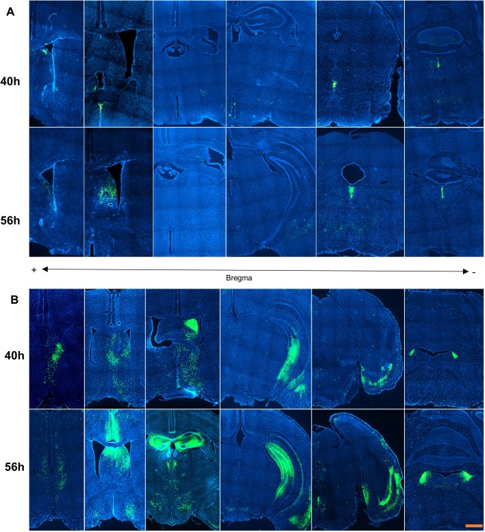
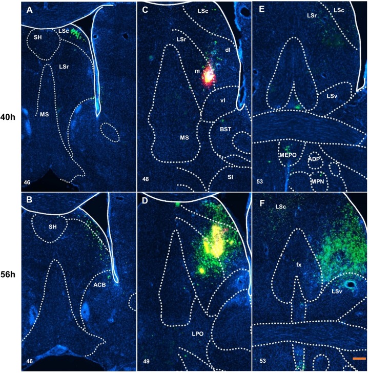
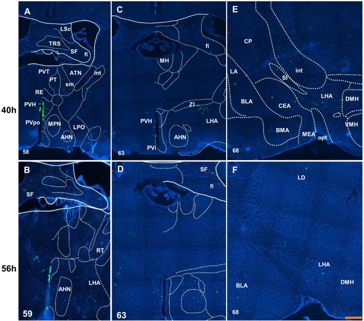
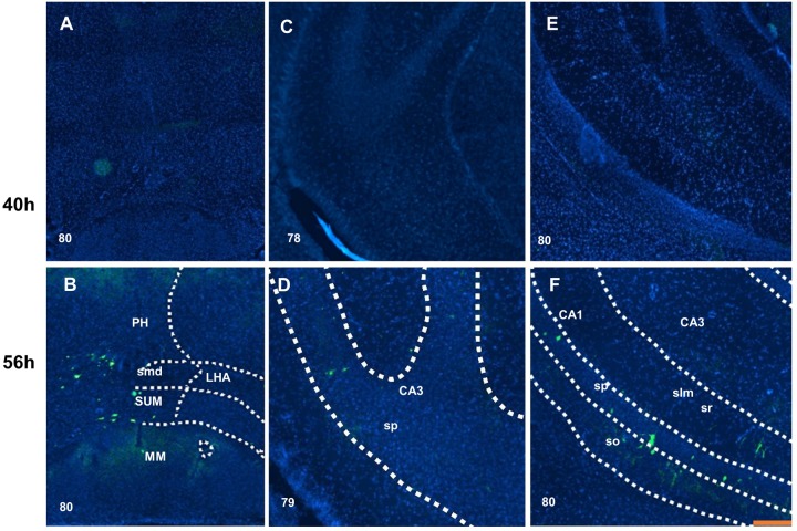
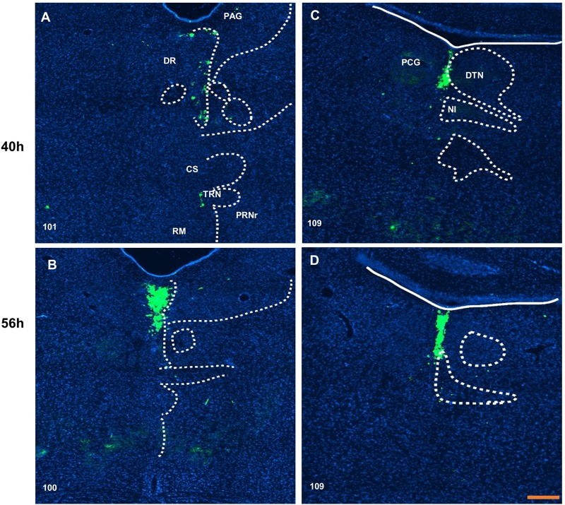
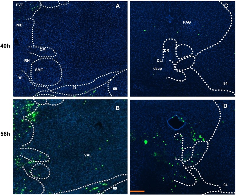
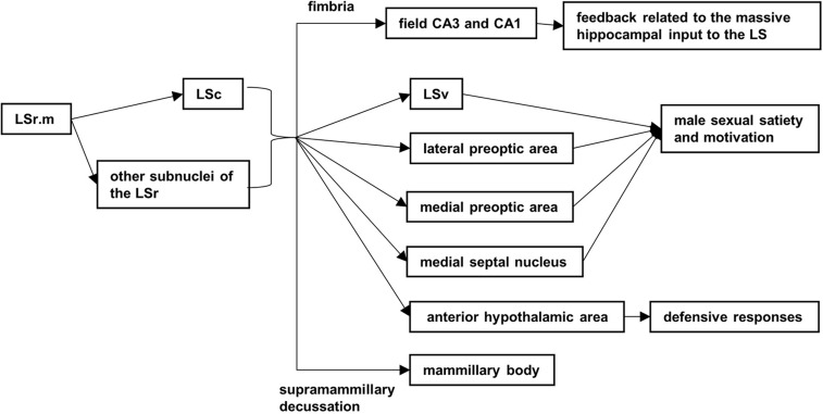
The lateral septal nucleus (LS) plays a critical role in emotionality, social behavior and feeding processes, through neural connections with the hippocampus and hypothalamus. We investigated the neural circuits of LS by using herpes simplex virus 1 strain H129 (H129) and pseudorabies virus stain Bartha (PRV). Virus H129 indicates that LS directly projects to some cerebral nuclei (nucleus accumbens, bed nuclei of the stria terminalis and amygdala), part of the hypothalamus (median preoptic, paraventricular, dorsomedial nucleus and lateral area), thalamus (medial habenula, the paraventricular, parataenial and reuniens nuclei, and the medial line nuclei) and the pontine central gray. Then the LS has secondary projections to the CA3 and CA1 field of the hippocampal formation, lateral and medial preoptic area, and the mammillary body. PRV tracing shows that LS are mainly receiving primary inputs from the amygdala, hippocampus, hypothalamic, thalamus, midbrain and hindbrain, and secondary inputs from dorsal and central linear nucleus raphe, the lateral part of the superior central nucleus raphe, the ventral anterior-lateral complex, the intermediodorsal nucleus, the central medial nucleus, the rhomboid nucleus, and the submedial nucleus of the thalamus. The neural circuit data revealed here could help to understand and further research on the function of LS.

Keywords: H129; Lateral septal nucleus; Neural circuits; Neural projection; PRV.


Conflict of interest statement

Competing interestsThe authors declare no competing or financial interests.

Figures

Fig. 1.
Fig. 1.
Overview of the projections from mice rostral part of lateral septal nuclei. The distribution of projections from mice lateral septal nuclei are summarized. A is the herpes simplex virus 1 strain H129 (HSV-EGFP). B is the pseudorabies virus strain Bartha (PRV-CMV-EGFP). The images in A are reproduced to show the distribution of labeled neurons in more detail in Figs 2, 3 and 5. The images in B are reproduced to show the distribution of labeled neurons in more detail in Fig. 6. Scale bar: 500 μm.
Fig. 2.
Fig. 2.
Distribution of virus H129 labeled neurons around injection sites. Labeled neurons were mainly scattered in the rostral and caudal part of lateral septal nuclei at 40 h after inoculation (A,C,E) and the bed nuclei of the stria terminalis. Then at 56 h after inoculation (B,D,F), the density of neurons was increased and there was weak input to the medial septal nucleus, lateral and medial preoptic area (D), also to the fornix and the contralateral lateral septal caudal part (F). Scale bar: 100 μm. The numbers in the left corner of the figure panels represent corresponding reference images in the Allen Mouse Brain Atlas. SH, septohippocampal nucleus; LSc, caudal part of the lateral septal nucleus; LSr, rostral part of lateral septal nucleus; MS, medial septal nucleus; dl, dorsolateral zone on LSr; m, medial zone on LSr; vl, ventrolateral zone on LSr; BST, bed nuclei of the stria terminalis; SI, substantia innominata; LSv, ventral part of the lateral septal nucleus; MEPO, median preoptic nucleus; ADP, anterodorsal preoptic nucleus; MPN, medial preoptic nucleus; ACB, nucleus accumbens; LPO, lateral preoptic area; fx, columns of the fornx.
Fig. 3.
Fig. 3.
Distribution of virus H129 labeled neurons in the anterior hypothalamus and amygdala. Only weak input can be found in the septofimbrial (A for 40 h and B for 56 h) and fimbria (C for 40 h and D for 56 h). The anterior hypothalamic nucleus shows a difference between at 40 h (A,C) and 56 h after inoculation (B,D). The weak and scattered inputs are shown at 40 h after inoculation (E) in basolateral amygdala nucleus and at 56 h after inoculation (F). Scale bar: 250 μm. The numbers in the left corner of the figure panels represent corresponding reference images in the Allen Mouse Brain Atlas. LSc, caudal part of the lateral septal nucleus; TRS, triangular nucleus of septum; SF, septofimbrial nucleus; fi, fimbria; PVT, paraventricular nucleus of the thalamus; ATN, anterior group of the dorsal thalamus; int, internal capsule; PT, parataenial nucleus; sm, stria medullaris; RE, nucleus of reuniens; PVH, paraventricular hypothalamic nucleus; PVpo, preoptic part of the periventricular hypothalamic nucleus; MPN, medial preoptic nucleus; LPO, lateral preoptic area; AHN, anterior hypothalamic nucleus; MH, medial habenula; ZI, zona incerta; LHA, lateral hypothalamic area; PVI, periventricular hypothalamic nucleus, intermediate part; CP, caudoputamen; LA, lateral amygdala nucleus; SI, substantia innominata; DMH, dorsomedial nucleus of the hypothalamus; BLA, basolateral amygdala nucleus; CEA, central amygdala nucleus; BMA, basomedial amygdala nucleus; MEA, medial amygdala nucleus; VMH, ventromedial hypothalamic nucleus; opt, optic tract; RT, reticular nucleus of the thalamus; LD, lateral dorsal nucleus of thalamus.
Fig. 4.
Fig. 4.
Distribution of virus H129-labeled neurons in the posterior hypothalamus and the ventral hippocampus. The posterior hypothalamic nucleus, supramammillary nucleus and mammillary nucleus received second-order projection at 56 h after inoculation, but not at 40 h (B). Some labeled neurons showed up on the pyramidal layer of CA3 field at 56 h (D) but not at 40 h after inoculation (C). In the field of CA1, moderate input was distributed from the stratum oriens to the pyramidal layer, stratum radiatum and stratum lacunosum-molecular (F) at 56 h but not at 40 h after inoculation (E). Scale bar: 200 μm. The numbers in the left corner of the figure panels represent corresponding reference images in the Allen Mouse Brain Atlas. PH, posterior hypothalamic nucleus; smd, supramammillary decussation; LHA, lateral hypothalamic area; SUM, the supramammillary nucleus; MM, medial mammillary nucleus; CA3, field CA3; sp, pyramidal layer; CA1, field CA1; slm, stratum lacunosum-molecular; sr, stratum radiatum; so, stratum oriens.
Fig. 5.
Fig. 5.
Distribution of virus H129-labeled neurons in the midbrain and pontine. The labeled neurons in dorsal nucleus raphe (A,B) and pontine central gray (C,D) show density change over time. Scale bar: 200 μm. The numbers in the left corner of the figure panels represent corresponding reference images in the Allen Mouse Brain Atlas. PAG, periaqueductal gray; DR, dorsal nucleus raphe; CS, superior central nucleus raphe; TRN, tegmental reticular nucleus; PRNr, pontine reticular nucleus; RM, nucleus raphe magnus; PCG, pontine central gray; DTN, dorsal tegmental nucleus; NI, nucleus incertus.
Fig. 6.
Fig. 6.
Projection of virus PRV-labeled neurons in the thalamus and midbrain. The density of labeled neurons shown in the intermediodorsal nucleus, central medial nucleus, submedial nucleus and rhomboid nucleus of the thalamus. The ventral anterior-lateral complex of the thalamus received signals at 56 h after inoculation (B), the central linear nucleus raphe also had input at 56 h after inoculation (D), but not at 40 h after inoculation (A,C). Scale bar: 200 μm. The numbers in the left corner of the figure panels represent corresponding reference images in the Allen Mouse Brain Atlas. PVT, paraventricular nucleus of the thalamus; IMD, intermediodorsal nucleus of the thalamus; CM, central medial nucleus of the thalamus; RH, rhomboid nucleus; SMT, submedial nucleus of the thalamus; RE, nucleus of reuniens; ZI, zona incerta; PAG, periaqueductal gray; DR, dorsal nucleus raphe; CLi, central linear nucleus raphe; dscp, superior cerebellar peduncle decussation; VAL, ventral anterior-lateral complex of the thalamus.
Fig. 7.
Fig. 7.
The possible anterograde secondary projections and relation functions.

Similar articles

Cited by

References

    1. Bielsky I. F., Hu S.-B., Ren X., Terwilliger E. F. and Young L. J. (2005). The V1a vasopressin receptor is necessary and sufficient for normal social recognition: a gene replacement study. Neuron 47, 503-513. 10.1016/j.neuron.2005.06.031 - DOI - PubMed
    1. Chee S. S. and Menard J. L. (2011). Lesions of the dorsal lateral septum do not affect neophagia in the novelty induced suppression of feeding paradigm but reduce defensive behaviours in the elevated plus maze and shock probe burying tests. Behav. Brain Res. 220, 362-366. 10.1016/j.bbr.2011.02.027 - DOI - PubMed
    1. Chen S., Yang M., Miselis R. R. and Aston-Jones G. (1999). Characterization of transsynaptic tracing with central application of pseudorabies virus. Brain Res. 838, 171-183. 10.1016/S0006-8993(99)01680-7 - DOI - PubMed
    1. Ebner K., Singewald G. M., Whittle N., Ferraguti F. and Singewald N. (2008). Neurokinin 1 receptor antagonism promotes active stress coping via enhanced septal 5-HT transmission. Neuropsychopharmacology 33, 1929-1941. 10.1038/sj.npp.1301594 - DOI - PubMed
    1. Ekstrand M. I., Enquist L. W. and Pomeranz L. E. (2008). The alpha-herpesviruses: molecular pathfinders in nervous system circuits. Trends Mol. Med. 14, 134-140. 10.1016/j.molmed.2007.12.008 - DOI - PMC - PubMed