. 2019 Jul 2;116(27):13200-13209.
doi: 10.1073/pnas.1902035116. Epub 2019 Jun 17.

Regulation of nuclear architecture, mechanics, and nucleocytoplasmic shuttling of epigenetic factors by cell geometric constraints

Affiliations

Regulation of nuclear architecture, mechanics, and nucleocytoplasmic shuttling of epigenetic factors by cell geometric constraints

Farid Alisafaei et al. Proc Natl Acad Sci U S A. .

Abstract

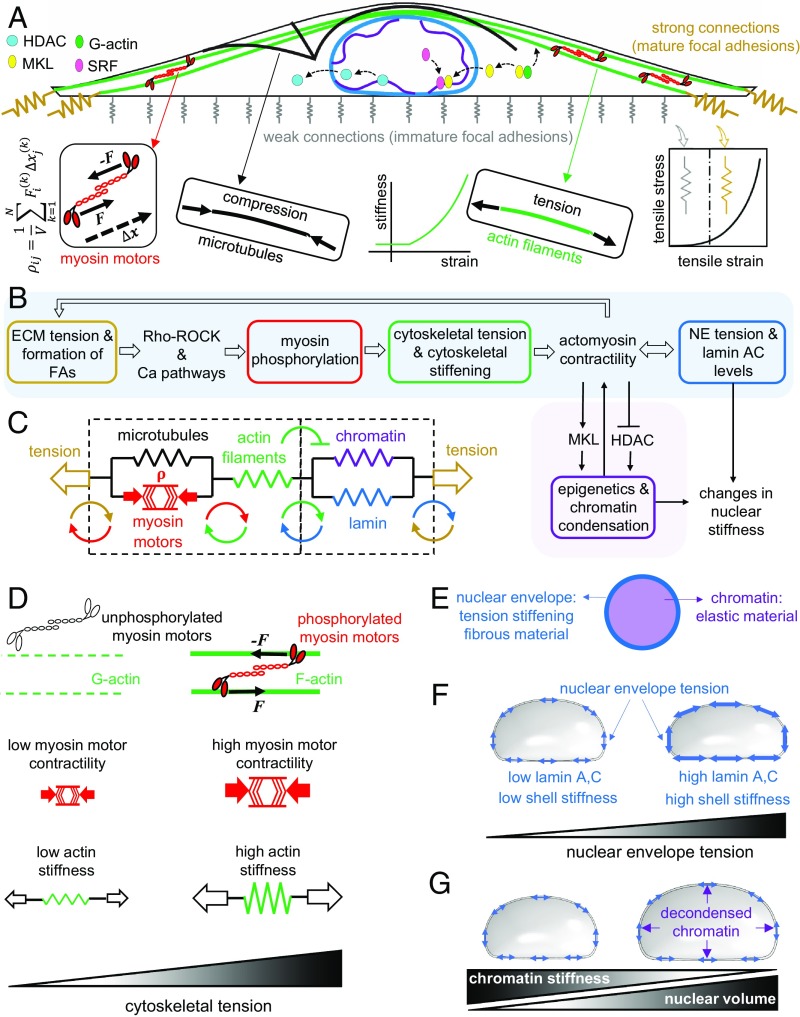
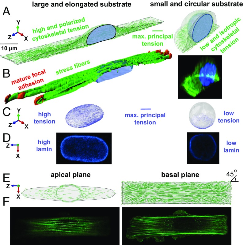
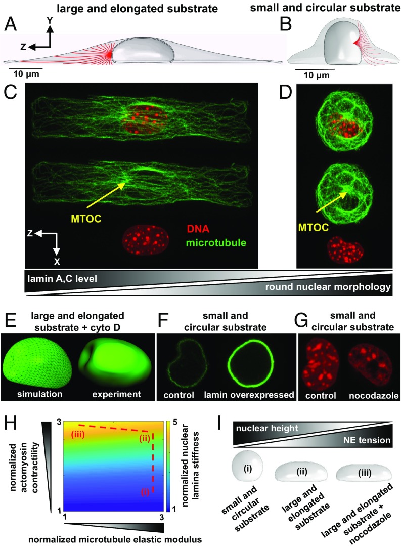
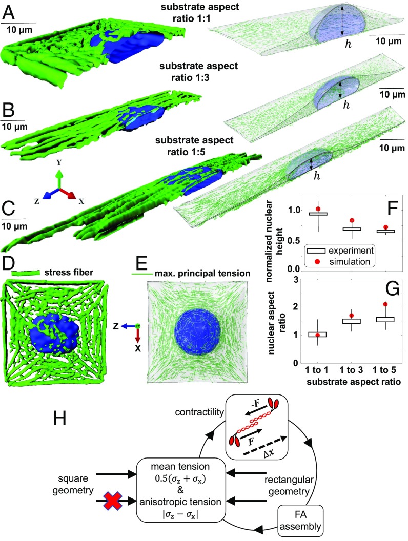
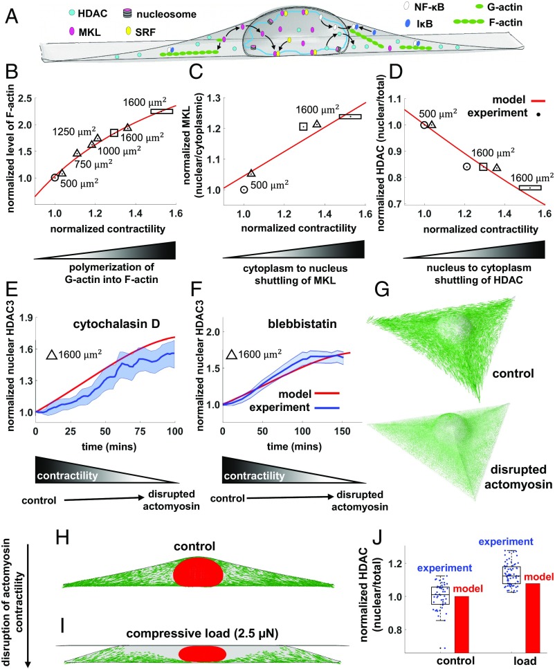
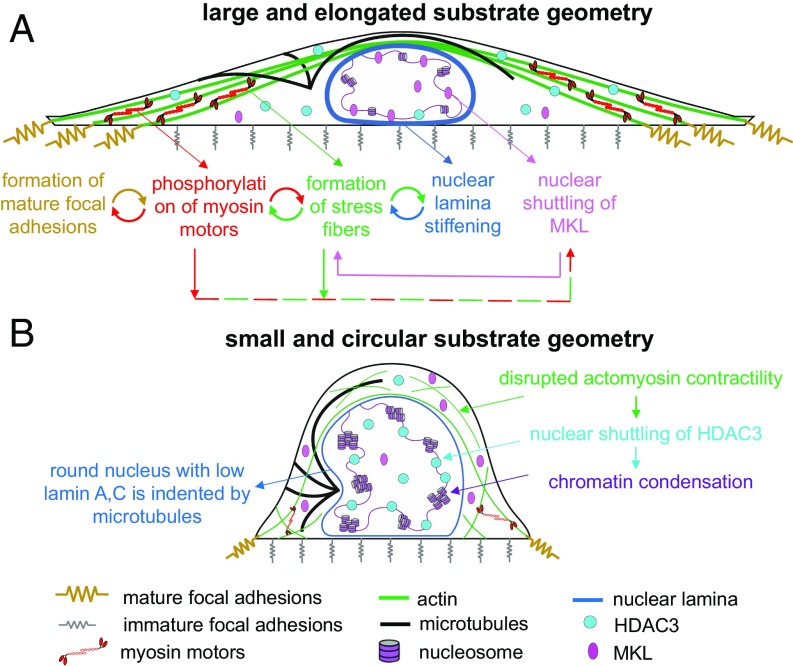
Cells sense mechanical signals from their microenvironment and transduce them to the nucleus to regulate gene expression programs. To elucidate the physical mechanisms involved in this regulation, we developed an active 3D chemomechanical model to describe the three-way feedback between the adhesions, the cytoskeleton, and the nucleus. The model shows local tensile stresses generated at the interface of the cell and the extracellular matrix regulate the properties of the nucleus, including nuclear morphology, levels of lamin A,C, and histone deacetylation, as these tensile stresses 1) are transmitted to the nucleus through cytoskeletal physical links and 2) trigger an actomyosin-dependent shuttling of epigenetic factors. We then show how cell geometric constraints affect the local tensile stresses and subsequently the three-way feedback and induce cytoskeleton-mediated alterations in the properties of the nucleus such as nuclear lamina softening, chromatin stiffening, nuclear lamina invaginations, increase in nuclear height, and shrinkage of nuclear volume. We predict a phase diagram that describes how the disruption of cytoskeletal components impacts the feedback and subsequently induce contractility-dependent alterations in the properties of the nucleus. Our simulations show that these changes in contractility levels can be also used as predictors of nucleocytoplasmic shuttling of transcription factors and the level of chromatin condensation. The predictions are experimentally validated by studying the properties of nuclei of fibroblasts on micropatterned substrates with different shapes and areas.

Keywords: cell geometry; cytoskeletal mechanics; mechanotransduction; nuclear mechanics.


Conflict of interest statement

The authors declare no conflict of interest.

Figures

Fig. 1.
Fig. 1.
A mechanochemical feedback model for cytoskeletal and nuclear mechanics. A contractile cell on a large and elongated adhesive substrate (A). The model is composed of 1) the focal adhesions, 2) the cytoskeleton, and 3) the nucleus. When the tensile stress exerted by the contractile cell to the adhesion layer exceeds a certain threshold, the stiffness of the connections to the substrate increases and mature focal adhesions are formed. The formation of mature focal adhesions (at the two ends for elongated substrate geometries) generates higher resistance against cell contraction and subsequently higher cytoskeletal tension. The tensile stresses arise at the mature focal adhesions activate the Ca2+ and the Rho–Rock signaling pathways which promote cell contractility through phosphorylation of myosin molecular motors (B). The increase in cell contractility leads to higher compression in microtubules and higher tension in actin filaments (C). In addition to the cell contractility ρij, the stiffness of actin filaments in our model increases with tension in an orientation-dependent manner to capture the fact that cells respond to tension by increasing their own stiffness through recruitment and alignment of actin filaments along the direction of the tensile stress (D). The increase in actomyosin contractility alters nuclear morphology and nuclear stiffness through 1) cytoskeletal physical links that transmit the stresses generated at the mature focal adhesions to the nucleus (sky blue path in B) and 2) a cascade of biochemical pathways that changes chromatin stiffness by nucleocytoplasmic shuttling of epigenetic factors (pink path in B). The nuclear envelope (E) is modeled as a filamentous network material (lamin network) which stiffens with tension to capture the fact that lamin A,C level and strain stiffening of the nuclear envelope increase with actomyosin contractility (F), while chromatin is treated as an elastic material (E) whose stiffness increases (decreases) with chromatin condensation (decondensation) in proportion to nuclear level of HDAC predicted by our model (G).
Fig. 2.
Fig. 2.
Cell geometric constraints regulate cell contractility, actin organization, and nuclear envelope lamina stiffness. (A and B) NIH 3T3 mouse fibroblast cells are cultured on fibronectin-coated micropatterned substrates with two extreme geometries: a rectangle with an aspect ratio of 1:5 and a substrate surface area of 1,600 µm2 (large and elongated substrate geometry), and a circle with a substrate surface area of 500 µm2 (small and circular substrate geometry). Cells on the rectangular substrate have higher and polarized 1) contractility, 2) cytoskeletal tension, and 3) cytoskeletal stiffness leading to a flattened and elongated nuclear morphology (C). As a result, tension is generated in the nuclear envelope lamina of rectangular cells correlated with their higher levels of lamin A,C and nuclear envelope stiffness (D). Stress fibers are formed along the long axis of the cell in the apical plane (E) and at 45° at the corners of the basal plane (F).
Fig. 3.
Fig. 3.
Nuclei with low levels of lamin A,C and round morphologies are indented by the MTOC. Microtubules in large and elongated cells buckle without being able to significantly indent the nucleus as the MTOC is pushed toward the cell boundary by the nucleus (A and C). In contrast, cells on small and circular substrates exhibit crescent-shaped nuclear morphologies as the MTOC pushes against the nucleus and forms a local indentation in the nucleus (B and D). Similar to the circular cell, the nucleus is indented by the MTOC when actin filaments are depolymerized in the rectangular cell (E). Overexpression of lamin A,C (F) and nocodazole treatment (G) rescue nuclear invagination in circular cells. Cell geometric constraints and polymerization of microtubules reduce nuclear lamina stiffness by decreasing actomyosin contractility (H); compared with small and circular cells (i), cells on large and elongated substrates (ii) show increased contractility which in turn leads to stiffening of the nuclear envelope lamina network in proportion to actomyosin-driven tension in the nuclear envelope. Depolymerization of microtubules in large and elongated cells (iii) further increases actomyosin contractility and subsequently nuclear lamina stiffness. The actomyosin-driven reduction in nuclear height tenses and stiffens the nuclear envelope lamina network (I).
Fig. 4.
Fig. 4.
Substrate aspect ratio induces alterations in prenuclear actin organizations and nuclear morphology. Fibroblasts on fibronectin-coated micropatterned substrates with the same substrate surface area of 1,600 µm2 but various aspect ratios (AC). Stress fibers (D) are formed in the direction of the maximum principal stress (E). Formations of apical stress fibers lead to flattening (F) and elongation (G) of the nucleus in the rectangular substrate geometries as actomyosin contractility increases with anisotropy in tensile stresses (H).
Fig. 5.
Fig. 5.
Cell geometric constraints induce chromatin condensation through nuclear translocation of HDAC driven by decreases in actomyosin contractility. The level of contractility, represented by ρmean=(ρ11+ρ22+ρ33)/3 in our model, regulates nucleocytoplasmic translocations of epigenetic and transcription factors (A). Cells on large and elongated substrates have higher contractility ρmean (compared with small and circular cells) and higher levels of polymerized F-actin (B). As G-actin is polymerized into F-actin, MKL unbinds from G-actin and shuttles to the nucleus, leading to higher nuclear accumulations of MKL in large and elongated cells (C). On the other hand, decreasing ρmean by constraining cells on smaller and unpolarized substrates (D) or by using actomyosin inhibitors (EG) leads to translocation of HDAC to the nucleus. Similar to geometric constraints and actomyosin inhibitors, compressive forces on fibroblasts cause disruption of actomyosin contractility (H and I) and translocation of HDAC to the nucleus (J).
Fig. 6.
Fig. 6.
A model summarizing how cell geometry regulates focal adhesion formation, cell contractility, actin organization, nuclear morphology, and nuclear stiffness. Cell geometry controls the dynamic reciprocities between the focal adhesions, the cytoskeleton, and the nucleus by regulating the local tensile stresses that are generated on the cell substrate. For example, cells on large and rectangular substrates (A) generate high, polarized (along the long axis of the cell), and localized tensile stresses (at the two ends), while cells on small and circular geometries (B) generate low, isotropic (independent of direction), and uniform tension on the cell periphery. These mechanical stresses 1) are transmitted to the nucleus through cytoskeletal physical links and 2) trigger an actomyosin-dependent shuttling of epigenetic factors, causing alterations in nuclear morphology, nuclear stiffness (both nuclear envelope and nuclear interior), and gene expression.

References

    1. Discher D. E., Janmey P., Wang Y. L., Tissue cells feel and respond to the stiffness of their substrate. Science 310, 1139–1143 (2005). - PubMed
    1. Makhija E., Jokhun D. S., Shivashankar G. V., Nuclear deformability and telomere dynamics are regulated by cell geometric constraints. Proc. Natl. Acad. Sci. U.S.A. 113, E32–E40 (2016). - PMC - PubMed
    1. Jain N., Iyer K. V., Kumar A., Shivashankar G. V., Cell geometric constraints induce modular gene-expression patterns via redistribution of HDAC3 regulated by actomyosin contractility. Proc. Natl. Acad. Sci. U.S.A. 110, 11349–11354 (2013). - PMC - PubMed
    1. Le Beyec J., et al. , Cell shape regulates global histone acetylation in human mammary epithelial cells. Exp. Cell Res. 313, 3066–3075 (2007). - PMC - PubMed
    1. Damodaran K., et al. , Compressive force induces reversible chromatin condensation and cell geometry dependent transcriptional response. Mol. Biol. Cell 10.1091/mbc.E18-04-0256 (2018). - DOI - PMC - PubMed

Publication types

LinkOut - more resources