. 2019 Jul 2;116(27):13293-13298.
doi: 10.1073/pnas.1905873116. Epub 2019 Jun 17.

The E3 ligase HOIL-1 catalyses ester bond formation between ubiquitin and components of the Myddosome in mammalian cells

Affiliations

The E3 ligase HOIL-1 catalyses ester bond formation between ubiquitin and components of the Myddosome in mammalian cells

Ian R Kelsall et al. Proc Natl Acad Sci U S A. .

Abstract

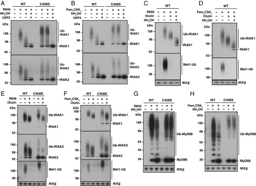
The linear ubiquitin assembly complex (LUBAC) comprises 3 components: HOIP, HOIL-1, and Sharpin, of which HOIP and HOIL-1 are both members of the RBR subfamily of E3 ubiquitin ligases. HOIP catalyses the formation of Met1-linked ubiquitin oligomers (also called linear ubiquitin), but the function of the E3 ligase activity of HOIL-1 is unknown. Here, we report that HOIL-1 is an atypical E3 ligase that forms oxyester bonds between the C terminus of ubiquitin and serine and threonine residues in its substrates. Exploiting the sensitivity of HOIL-1-generated oxyester bonds to cleavage by hydroxylamine, and macrophages from knock-in mice expressing the E3 ligase-inactive HOIL-1[C458S] mutant, we identify IRAK1, IRAK2, and MyD88 as physiological substrates of the HOIL-1 E3 ligase during Toll-like receptor signaling. HOIL-1 is a monoubiquitylating E3 ubiquitin ligase that initiates the de novo synthesis of polyubiquitin chains that are attached to these proteins in macrophages. HOIL-1 also catalyses its own monoubiquitylation in cells and most probably the monoubiquitylation of Sharpin, in which ubiquitin is also attached by an oxyester bond. Our study establishes that oxyester-linked ubiquitylation is used as an intracellular signaling mechanism.

Keywords: IRAK; LUBAC; NEMO; TRAF6; Toll-like receptor.


Conflict of interest statement

The authors declare no conflict of interest.

Figures

Fig. 1.
Fig. 1.
A hydroxylamine-sensitive modification on HOIL-1. (A) Immunoblots of BMDM extracts (20 μg of protein) derived from WT and HOIL-1[C458S] knock-in mice. (B) LUBAC was immunoprecipitated with anti-HOIP from WT BMDM extracts, then treated with λPPase, USP2, or hydroxylamine, followed by immunoblotting with the antibodies indicated. (C) HaCaT cell lysates (30 μg of protein) were treated for 30 min without (−) or with (+) 1.5 M hydroxylamine, subjected to SDS/PAGE without (−) or with (+) 50 mM DTT and immunoblotted with the antibodies indicated. The mUb-H2B antibody detects Histone H2B monoubiquitylated at Lys120. (D) LUBAC was immunoprecipitated from HaCaT cell lysates as in B and incubated with λPPase in the presence or absence of hydroxylamine, USP2, or Otulin, followed by immunoblotting with the antibodies indicated.
Fig. 2.
Fig. 2.
Characterization of the ubiquitin ligase activity of HOIL-1. (A) Autoubiquitylation was studied using bacterially expressed His6-HOIL-1 and UbcH7. HOIL-1, ATP, UBE1, or UbcH7 were omitted where indicated. The hydroxylamine sensitivity of ubiquitylated HOIL-1 was examined by incubating completed reactions with (+) or without (−) 1.5 M NH2OH. The proteins were visualized by immunoblotting with anti-HOIL-1 (Top) or anti-ubiquitin (Middle) or by staining with Coomassie blue (Bottom). The asterisk (*) denotes contaminating bacterial protein in the His6-HOIL-1 prep. (B) As in A, except that reactions contained WT His6-HOIL-1 or the indicated His6-HOIL-1 mutants. (C) As in A, except that reactions were carried out using WT Ub, Ub with every lysine mutated to arginine (WNK-Ub), or N-terminally biotinylated WNK-Ub (BWNK-Ub). (D) Tandem mass (MS/MS) spectrum of autoubiquitylated HOIL-1 reveals Ser365 as a site of ester-linked ubiquitylation. y ions used for scoring are in blue and b ions in red. The asterisk (*) on the peptide sequence indicates the site of diglycine attachment. b0 on the spectrum plot indicates neutral loss of H2O from a b ion; y++ indicates a doubly charged y ion. (E) Schematic of HOIL-1 domain architecture with identified sites of Ser/Thr ubiquitylation indicated. IBR, in between RING domain; NZF, NPL4 zinc finger domain; RING, really interesting new gene; UBL, ubiquitin-like domain. (F) Annotated tryptic MS/MS spectrum of a HOIL-1–generated ubiquitin dimer reveals Thr12 as a site of ubiquitin ligation. Other details are as in D, except that y* on the spectrum plot indicates the neutral loss of NH3 from a y ion.
Fig. 3.
Fig. 3.
Ubiquitylated IRAK1 and IRAK2 formed during TLR signaling contain HOIL-1–catalyzed, hydroxylamine-cleavable bonds. (A) Primary WT BMDM were stimulated with 1 μg/mL R848 or Pam3CSK4 for the times indicated and lysed. Ubiquitylated proteins were captured on Halo-NEMO beads, incubated for 30 min at 37 °C with λPPase and immunoblotted with antibodies recognizing IRAK1 or IRAK2. IKKβ, which binds to NEMO in a ubiquitin-independent manner, was used as a loading control. (B) WT BMDM were stimulated with R848 and ubiquitylated proteins were captured from extracts on Halo-NEMO beads and treated with λPPase as in A. Beads were then incubated with 0.5 M hydroxylamine at pH 7.5 or 9.0 for the times indicated. Immunoblotting was as in A. (C and D) BMDM from WT or HOIL-1[C458S] mice were stimulated for 10 min with R848 (C) or 20 min with Pam3CSK4 (D) and ubiquitylated proteins captured on Halo-NEMO beads, incubated for 60 min without (−) or with (+) 0.5 M hydroxylamine at pH 9.0 and immunoblotted as in A.
Fig. 4.
Fig. 4.
The ubiquitin chains attached to IRAK1, IRAK2, and MyD88 during TLR ligation are initiated by both isopeptide and oxyester bonds. (A and B) BMDM from WT or HOIL-1[C458S] knock-in mice were stimulated for 10 min with R848 (A) or for 20 min with Pam3CSK4 (B) and ubiquitylated proteins captured on Halo-NEMO beads as in Fig. 3 C and D. Following incubation for 60 min at pH 9.0 without (−) or with (+) 0.5 M hydroxylamine, the beads were incubated for a further 60 min at pH 7.5 without (−) or with (+) 1 μM USP2. Immunoblotting was performed with antibodies recognizing IRAK1 or IRAK2. (C and D) WT BMDM were stimulated for 10 min with R848 (C) or 20 min with Pam3CSK4 (D) and processed as in Fig. 3 C and D, except that the Halo-NEMO beads were first incubated for 60 min at pH 7.5 without (−) or with (+) 1 μM Otulin and then for 60 min at pH 9.0 without (−) or with (+) 0.5 M hydroxylamine. Immunoblotting was performed with antibodies recognizing IRAK1 or Met1-Ub chains. (E and F) As in C and D except that both WT and HOIL-1[C458S] BMDM were used and incubation with hydroxylamine was omitted. (G and H) As in C and D, except that immunoblotting was with anti-MyD88.

References

    1. Kirisako T., et al. , A ubiquitin ligase complex assembles linear polyubiquitin chains. EMBO J. 25, 4877–4887 (2006). - PMC - PubMed
    1. Gerlach B., et al. , Linear ubiquitination prevents inflammation and regulates immune signalling. Nature 471, 591–596 (2011). - PubMed
    1. Tokunaga F., et al. , SHARPIN is a component of the NF-κB-activating linear ubiquitin chain assembly complex. Nature 471, 633–636 (2011). - PubMed
    1. Ikeda F., et al. , SHARPIN forms a linear ubiquitin ligase complex regulating NF-κB activity and apoptosis. Nature 471, 637–641 (2011). - PMC - PubMed
    1. Lo Y. C., et al. , Structural basis for recognition of diubiquitins by NEMO. Mol. Cell 33, 602–615 (2009). - PMC - PubMed

Publication types

LinkOut - more resources