Review
. 2019 Jun 17;132(12):jcs226928.
doi: 10.1242/jcs.226928.

Molecular form and function of the cytokinetic ring

Affiliations
Review

Molecular form and function of the cytokinetic ring

MariaSanta C Mangione et al. J Cell Sci. .

Abstract

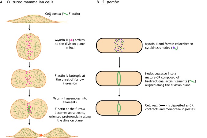
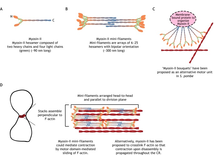
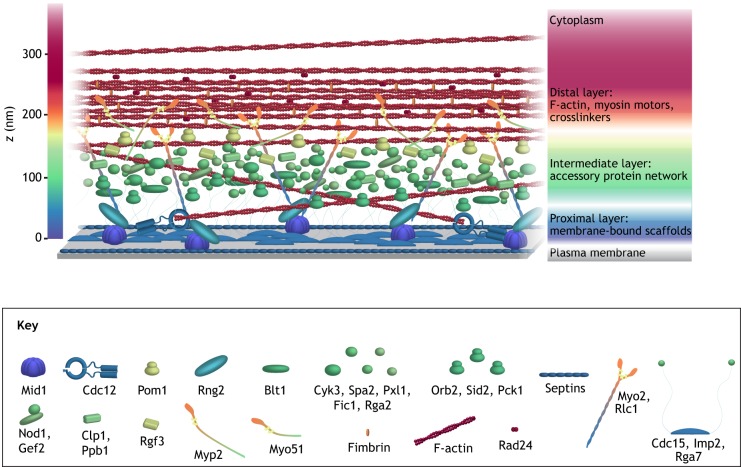
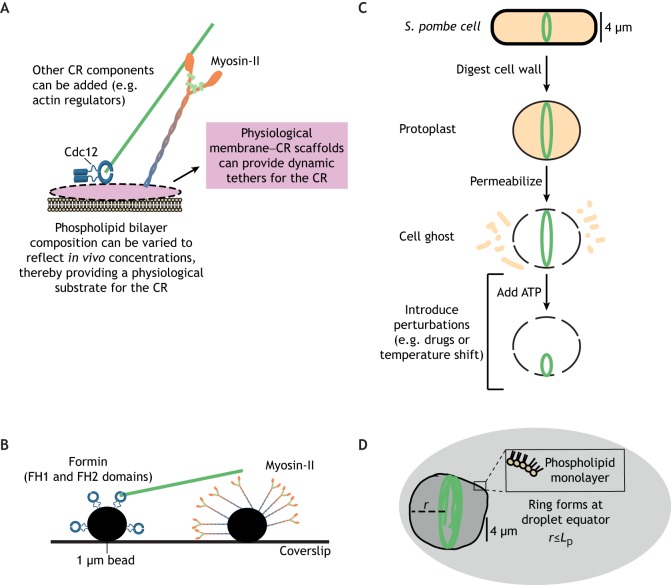
Animal cells, amoebas and yeast divide using a force-generating, actin- and myosin-based contractile ring or 'cytokinetic ring' (CR). Despite intensive research, questions remain about the spatial organization of CR components, the mechanism by which the CR generates force, and how other cellular processes are coordinated with the CR for successful membrane ingression and ultimate cell separation. This Review highlights new findings about the spatial relationship of the CR to the plasma membrane and the arrangement of molecules within the CR from studies using advanced microscopy techniques, as well as mechanistic information obtained from in vitro approaches. We also consider advances in understanding coordinated cellular processes that impact the architecture and function of the CR.

Keywords: Actomyosin ring; Amoeba; Animal cells; Cell division; Contractile ring; Cytokinesis; Cytokinetic ring; Fungi; Metazoa; Yeast.


Conflict of interest statement

Competing interestsThe authors declare no competing or financial interests.

Figures

Fig. 1.
Fig. 1.
Elemental steps of CR formation and contraction. CR formation and contraction is illustrated for (A) cultured mammalian cells and (B) S. pombe. Although not depicted, F-actin and myosin-II exhibit similar dynamics in animal embryos (e.g. Caenorhabditis elegans and sea urchin) and amoeboid cells.
Fig. 2.
Fig. 2.
Molecular structure of myosin-II. (A) Double-headed myosin-II is a hexamer. (B) Animal and amoeboid myosin-II can oligomerize via C-terminal tails into bipolar filaments. (C) Radial arrangement of myosin-II predicted to act as a motor unit in S. pombe, which does not form mini-filaments. Actin is red. (D) Cartoon of myosin-II arrangement in animal cell CRs. Actin is red.
Fig. 3.
Fig. 3.
A model for CR arrangement in S. pombe. Scale model of S. pombe CR architecture based on experimentally determined distances of CR proteins from the plasma membrane. The model does not incorporate stoichiometry. Panels are modified from McDonald et al., 2017, where they were published under a CC-BY 4.0 license (https://creativecommons.org/licenses/by/4.0/).
Fig. 4.
Fig. 4.
In vitro methods to reconstitute the CR. (A) Schematic showing the method of reconstituting CRs on a phospholipid bilayer. Myosin-II and the formin Cdc12 from S. pombe are included as sample proteins. (B) Schematic of in vitro reconstitution of SCPR model of CR formation using 1 µm beads coated with FH1-FH2 fragments or myosin-II. Myosin-II is ∼90 nm (Friend et al., 2018) and is presented at ∼100 times its size relative to the bead. FH1–FH2 fragments are presented at ∼200× their size relative to the bead. (C) Method for preparing cell ghosts from S. pombe cells. Green represents a fluorescent CR protein. (D) Just after the initiation of actin filament formation, phospholipids are added to a solution containing G-actin and other actin-binding proteins so that F-actin rings (green) form inside of a phospholipid monolayer. The maximum radius (r) of these rings is limited by the persistence length (Lp) of the actin filament.

Similar articles

Cited by

References

    1. Akamatsu M., Berro J., Pu K.-M., Tebbs I. R. and Pollard T. D. (2014). Cytokinetic nodes in fission yeast arise from two distinct types of nodes that merge during interphase. J. Cell Biol. 204, 977-988. 10.1083/jcb.201307174 - DOI - PMC - PubMed
    1. Atilla-Gokcumen G. E., Muro E., Relat-Goberna J., Sasse S., Bedigian A., Coughlin M. L., Garcia-Manyes S. and Eggert U. S. (2014). Dividing cells regulate their lipid composition and localization. Cell 156, 428-439. 10.1016/j.cell.2013.12.015 - DOI - PMC - PubMed
    1. Balasubramanian M. K. (2016). Exploring the diversity of cytokinesis. Semin. Cell Dev. Biol. 53, 1 10.1016/j.semcdb.2016.04.003 - DOI - PubMed
    1. Beach J. R., Shao L., Remmert K., Li D., Betzig E. and Hammer J. A. (2014). Nonmuscle Myosin II Isoforms Coassemble in Living Cells. Curr. Biol. 24, 1160-1166. 10.1016/j.cub.2014.03.071 - DOI - PMC - PubMed
    1. Bertin A., McMurray M. A., Thai L., Garcia G. III, Votin V., Grob P., Allyn T., Thorner J. and Nogales E. (2010). Phosphatidylinositol-4,5-bisphosphate promotes budding yeast septin filament assembly and organization. J. Mol. Biol. 404, 711-731. 10.1016/j.jmb.2010.10.002 - DOI - PMC - PubMed

Publication types

Substances