. 2019 Aug 1;203(3):736-748.
doi: 10.4049/jimmunol.1900228. Epub 2019 Jun 17.

The Pyroptotic Cell Death Effector Gasdermin D Is Activated by Gout-Associated Uric Acid Crystals but Is Dispensable for Cell Death and IL-1β Release

Affiliations

The Pyroptotic Cell Death Effector Gasdermin D Is Activated by Gout-Associated Uric Acid Crystals but Is Dispensable for Cell Death and IL-1β Release

Maryam Rashidi et al. J Immunol. .

Abstract

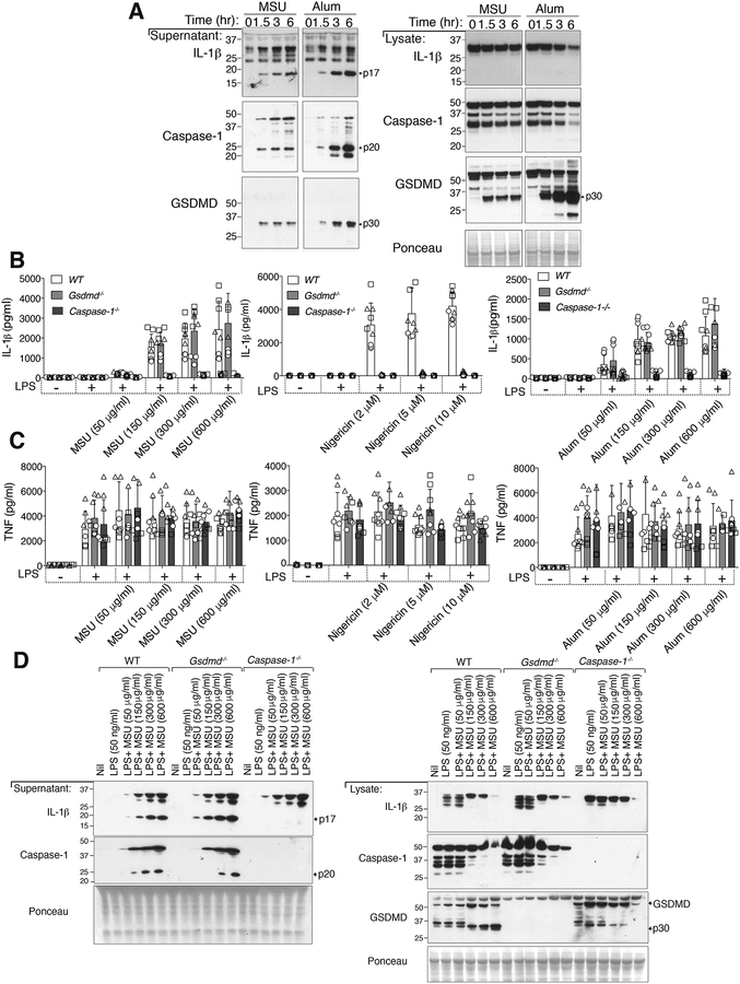
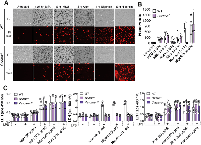
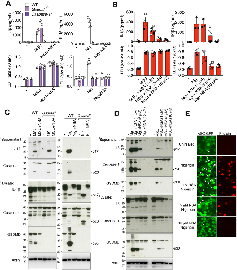
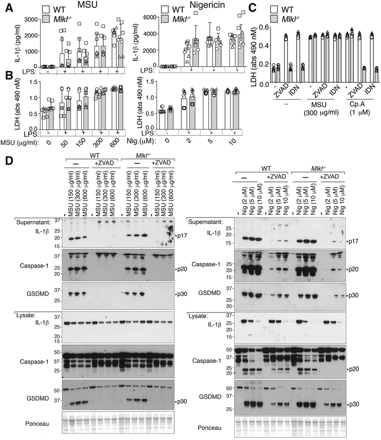
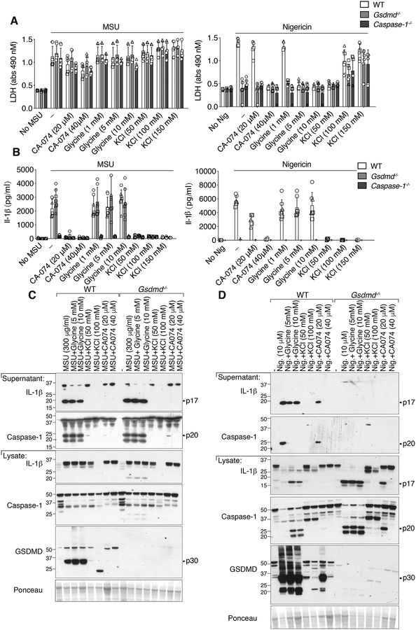
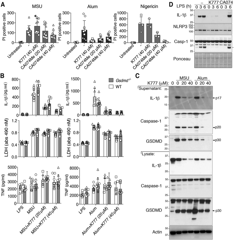
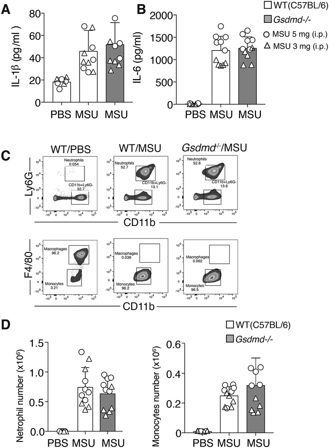
The pyroptotic cell death effector gasdermin D (GSDMD) is required for murine models of hereditary inflammasome-driven, IL-1β-dependent, autoinflammatory disease, making it an attractive therapeutic target. However, the importance of GSDMD for more common conditions mediated by pathological IL-1β activation, such as gout, remain unclear. In this study, we address whether GSDMD and the recently described GSDMD inhibitor necrosulfonamide (NSA) contribute to monosodium urate (MSU) crystal-induced cell death, IL-1β release, and autoinflammation. We demonstrate that MSU crystals, the etiological agent of gout, rapidly activate GSDMD in murine macrophages. Despite this, the genetic deletion of GSDMD or the other lytic effector implicated in MSU crystal killing, mixed lineage kinase domain-like (MLKL), did not prevent MSU crystal-induced cell death. Consequently, GSDMD or MLKL loss did not hinder MSU crystal-mediated release of bioactive IL-1β. Consistent with in vitro findings, IL-1β induction and autoinflammation in MSU crystal-induced peritonitis was not reduced in GSDMD-deficient mice. Moreover, we show that the reported GSDMD inhibitor, NSA, blocks inflammasome priming and caspase-1 activation, thereby preventing pyroptosis independent of GSDMD targeting. The inhibition of cathepsins, widely implicated in particle-induced macrophage killing, also failed to prevent MSU crystal-mediated cell death. These findings 1) demonstrate that not all IL-1β-driven autoinflammatory conditions will benefit from the therapeutic targeting of GSDMD, 2) document a unique mechanism of MSU crystal-induced macrophage cell death not rescued by pan-cathepsin inhibition, and 3) show that NSA inhibits inflammasomes upstream of GSDMD to prevent pyroptotic cell death and IL-1β release.


Figures

Figure 1.
Figure 1.. MSU crystals and alum activate IL-1β and GSDMD, but GSDMD is dispensable for IL-1β release.
(A) Bone marrow-derived macrophages (BMDMs) from WT (C57BL/6) mice were pre-incubated for 2.5 hr with LPS (50 ng/ml) and then treated with MSU crystals or alum (300 μg/ml) for the indicated times. Cell supernatant and total cell lysates were analyzed by immunoblot. Ponceau stain depicts protein loading. (B) IL-1β and (C) TNF levels in the cell supernatant of BMDMs from WT (C57BL/6), Gsdmd −/− and Caspase-1−/− mice pre-incubated for 2.5 hr with LPS (50 ng/ml) and then treated with the indicated concentrations of MSU crystals (6 hr), nigericin (1 hr) and alum (6 hr). (D) BMDMs from WT (C57BL/6), Gsdmd/− and Caspase-1−/− mice were pre-incubated with LPS (50 ng/ml, 2.5 hr) and then treated with the indicated concentrations of MSU crystals (6 hr) and the cell supernatant and total cell lysates analyzed by immunoblot. Ponceau staining depicts protein loading. (B and C) Mean ± SD of 7–9 replicates pooled from three independent experiments (symbols). (A and D) One of two independent experiments.
Figure 2.
Figure 2.. GSDMD is not required for MSU crystal- and alum-induced cell death.
(A and B) Imaging of BMDMs from WT and Gsdmd/− mice pre-incubated for 2.5 hr with LPS (50 ng/ml) and then treated with MSU crystals (300 μg/ml), nigericin (10 μM) or alum (300 μg/ml) for the indicated times. Cell death was assessed by propidium iodide (PI) uptake (red fluorescence). Representative bright field (BF) and PI images are shown in (A) and the number of PI positive cells from three independent experiments (symbols) quantified (mean ± SD) in (B) using Fiji imaging software. (C) LDH levels in the cell supernatant of BMDMs from WT (C57BL/6), Gsdmd/− and Caspase-1−/− mice pre-incubated with LPS (50 ng/ml, 2.5 hr) and then treated with the indicated concentrations of MSU crystals (6 hr) or nigericin (1 hr). Mean ± SD of 9 replicates (symbols) pooled from three independent experiments.
Figure 3.
Figure 3.. Necrosulfonamide inhibits NLRP3 activation and pyroptosis independent of GSDMD targeting.
(A) IL-1β (top panel) and LDH (bottom panel) levels in the cell supernantant of BMDMs from WT (C57BL/6), Gsdmd−/− and Caspase-1−/− mice primed for 2.5 hr with LPS (50 ng/ml), with necrosulfonamide (NSA) (10 μM) added in the last 30 min of priming, then treated with MSU crystals (300 μg/ml, 6 hr) or nigericin (10 μM, 1 hr). (B) IL-1β and LDH levels in the supernatant of BMDMs from WT (C57BL/6) mice primed with LPS (50 ng/ml, 2.5 hr), with the indicated concentration of NSA added in the last 30 min of priming, followed by treatment with MSU crystals (300 μg/ml, 6 hr) or nigericin (10 μM, 1 hr). (C) WT (C57BL/6) and Gsdmd−/− BMDMs were treated as in (A) and supernatant and total cell lysates analyzed by immunoblot. (D) WT (C57BL/6) BMDMs were treated as in (B) and supernatant and total cell lysates analyzed by immunoblot. Ponceau staining depicts protein loading. (E) Immortalized BMDMs (iBMDMs) expressing ASC-GFP and FLAG-NLRP3 were analyzed by fluorescent microscopy for ASC speck formation (green fluorescence) and PI uptake (red fluorescence) after treatment with the indicated concentrations of NSA for 30–40 min and then nigericin (10 μM, 80 min). ASC specks are indicated with white arrows. (A) Mean ± SD of 4–5 replicates (symbols) pooled from two independent experiments. (B) Mean ± SD of BMDMs from 3–6 mice (symbols) pooled from two independent experiments. (C, D and E) Data are representative of three independent experiments.
Figure 4.
Figure 4.. MLKL is not required for MSU crystal-induced death or NLRP3 inflammasome activation in macrophages.
(A) IL-1β and (B) LDH levels in the cell supernatant of BMDMs from WT (C57BL/6) and Mlkl−/− mice primed with LPS (50 ng/ml, 2.5 hr) and then treated with the indicated concentrations of MSU crystals (6 hr) or nigericin (1 hr). (C) LDH levels in the cell supernatant of BMDMs from WT (C57BL/6) and Mlkl−/− mice primed with LPS (50 ng/ml, 2.5 hr), with Z-VAD-fmk (40 μM) or IDN-6556 (20 μM) added in the last 30 min of priming, then treated with MSU crystals (300 μg/ml or Smac-mimetic compound (Cp.A, 1 μM) for 6 hr (D) BMDMs from WT (C57BL/6) and Mlkl−/− mice primed with LPS (50 ng/ml, 2.5 hr), with Z-VAD-fmk (20 μM) added in the last 30 min of priming, were then treated with the indicated concentrations of MSU crystals (6 hr) or nigericin (1 hr). Cell supernatants and total cell lysates were analyzed by immunoblot, as indicated. Ponceau staining depicts protein loading. (A, B, C) Mean ± SD of 8 (A, B) or 6 (C) replicates pooled from three (A, B) or two (C) independent experiments. (D) Data are representative of two independent experiments.
Figure 5.
Figure 5.. MSU crystal-induced membrane lysis is not significantly reduced by the osmoprotectant glycine, the prevention of potassium ion efflux, or the inhibition of cathepsin activity.
(A) LDH and (B) IL-1β levels in the cell supernatant of BMDMs from WT (C57BL/6), Gsdmd−/− and Caspase-1−/− mice primed with LPS (50 ng/ml, 2.5 hr), with the indicated concentrations of CA-074-Me, glycine and potassium chloride (KCl) added in the last 30 min of priming, and then treated with MSU crystals (300 μg/ml, 6 hr) or nigericin (10 μM, 1 hr). (C and D) BMDMs from WT (C57BL/6) and Gsdmd−/− mice were treated as (A) and cell supernatant and total cell lysates analyzed by immunoblot, as indicated. Ponceau staining depicts protein loading. (A, B) Mean ± SD of 7–9 replicates pooled from three independent experiments. (C, D) Data representative of two independent experiments.
Figure 6.
Figure 6.. The cathepsin inhibitors, K777 and CA-074-Me, do not prevent MSU crystal-induced death but do reduce inflammasome priming.
(A) BMDMs pre-treated with the indicated cathepsin inhibitors for 40–60 min were then stimulated with MSU (300 μg/ml), alum (300 μg/ml) or nigericin (10 μM) for 4–5 hrs, stained with propidium iodide (PI), and imaged. PI uptake was quantified using Fiji. See Figure S4 for representative images. (B) BMDMs from WT and Gsdmd−/− mice primed with LPS (50 ng/ml, 2.5 hr), and treated in the last 30 min with K777 (20 μM or 40 μM), were then stimulated with MSU (300 μg/ml) or alum (300 μg/ml) for 6 hr. Levels of IL-1β, TNF and LDH in the cell supernatant were measured. (C) BMDMs were treated as in (B) and cell supernatant and total cell lysates analyzed by immunoblot. (D) BMDMs were pre-treated with the indicated cathepsin inhibitor (40 μM) for 40–60 min then stimulated with LPS (100 ng/ml) and total cell lysates examined by immunoblot. *IL-1β band. (A) Mean ± SD of two (nigericin) or three (MSU and alum) pooled independent experiments. Similar symbols represent multiple images per experiment. Different symbols represent independent experiments. (B) Mean ± SD of 6–9 replicates (symbols) pooled from three independent experiments. (C and D) Data are representative of two (C) or three (D) independent experiments.
Figure 7.
Figure 7.. GSDMD is dispensable for MSU crystal-induced IL-1β release and neutrophil and monocyte influx in vivo.
C57BL/6 and Gsdmd−/− mice were injected i.p. with carrier (PBS) or MSU crystals (3 mg [triangles] or 5 mg [circles]) and (A) IL-1β and (B) IL-6 levels measured in the peritoneal lavage fluid supernatant after 4 hr. (C) Gating strategy for neutrophil and monocyte quantification in peritoneal lavage fluid. (D) Quantification of peritoneal influx of neutrophils and monocytes in C57BL/6 and Gsdmd−/− mice i.p. injected with carrier (PBS) or MSU crystals (3 mg [triangles] or 5 mg [circles]) for 4 hr (A, B and D) Mean ± SD, n=11 mice per group (symbols) pooled from two independent experiments.

Similar articles

Cited by

References

    1. Kuo CF, Grainge MJ, Zhang W, and Doherty M. 2015. Global epidemiology of gout: prevalence, incidence and risk factors. Nat Rev Rheumatol 11: 649–662. - PubMed
    1. Edwards NL 2011. Quality of care in patients with gout: why is management suboptimal and what can be done about it? Curr Rheumatol Rep 13: 154–159. - PubMed
    1. Mulay SR, and Anders HJ. 2016. Crystallopathies. N Engl J Med 374: 2465–2476. - PubMed
    1. Martinon F, Pétrilli V, Mayor A, Tardivel A, and Tschopp J. 2006. Gout-associated uric acid crystals activate the NALP3 inflammasome. Nature 440: 237–241. - PubMed
    1. So AK, and Martinon F. 2017. Inflammation in gout: mechanisms and therapeutic targets. Nat Rev Rheumatol 13: 639–647. - PubMed

Publication types

MeSH terms