. 2019 Aug 2;294(31):11934-11943.
doi: 10.1074/jbc.RA119.008889. Epub 2019 Jun 16.

The steroid side-chain-cleaving aldolase Ltp2-ChsH2DUF35 is a thiolase superfamily member with a radically repurposed active site

Affiliations

The steroid side-chain-cleaving aldolase Ltp2-ChsH2DUF35 is a thiolase superfamily member with a radically repurposed active site

Rebecca Aggett et al. J Biol Chem. .

Abstract

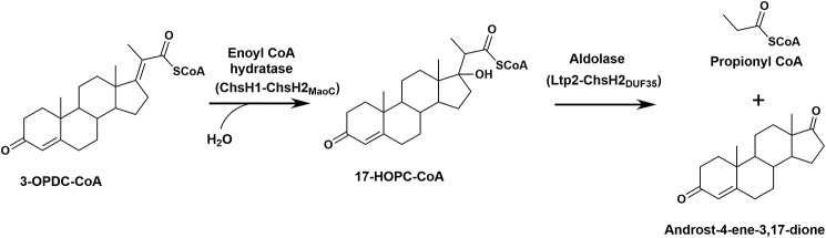
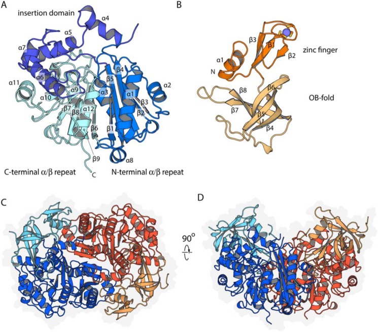
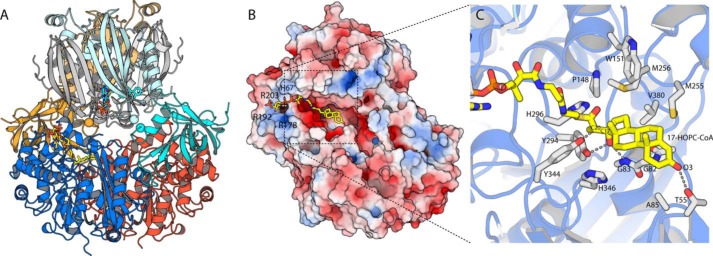
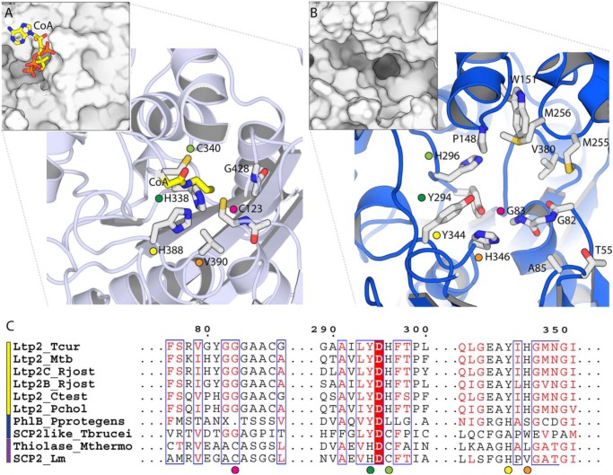
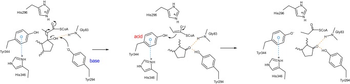
An aldolase from the bile acid-degrading actinobacterium Thermomonospora curvata catalyzes the C-C bond cleavage of an isopropyl-CoA side chain from the D-ring of the steroid metabolite 17-hydroxy-3-oxo-4-pregnene-20-carboxyl-CoA (17-HOPC-CoA). Like its homolog from Mycobacterium tuberculosis, the T. curvata aldolase is a protein complex of Ltp2 with a DUF35 domain derived from the C-terminal domain of a hydratase (ChsH2DUF35) that catalyzes the preceding step in the pathway. We determined the structure of the Ltp2-ChsH2DUF35 complex at 1.7 Å resolution using zinc-single anomalous diffraction. The enzyme adopts an αββα organization, with the two Ltp2 protomers forming a central dimer, and the two ChsH2DUF35 protomers being at the periphery. Docking experiments suggested that Ltp2 forms a tight complex with the hydratase but that each enzyme retains an independent CoA-binding site. Ltp2 adopted a fold similar to those in thiolases; however, instead of forming a deep tunnel, the Ltp2 active site formed an elongated cleft large enough to accommodate 17-HOPC-CoA. The active site lacked the two cysteines that served as the nucleophile and general base in thiolases and replaced a pair of oxyanion-hole histidine residues with Tyr-246 and Tyr-344. Phenylalanine replacement of either of these residues decreased aldolase catalytic activity at least 400-fold. On the basis of a 17-HOPC-CoA -docked model, we propose a catalytic mechanism where Tyr-294 acts as the general base abstracting a proton from the D-ring hydroxyl of 17-HOPC-CoA and Tyr-344 as the general acid that protonates the propionyl-CoA anion following C-C bond cleavage.

Keywords: C–C bond cleavage; actinobacteria; aldolase; bile acid; biodegradation; cholesterol; protein evolution; steroid; thiolase; β-oxidation.


Conflict of interest statement

The authors declare that they have no conflicts of interest with the contents of this article

Figures

Figure 1.
Figure 1.
Reactions catalyzed by the hydratase and aldolase in the last round of the β-oxidation of a steroid side chain. The metabolite 3-OPDC-CoA thioester is hydrated to 17-HOPC-CoA thioester that subsequently undergoes C–C bond cleavage catalyzed by an aldolase.
Figure 2.
Figure 2.
Structure of Ltp2–ChsH2DUF35. A, secondary structure organization of Ltp2, with secondary structure labeled. B, secondary structural organization of the ChsH2DUF35 domain. The zinc ion is depicted as a blue sphere, with the coordinating cysteine residues shown as sticks. C, organization of Ltp2–ChsH2DUF35 heterotetramer. The Ltp2 chains are colored dark orange and blue, and the DUF35 domains of ChsH2 are colored light orange and light blue. A faint gray silhouette depicts the extent of the protein surface. D, Ltp2–ChsH2DUF35 complex depiction is 90° from C.
Figure 3.
Figure 3.
Modeling the Ltp2 multiprotein and substrate complexes. A, Rosettadock model of the Ltp2–ChsH2DUF35 complex (colored as in Fig. 2) docked onto the M. tuberculosis ChsH1–ChsH2MaoC complex (ChsH2 in pale cyan and pale orange, and ChsH1 in gray shades). Docking is constrained by the high probability of the proteins docking with their 2-fold axis aligned, plus the limited length of the linker joining the C terminus of the ChsH2MaoC and N terminus of the ChsH2DUF35 (marked with spheres). Both proteins have a distinctly saddle-like shape, drastically restricting the plausible conformations. Note that this complex omits two extended loops (joining the ChsH2 domains and covering the Ltp2-active site). B, electrostatic surface (generated using APBS) for Ltp2 with 17-HOPC-CoA manually docked. C, close-up of the model, showing details of 17-HOPC-CoA binding (yellow sticks) and key proposed binding and catalytic residues (white sticks; note: protein residues are shown in their experimentally observed positions and were not shifted during docking).
Figure 4.
Figure 4.
Comparison of the active site of Ltp2 aldolase with an SCP-2 thiolase. A, active-site structure of the SCP-2 thiolase from L. mexicana (PDB code 3zbg). Larger panel shows key active-site residues, and the smaller inset panel shows a surface view of the same region. CoA (yellow) was positioned from 3zbn. Colored circles denote equivalent residues in A, B, and C. B, equivalent active-site region in Ltp2. C, multiple sequence alignment of Ltp2 from T. curvata and its homologs. The first six sequences (yellow bar) are Ltp2 aldolases; the following two (blue bar) are thiolase-like proteins of unknown activity; and the last two sequences are thiolases. Amino acid sequences of proteins are as follows: T. curvata Ltp2 (Ltp2_Tcur); M. tuberculosis Ltp2 (Ltp2_Mtb); R. jostii RHA1 cholesterol Ltp2 (Ltp2C_Rjost); bile acid Ltp2 (Ltp2B_Rjost); Comamonas testosteroni KF1 Ltp2 (Ltp2_Ctest); Pseudomonas sp. Chol1 Ltp2 (Ltp2_Pchol); PhlB from P. protogens (PhlB_Pprotegens); thiolase-type SCP-2-like from T. brucei (SCP2like_Tbrucei); M. thermolithotrophicus acetoacetyl-CoA thiolase-type SCP-2 (Thiolase_Mthermo); and L. mexicana thiolase-type SCP-2 (SCP2_Lm). Sequences are aligned using Clustal Omega and displayed using ESPRIPT (41, 42).
Figure 5.
Figure 5.
Proposed retro-aldol cleavage mechanism of Ltp2–ChsH2DUF35. This prediction was made based on the modeling of the 17-HOPC-CoA into the active site and the results obtained from site-directed mutagenesis studies. Tyr-294 deprotonates the C17 hydroxyl group of 17-HOPC-CoA leading to C–C bond cleavage. The second tyrosine residue, Tyr-344, acts as an acid, donating a proton to the enolate anion of the CoA thioester, forming propionyl-CoA and androst-4-ene-3,17-dione. Either Tyr-294 or Tyr-344 could be activated by the nearby histidine residues.

References

    1. Bergstrand L. H., Cardenas E., Holert J., Van Hamme J. D., and Mohn W. W. (2016) Delineation of steroid-degrading microorganisms through comparative genomic analysis. MBio 7, e00865–16 10.1128/mBio.00865-16 - DOI - PMC - PubMed
    1. Van der Geize R., Yam K., Heuser T., Wilbrink M. H., Hara H., Anderton M. C., Sim E., Dijkhuizen L., Davies J. E., Mohn W. W., and Eltis L. D. (2007) A gene cluster encoding cholesterol catabolism in a soil actinomycete provides insight into Mycobacterium tuberculosis survival in macrophages. Proc. Natl. Acad. Sci. U.S.A. 104, 1947–1952 10.1073/pnas.0605728104 - DOI - PMC - PubMed
    1. García J. L., Uhía I., and Galán B. (2012) Catabolism and biotechnological applications of cholesterol degrading bacteria. Microb. Biotechnol. 5, 679–699 10.1111/j.1751-7915.2012.00331.x - DOI - PMC - PubMed
    1. Dheda K., Gumbo T., Maartens G., Dooley K. E., McNerney R., Murray M., Furin J., Nardell E. A., London L., Lessem E., Theron G., van Helden P., Niemann S., Merker M., Dowdy D., et al. (2017) The epidemiology, pathogenesis, transmission, diagnosis, and management of multidrug-resistant, extensively drug-resistant, and incurable tuberculosis. Lancet Respir. Med. 5, 291–360 10.1016/S2213-2600(17)30079-6 - DOI - PubMed
    1. Ouellet H., Johnston J. B., and de Montellano P. R. (2011) Cholesterol catabolism as a therapeutic target in Mycobacterium tuberculosis. Trends Microbiol. 19, 530–539 10.1016/j.tim.2011.07.009 - DOI - PMC - PubMed

Publication types

MeSH terms

Supplementary concepts

LinkOut - more resources