

Nicotine protects fetus against LPS-induced fetal growth restriction through ameliorating placental inflammation and vascular development in late pregnancy in rats
- PMID: 31209145
- PMCID: PMC6603276
- DOI: 10.1042/BSR20190386
Nicotine protects fetus against LPS-induced fetal growth restriction through ameliorating placental inflammation and vascular development in late pregnancy in rats
Abstract
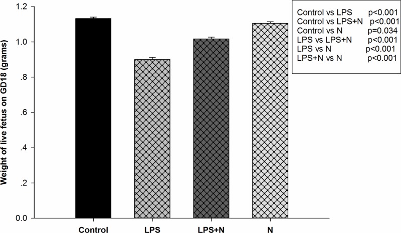
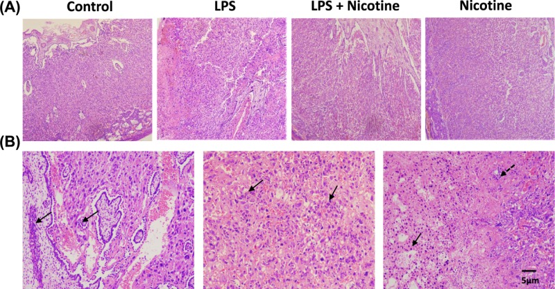
Our previous work has shown that nicotine suppressed lipopolysaccharide (LPS)-induced placental inflammation by inhibiting cytokine release as well as infiltration of leukocytes into the placenta through the cholinergic anti-inflammatory pathway. Nicotine also increased fetal survival and restored pup weight. In the present study, we aim to further investigate if fetal growth restriction (FGR) occurs with LPS treatment, and evaluate the protective effects of nicotine on fetuses in late gestation of rats. Pregnant Sprague-Dawley rats were divided into control group, nicotine group, LPS group and LPS + nicotine group. Rats were first pretreated with nicotine or vehicle by subcutaneous injection on gestation day (GD)14 and GD15, followed by LPS or vehicle intraperitoneal injection on GD16, and were killed on GD18. Loss of fetuses, number and weights of live fetuses and weights of placentas were recorded. Placentas were collected to evaluate placental pathology and determine inflammatory cytokines and vascular endothelial growth factor (VEGF) levels. We found that LPS treatment increased levels of placental inflammatory cytokines and placental pathological damage, decreased levels of VEGF, reduced number of live fetuses and induced FGR. Pretreatment with nicotine reversed LPS-induced high levels of placental inflammatory cytokines, low levels of placental VEGF and placental pathological damage, then rescued the number and weights of live fetuses. These data demonstrated that activation of the cholinergic anti-inflammatory pathway by nicotine protected fetus against LPS-induced FGR through ameliorating placental inflammation and vascular development in late pregnancy in rats. It may be an alternative therapeutic strategy for inflammation- induced FGR in late pregnancy.
Keywords: fetal growth restriction; lipopolysaccharides; nicotine; pregnancy.
© 2019 The Author(s).
Conflict of interest statement
The authors declare that there are no competing interests associated with the manuscript.
Figures


Similar articles
-
Nicotine inhibits LPS-induced cytokine production and leukocyte infiltration in rat placenta.Placenta. 2016 Mar;39:77-83. doi: 10.1016/j.placenta.2016.01.015. Epub 2016 Jan 19. Placenta. 2016. PMID: 26992678
-
Inflammation-induced fetal growth restriction in rats is associated with increased placental HIF-1α accumulation.PLoS One. 2017 Apr 19;12(4):e0175805. doi: 10.1371/journal.pone.0175805. eCollection 2017. PLoS One. 2017. PMID: 28423052 Free PMC article.
-
Nicotine, an α7 nAChR agonist, reduces lipopolysaccharide-induced inflammatory responses and protects fetuses in pregnant rats.Am J Obstet Gynecol. 2014 Nov;211(5):538.e1-7. doi: 10.1016/j.ajog.2014.04.026. Epub 2014 Apr 23. Am J Obstet Gynecol. 2014. PMID: 24769008
-
Placental pathology in early-onset and late-onset fetal growth restriction.Fetal Diagn Ther. 2014;36(2):117-28. doi: 10.1159/000359969. Epub 2014 Feb 21. Fetal Diagn Ther. 2014. PMID: 24577279 Review.
-
Angiogenic factors in placentas from pregnancies complicated by fetal growth restriction (review).Mol Med Rep. 2012 Jul;6(1):23-7. doi: 10.3892/mmr.2012.898. Epub 2012 May 2. Mol Med Rep. 2012. PMID: 22552373 Review.
Cited by
-
LPS Administration during Fertilization Affects Epigenetic Inheritance during Embryonic Development.Animals (Basel). 2023 Mar 23;13(7):1135. doi: 10.3390/ani13071135. Animals (Basel). 2023. PMID: 37048391 Free PMC article.
-
NCOA2-induced secretion of leptin leads to fetal growth restriction via the NF-κB signaling pathway.Ann Transl Med. 2023 Feb 28;11(4):166. doi: 10.21037/atm-22-6444. Epub 2023 Feb 13. Ann Transl Med. 2023. PMID: 36923094 Free PMC article.
-
Nicotine in Inflammatory Diseases: Anti-Inflammatory and Pro-Inflammatory Effects.Front Immunol. 2022 Feb 18;13:826889. doi: 10.3389/fimmu.2022.826889. eCollection 2022. Front Immunol. 2022. PMID: 35251010 Free PMC article. Review.
-
Dietary Folic Acid Supplementation Attenuates Maternal High-Fat Diet-Induced Fetal Intrauterine Growth Retarded via Ameliorating Placental Inflammation and Oxidative Stress in Rats.Nutrients. 2023 Jul 24;15(14):3263. doi: 10.3390/nu15143263. Nutrients. 2023. PMID: 37513681 Free PMC article.
-
Smoking and Smoking Cessation in the Risk for Fetal Growth Restriction and Low Birth Weight and Additive Effect of Maternal Obesity.J Clin Med. 2020 Oct 29;9(11):3504. doi: 10.3390/jcm9113504. J Clin Med. 2020. PMID: 33138256 Free PMC article.
References
-
- Berle J.O., Mykletun A., Daltveit A.K., Rasmussen S. and Dahl A.A. (2006) Outcomes in adulthood for children with foetal growth retardation. A linkage study from the Nord-Trondelag Health Study (HUNT) and the Medical Birth Registry of Norway. Acta Psychiatr. Scand. 113, 501–50910.1111/j.1600-0447.2005.00704.x - DOI - PubMed
Publication types
MeSH terms
Substances
LinkOut - more resources
Full Text Sources
Miscellaneous