. 2019 Aug 7;39(32):6315-6324.
doi: 10.1523/JNEUROSCI.0503-19.2019. Epub 2019 Jun 17.

Sleep as a Potential Biomarker of Tau and β-Amyloid Burden in the Human Brain

Affiliations

Sleep as a Potential Biomarker of Tau and β-Amyloid Burden in the Human Brain

Joseph R Winer et al. J Neurosci. .

Abstract

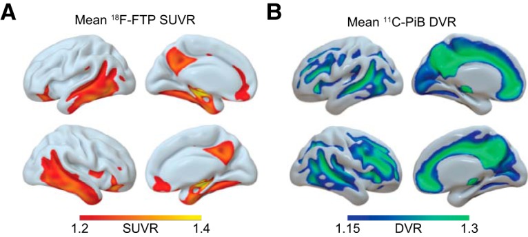
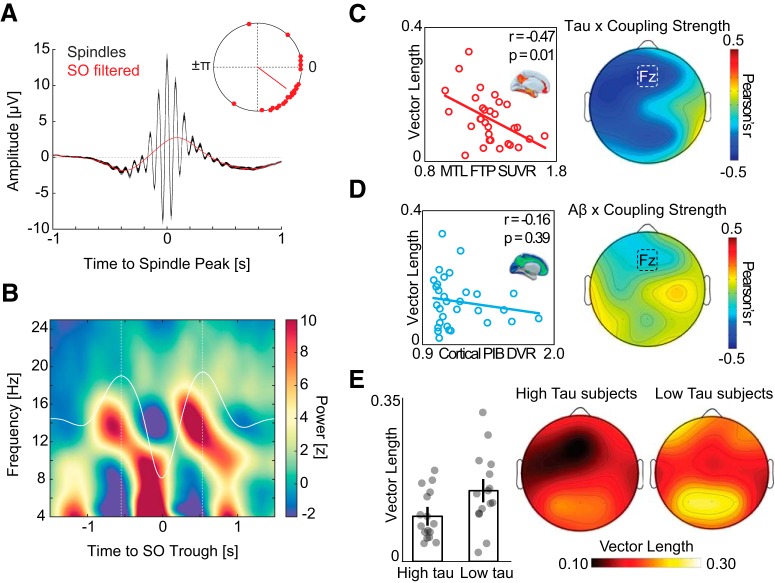
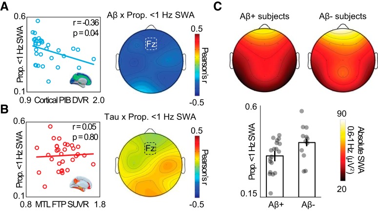
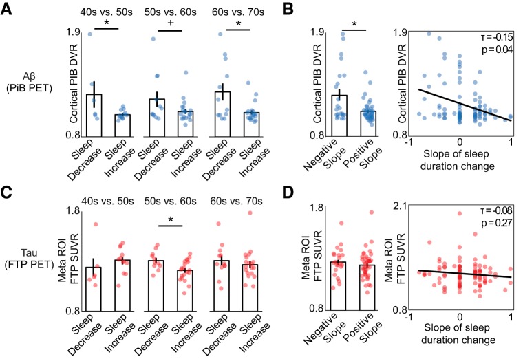
Recent proposals suggest that sleep may be a factor associated with accumulation of two core pathological features of Alzheimer's disease (AD): tau and β-amyloid (Aβ). Here we combined PET measures of Aβ and tau, electroencephalogram sleep recordings, and retrospective sleep evaluations to investigate the potential utility of sleep measures in predicting in vivo AD pathology in male and female older adults. Regression analyses revealed that the severity of impaired slow oscillation-sleep spindle coupling predicted greater medial temporal lobe tau burden. Aβ burden was not associated with coupling impairment but instead predicted the diminished amplitude of <1 Hz slow-wave-activity, results that were statistically dissociable from each other. Additionally, comparisons of AD pathology and retrospective, self-reported changes in sleep duration demonstrated that changes in sleep across the lifespan can predict late-life Aβ and tau burden. Thus, quantitative and qualitative features of human sleep represent potential noninvasive, cost-effective, and scalable biomarkers (current and future forecasting) of AD pathology, and carry both therapeutic and public health implications.SIGNIFICANCE STATEMENT Several studies have linked sleep disruption to the progression of Alzheimer's disease (AD). Tau and β-amyloid (Aβ), the primary pathological features of AD, are associated with both objective and subjective changes in sleep. However, it remains unknown whether late life tau and Aβ burden are associated with distinct impairments in sleep physiology or changes in sleep across the lifespan. Using polysomnography, retrospective questionnaires, and tau- and Aβ-specific PET, the present study reveals human sleep signatures that dissociably predict levels of brain tau and Aβ in older adults. These results suggest that a night of polysomnography may aid in evaluating tau and Aβ burden, and that treating sleep deficiencies within decade-specific time windows may serve in delaying AD progression.

Keywords: Alzheimer's disease; PET; aging; beta-amyloid; sleep; tau.


Figures

Figure 1.
Figure 1.
Mean PET binding demonstrates tau and Aβ aggregation. 18F-FTP tau and 11C-PiB Aβ PET show distinct binding patterns in healthy older adults. Using SPM12, PET images for all subjects were normalized to a common template; then a mean image was created for each radiotracer. A, Mean 18F-FTP SUVR for n = 101 PET scans, representing tau distribution in healthy older adults. B, Mean 11C-PiB DVR for n = 100 PET scans, representing Aβ distribution in healthy older adults.
Figure 2.
Figure 2.
Associations between SO-spindle coupling, tau, and Aβ. A, Peak-locked sleep spindle average across all detected events in NREM sleep (black). Low-pass filtered events (red) highlight that sleep spindles preferentially peaked before the SO “up-state.” Top right, Mean SO phase where sleep spindle power peaks. Red dots represent individual subjects. B, Baseline-corrected grand-average SO-locked time-frequency representation. Dashed white lines indicate the two largest SO peaks. Sleep spindle activity (12–16 Hz) is greatest before SO peak. C, Left, Negative association between SO-spindle coupling strength (resultant vector length) and MTL tau PET at Fz electrode. Right, Topography of correlation between SO-spindle coupling strength and MTL tau PET across all EEG electrodes. D, Left, No association between strength of SO-spindle coupling and cortical Aβ PET. Right, Topography of correlation across all EEG electrodes. E, Left, Bar plots represent mean vector length in high and low tau groups at electrode Fz. Error bars indicate SEM. Right, Topography of SO-spindle coupling strength in subjects with low versus high MTL tau burden.
Figure 3.
Figure 3.
Associations between NREM SWA, tau, and Aβ. A, Left, Negative association between proportion of 0.6–1 Hz SWA and cortical Aβ PET at electrode Fz. Right, Topography of correlation across all EEG electrodes. B, Left, No association between proportion of 0.6–1 Hz SWA and MTL tau PET. Right, Topography of correlation between proportion of 0.6–1 Hz SWA and MTL tau PET across all EEG electrodes. C, Top, Topography of 0.6–1 Hz SWA in Aβ-negative and Aβ-positive subjects. Bottom, Bar plots represent proportion of 0.6–1 Hz SWA in Aβ-negative and Aβ-positive subjects at electrode Fz. Error bars indicate SEM.
Figure 4.
Figure 4.
Associations between retrospective change in sleep duration, tau, and Aβ. A, Bar plots represent mean late-life Aβ burden (cortical PiB DVR) for subjects with self-reported decreasing sleep duration versus subjects with increasing sleep duration in each decade from age 40 to 70. Aβ burden was significantly higher in subjects whose sleep duration decreased both for the 50s and 70s decades (p < 0.05), with a trend in the same direction for the 60s (p < 0.1). B, Left, Bar plot represents that subjects with a negative sleep duration slope, indicating their sleep duration decreased over their lifespan, had significantly greater Aβ burden in late life compared with those with positive slope (p < 0.05). Right, Negative association between slope of sleep duration change and late-life Aβ burden, demonstrating that greater loss of sleep duration was predictive of greater Aβ. C, Mean late-life tau burden (meta ROI FTP SUVR) for subjects with self-reported decreasing sleep duration versus subjects with increasing sleep duration for each decade. Late-life tau burden was significantly higher for individuals with decreasing sleep duration in their 60s (p < 0.05). D, Left, Tau burden is not significantly different in subjects with negative sleep duration slope relative to positive slope subjects. Right, No association between slope of sleep duration change and late-life tau burden. *Significant t value (p < 0.05). +Trending t value (p < 0.10). Error bars indicate SEM.

Similar articles

Cited by

References

    1. Adams JN, Lockhart SN, Li L, Jagust WJ (2018) Relationships between tau and glucose metabolism reflect Alzheimer's disease pathology in cognitively normal older adults. Cereb Cortex 29:1997–2009. 10.1093/cercor/bhy078 - DOI - PMC - PubMed
    1. Ahnaou A, Moechars D, Raeymaekers L, Biermans R, Manyakov NV, Bottelbergs A, Wintmolders C, Van Kolen K, Van De Casteele T, Kemp JA, Drinkenburg WH (2017) Emergence of early alterations in network oscillations and functional connectivity in a tau seeding mouse model of Alzheimer's disease pathology. Sci Rep 7:14189. 10.1038/s41598-017-13839-6 - DOI - PMC - PubMed
    1. Arriagada PV, Growdon JH, Hedley-Whyte ET, Hyman BT (1992) Neurofibrillary tangles but not senile plaques parallel duration and severity of Alzheimer's disease. Neurology 42:631–639. 10.1212/WNL.42.3.631 - DOI - PubMed
    1. Baker SL, Maass A, Jagust WJ (2017) Considerations and code for partial volume correcting [18F]-AV-1451 tau PET data. Data Brief 15:648–657. 10.1016/j.dib.2017.10.024 - DOI - PMC - PubMed
    1. Berens P. (2009) CircStat: a MATLAB toolbox for circular statistics. J Stat Softw 31:1–21.

Publication types

MeSH terms