

Alda-1 attenuates hyperoxia-induced mitochondrial dysfunction in lung vascular endothelial cells
- PMID: 31209184
- PMCID: PMC6628993
- DOI: 10.18632/aging.102012
Alda-1 attenuates hyperoxia-induced mitochondrial dysfunction in lung vascular endothelial cells
Abstract
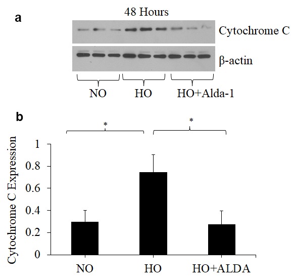
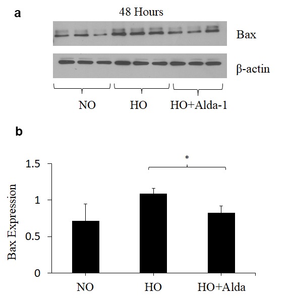
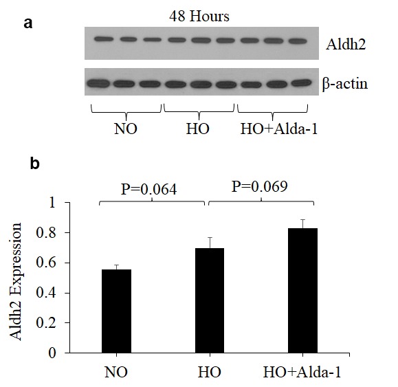
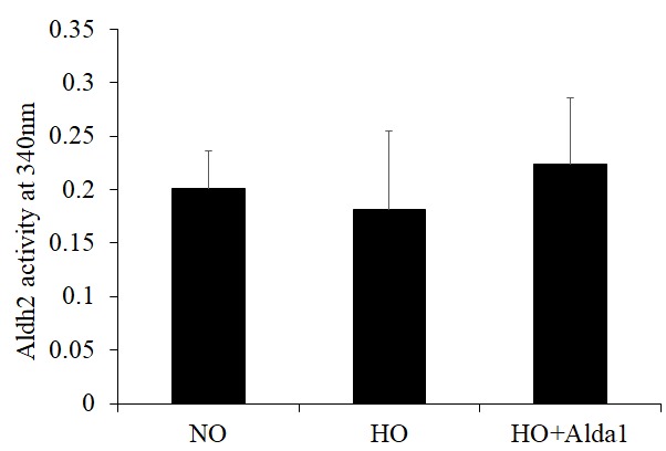
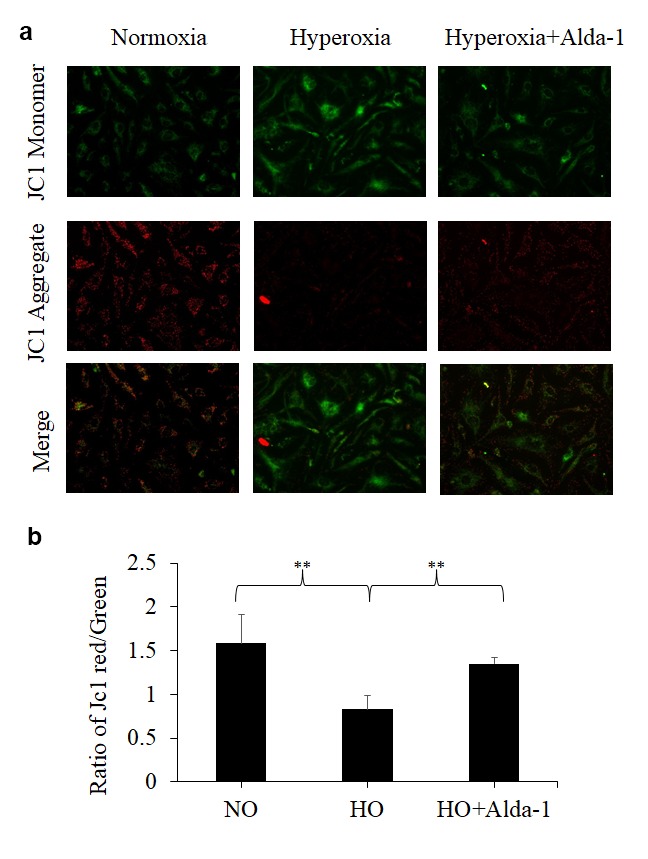
Acute lung injury (ALI) is a major cause of morbidity and mortality worldwide, especially in aged populations. Mitochondrial damage is one of the key features of ALI. Hyperoxia-induced lung injury model in mice has been widely used for ALI study because it features many ALI phenotypes including, but not limited to, mitochondrial and vascular endothelial cell damage. Recently, accumulating evidence has shown that mitochondrial aldehyde dehydrogenase 2 (ALDH2) has a protective effect against oxidative stress mediated cell damage in epithelial cells. However, it is not known whether ALDH2 protects against oxidative stress in vascular endothelial cells. In this current study, we attempted to find the capacity of Alda-1 [(N-(1,3benzodioxol-5-ylmethyl)-2,6- dichloro-benzamide), an ALDH2 activator] to protect against oxidative stress in human microvascular endothelial cells (HMVEC). HMVEC pretreated with Alda-1 prior to hyperoxic exposure vs non-treated controls showed i) lower 4-hydroxynonenal (4-HNE) levels, ii) significantly decreased expressions of Bax and Cytochrome C, iii) partially restored activity and expression of ALDH2 and iv) significantly improved mitochondrial membrane potential. These results suggest that ALDH2 protein in lung vascular endothelial cells is a promising therapeutic target for the treatment of ALI and that Alda-1 is a potential treatment option.
Keywords: ALDH2; Alda-1; HMVEC; acute lung injury; hyperoxia.
Conflict of interest statement
Figures

Similar articles
-
Alda-1 Attenuates Hyperoxia-Induced Acute Lung Injury in Mice.Front Pharmacol. 2021 Jan 8;11:597942. doi: 10.3389/fphar.2020.597942. eCollection 2020. Front Pharmacol. 2021. PMID: 33597876 Free PMC article.
-
Pharmacological Activation Of Aldehyde Dehydrogenase 2 Protects Against Heatstroke-Induced Acute Lung Injury by Modulating Oxidative Stress and Endothelial Dysfunction.Front Immunol. 2021 Oct 26;12:740562. doi: 10.3389/fimmu.2021.740562. eCollection 2021. Front Immunol. 2021. PMID: 34764958 Free PMC article.
-
Alda-1, an ALDH2 activator, protects against hepatic ischemia/reperfusion injury in rats via inhibition of oxidative stress.Free Radic Res. 2018 Jun;52(6):629-638. doi: 10.1080/10715762.2018.1459042. Epub 2018 Apr 13. Free Radic Res. 2018. PMID: 29589772
-
Role of aldehyde dehydrogenase 2 in ischemia reperfusion injury: An update.World J Gastroenterol. 2018 Jul 21;24(27):2984-2994. doi: 10.3748/wjg.v24.i27.2984. World J Gastroenterol. 2018. PMID: 30038465 Free PMC article. Review.
-
Aldehyde dehydrogenase-2 as a therapeutic target.Expert Opin Ther Targets. 2019 Nov;23(11):955-966. doi: 10.1080/14728222.2019.1690454. Epub 2019 Nov 16. Expert Opin Ther Targets. 2019. PMID: 31697578 Review.
Cited by
-
Mycobacterium avium subsp. paratuberculosis Proteome Changes Profoundly in Milk.Metabolites. 2021 Aug 20;11(8):549. doi: 10.3390/metabo11080549. Metabolites. 2021. PMID: 34436489 Free PMC article.
-
Alda-1 ameliorates air embolism-induced acute lung injury.Int J Immunopathol Pharmacol. 2023 Jan-Dec;37:3946320231223005. doi: 10.1177/03946320231223005. Int J Immunopathol Pharmacol. 2023. PMID: 38113877 Free PMC article.
-
Alda-1 Attenuates Hyperoxia-Induced Acute Lung Injury in Mice.Front Pharmacol. 2021 Jan 8;11:597942. doi: 10.3389/fphar.2020.597942. eCollection 2020. Front Pharmacol. 2021. PMID: 33597876 Free PMC article.
-
[Acetaldehyde dehydrogenase 2 ameliorates lung endothelial barrier and balances mitochondrial dynamics in mice with acute lung injury].Nan Fang Yi Ke Da Xue Xue Bao. 2023 Aug 20;43(8):1388-1395. doi: 10.12122/j.issn.1673-4254.2023.08.16. Nan Fang Yi Ke Da Xue Xue Bao. 2023. PMID: 37712276 Free PMC article. Chinese.
-
Disruption of the Molecular Regulation of Mitochondrial Metabolism in Airway and Lung Epithelial Cells by Cigarette Smoke: Are Aldehydes the Culprit?Cells. 2023 Jan 12;12(2):299. doi: 10.3390/cells12020299. Cells. 2023. PMID: 36672235 Free PMC article. Review.
References
-
- Cox R Jr, Phillips O, Fukumoto J, Fukumoto I, Parthasarathy PT, Arias S, Cho Y, Lockey RF, Kolliputi N. Enhanced Resolution of Hyperoxic Acute Lung Injury as a result of Aspirin Triggered Resolvin D1 Treatment. Am J Respir Cell Mol Biol. 2015; 53:422–35. 10.1165/rcmb.2014-0339OC - DOI - PMC - PubMed
Publication types
MeSH terms
Substances
Grants and funding
LinkOut - more resources
Full Text Sources
Research Materials
Miscellaneous
