. 2019 Jun 18;10(6):449.
doi: 10.1038/s41419-019-1678-y.

Hepatic ferroptosis plays an important role as the trigger for initiating inflammation in nonalcoholic steatohepatitis

Affiliations

Hepatic ferroptosis plays an important role as the trigger for initiating inflammation in nonalcoholic steatohepatitis

Shinya Tsurusaki et al. Cell Death Dis. .

Abstract

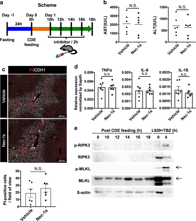
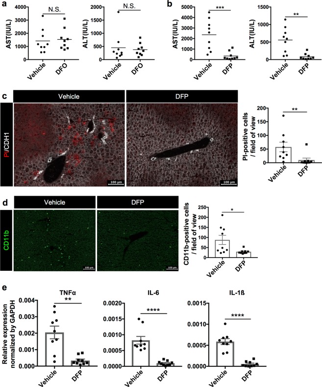
Nonalcoholic steatohepatitis (NASH) is a metabolic liver disease that progresses from simple steatosis to the disease state of inflammation and fibrosis. Previous studies suggest that apoptosis and necroptosis may contribute to the pathogenesis of NASH, based on several murine models. However, the mechanisms underlying the transition of simple steatosis to steatohepatitis remain unclear, because it is difficult to identify when and where such cell deaths begin to occur in the pathophysiological process of NASH. In the present study, our aim is to investigate which type of cell death plays a role as the trigger for initiating inflammation in fatty liver. By establishing a simple method of discriminating between apoptosis and necrosis in the liver, we found that necrosis occurred prior to apoptosis at the onset of steatohepatitis in the choline-deficient, ethionine-supplemented (CDE) diet model. To further investigate what type of necrosis is involved in the initial necrotic cell death, we examined the effect of necroptosis and ferroptosis inhibition by administering inhibitors to wild-type mice in the CDE diet model. In addition, necroptosis was evaluated using mixed lineage kinase domain-like protein (MLKL) knockout mice, which is lacking in a terminal executor of necroptosis. Consequently, necroptosis inhibition failed to block the onset of necrotic cell death, while ferroptosis inhibition protected hepatocytes from necrotic death almost completely, and suppressed the subsequent infiltration of immune cells and inflammatory reaction. Furthermore, the amount of oxidized phosphatidylethanolamine, which is involved in ferroptosis pathway, was increased in the liver sample of the CDE diet-fed mice. These findings suggest that hepatic ferroptosis plays an important role as the trigger for initiating inflammation in steatohepatitis and may be a therapeutic target for preventing the onset of steatohepatitis.


Conflict of interest statement

The authors declare that they have no conflict of interest.

Figures

Fig. 1
Fig. 1. Pathological features of steatohepatitis in the CDE diet model.
The lipid accumulation, inflammatory responses, and fibrosis in the liver were evaluated after choline-deficient, ethionine-supplemented (CDE) diet feeding. Six mice were used for analyses at each time point. a Representative images of lipid accumulation in the liver by Oil Red O staining after 0, 1, and 2 days post CDE diet feeding. b Measurement of serum liver injury markers, aspartate aminotransferase (AST) and alanine aminotransferase (ALT) after the CDE feeding (n = 6; **P< 0.01, ****P< 0.0001). c Fluorescence images of infiltration of immune cells by immunohistochemistry of liver sections for CD11b. The number of CD11b-positive cells was evaluated in 20 non-overlapping fields of view for each biological sample. The data are shown as the mean ± SEM (n = 6; **P< 0.01). d Gene expression analysis of inflammatory cytokines by quantitative reverse transcriptase PCR (RT-PCR). All data are normalized to Gapdh and shown as the means ± SEM (n = 6, *P< 0.05, **P< 0.01). e Representative images of liver fibrosis by Picro-Sirius Red staining. f Expression analysis of fibrosis-related genes by quantitative RT-PCR. All data are normalized to Gapdh and shown as the means ± SEM (n = 6; *P< 0.05, **P< 0.01, ***P < 0.001). Scale bar = 100 µm
Fig. 2
Fig. 2. Evaluation of apoptotic and necrotic cells in the liver by immunohistochemistry for CC3 and in vivo necrosis assay.
a Measurement of serum liver injury markers after 24 h of control (vehicle) and CCl4 treatment (n = 4; *P< 0.05). b HE staining of sections from control and CCl4-treated livers. c Fluorescence images of PI- and CC3-stained cells in control and CCl4-treated livers. d Fluorescence images of PI- and CC3-stained cells in the liver after 2 days of normal diet or the CDE diet feeding. Scale bar = 100 µm. PI propidium iodide, CCl4 carbon tetrachloride, CC3 cleaved caspase-3
Fig. 3
Fig. 3. Detection of apoptosis and necrosis at an early stage of steatohepatitis in the CDE diet model.
a Experimental design for identifying apoptosis and necrosis at the onset of steatohepatitis. b Western blot analysis of CC3 protein in the liver extracts after the CDE diet feeding. ß-Actin was used as an internal control. c Detection of apoptotic and necrotic cells in the liver after 14 to 18 h post CDE diet feeding by immunohistochemistry for CC3 and the in vivo necrosis assay. In images of apoptosis detection, CK19 is shown as white pseudocolor. d Measurement of serum liver injury markers after CDE diet feeding. (n = 5; *P< 0.05, ***P < 0.001). e Gene expression analysis of TNFα after the CDE diet feeding by quantitative RT-PCR. All data are normalized to Gapdh and shown as the mean ± SEM (n = 5; *P< 0.05, **P< 0.01). Scale bar = 100 µm
Fig. 4
Fig. 4. Evaluation of the effect of necroptosis inhibition in the CDE diet model.
a Experimental scheme for the administration of inhibitory agent. b Serum liver injury marker levels of vehicle or Nec-1s (5 mg/kg) treated mice after 18 h of the CDE diet feeding (n = 6). NS not significant. c Detection of necrotic cells in the CDE-fed mouse treated with vehicle or Nec-1s. Scale bar = 100 µm. The number of PI-positive cells was evaluated in 10 non-overlapping fields of view for each biological sample. The data are shown as the means ± SEM (n = 6). NS not significant. d Gene expression analysis of inflammatory cytokines by quantitative RT-PCR. All data are normalized to Gapdh and shown as the means ± SEM (n = 6). NS not significant. e Western blot analysis of phosphorylated RIPK3 (p-RIPK3), RIPK3, phosphorylated MLKL (p-MLKL), MLKL, and β-actin in the liver extracts after the CDE diet feeding. L929 cells treated with TNFα, BV, and ZVAD-FMK (L929 + TBZ) were used as control for necroptotic cell death
Fig. 5
Fig. 5. Evaluation of necrotic cell and inflammation in the liver of CDE-fed WT and Mlkl KO mice.
a Serum liver injury marker levels of WT and Mlkl KO mice after 18 h of the CDE diet feeding (n = 5). NS not significant. b Detection of necrotic cells in the liver of WT and Mlkl KO mice at 18 h post CDE feeding by the in vivo necrosis assay. The number of PI-positive cells was evaluated in 10 non-overlapping fields of view for each biological sample. The data are shown as the means ± SEM (n = 5). NS not significant. c Gene expression analysis of inflammatory cytokines by quantitative RT-PCR. All data are normalized to Gapdh and shown as the means ± SEM (n = 5; *P< 0.05)
Fig. 6
Fig. 6. Evaluation of the effect of ferroptosis inhibitor, Trolox, in the CDE diet model.
a Serum liver injury marker levels in control (vehicle) or Trolox (100 mg/kg) treated mice (n = 9). b Detection of necrosis in the CDE-fed mice treated with vehicle or Trolox by the in vivo necrosis assay. The number of PI-positive cells was evaluated in 10 non-overlapping fields of view for each biological sample. The data are shown as the means ± SEM (n = 9; *P< 0.05). c Immunohistochemical analysis of CDE-fed mouse with vehicle or Trolox for CD11b. The number of CD11b-positive cells was evaluated in 10 non-overlapping fields of view for each mouse. The data are shown as the means ± SEM (n = 9; *P< 0.05). d Gene expression analysis of inflammatory cytokines by quantitative RT-PCR (n = 9). All data are normalized to Gapdh and shown as the means ± SEM (n = 9; *P< 0.05, **P< 0.01). e Measurement of oxidized PEs in the liver. The level of each oxidized PE in the liver sample was compared among normal diet-fed mice, CDE diet-fed mice, and Trolox-treated CDE diet-fed mice (n = 4). Scale bar = 100 µm
Fig. 7
Fig. 7. Evaluation of the effect of iron chelators in the CDE diet model.
a Serum liver injury marker levels in the CDE-fed mice treated with vehicle or DFO (100 mg/kg) (n = 9). NS not significant. The serum data are shown as dot plots and mean. b Serum liver injury marker levels of the CDE-fed mice treated with vehicle or DFP (100 mg/kg) (n = 9; **P< 0.01, ***P < 0.001). c Detection of necrotic cells in the CDE-fed mice treated with vehicle or DFP. The number of PI-positive cells was evaluated in 10 non-overlapping fields of view for each biological sample. The data are shown as the means ± SEM (n = 9; **P< 0.01). d Immunohistochemical analysis of CDE-fed mouse treated with vehicle or DFP for CD11b. The number of CD11b-positive cells was evaluated in 10 non-overlapping fields of view for each biological sample. The data are shown as the mean ± SEM (n = 9; *P < 0.05). e Gene expression analysis of inflammatory cytokines by quantitative RT-PCR. The gene expression data are normalized to Gapdh and shown as the means ± SEM (n = 9; **P< 0.01, ****P< 0.0001). Scale bar = 100 µm

Similar articles

Cited by

References

    1. Matteoni C, et al. Nonalcoholic fatty liver disease: a spectrum of clinical and pathological severity. Gastroenterology. 1999;116:1413–1419. doi: 10.1016/S0016-5085(99)70506-8. - DOI - PubMed
    1. Yatsuji S, et al. Clinical features and outcomes of cirrhosis due to non-alcoholic steatohepatitis compared with cirrhosis caused by chronic hepatitis C. J. Gastroenterol. Hepatol. 2009;24:248–254. doi: 10.1111/j.1440-1746.2008.05640.x. - DOI - PubMed
    1. Takaki A, Kawai D, Yamamoto K. Molecular mechanisms and new treatment strategies for non-alcoholic steatohepatitis (NASH) Int. J. Mol. Sci. 2014;15:7352–7379. doi: 10.3390/ijms15057352. - DOI - PMC - PubMed
    1. Barreyro FJ, et al. The pan-caspase inhibitor Emricasan (IDN-6556) decreases liver injury and fibrosis in a murine model of non-alcoholic steatohepatitis. Liver Int. 2015;35:953–966. doi: 10.1111/liv.12570. - DOI - PubMed
    1. Gautheron J, et al. A positive feedback loop between RIP3 and JNK controls non-alcoholic steatohepatitis. EMBO Mol. Med. 2014;6:1062–1074. doi: 10.15252/emmm.201403856. - DOI - PMC - PubMed

Publication types

MeSH terms