. 2019 Jun 17;9(1):8662.
doi: 10.1038/s41598-019-45221-z.

Prenatal diagnosis of hypospadias with 2-dimensional and 3-dimensional ultrasonography

Affiliations

Prenatal diagnosis of hypospadias with 2-dimensional and 3-dimensional ultrasonography

Xiaohua Li et al. Sci Rep. .

Abstract

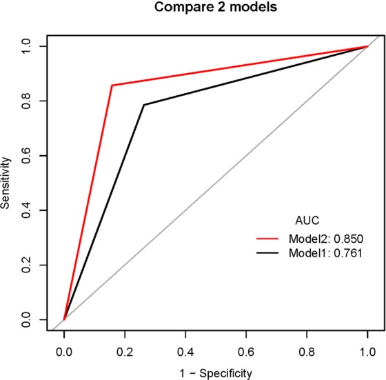
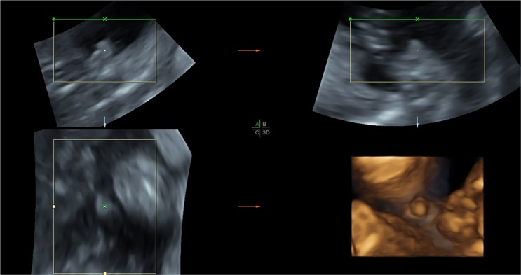
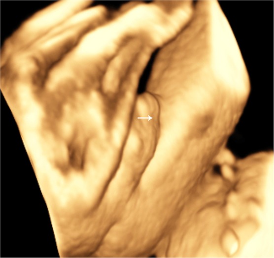
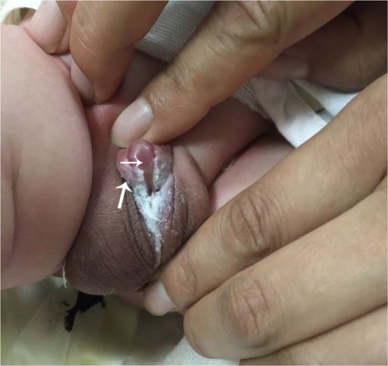
To compare the prenatal diagnostic performance as well as appearance of ultrasonic details between 2-dimensional ultrasonography (2DUS) combined with 3-dimensional ultrasonography (3DUS) and 2DUS alone for hypospadias. A total of 47 fetuses were enrolled and examined by 2DUS and then 3DUS. Postnatal follow-up data were obtained and 28 cases were confirmed of hypospadias. Although not statistically significant, there was a trend toward higher AUC (0.85 vs. 0.76; p = 0.08), ACC (85.1 vs. 76.6%; p = 0.22), SEN (85.7 vs. 78.6%; p = 0.63), and SPE (84.2 vs. 73.7%; p = 0.50) for 2DUS combined with 3DUS compared with 2DUS alone. The agreement between both methods was moderate [kappa = 0.592]. Both modalities showed accurately the short penis and blunt tip of the penis. 2DUS in combination with 3DUS showed more cases in other detailed features, such as "chordee", a "hooded" incomplete prepuce, and so on. Overall 2DUS combined with 3DUS showed a trend toward higher performance compared with 2DUS alone for the diagnosis of hypospadias, although the difference was not statistically significant. 3DUS is a useful complement for 2DUS in the diagnosis of fetal hypospadias and may provide more detailed information related to its diagnosis and prognosis.


Conflict of interest statement

The authors declare no competing interests.

Figures

Figure 1
Figure 1
Comparison of the area under receiver operating characteristics curves (ROC) for 2DUS combined with 3DUS and 2DUS alone. There was a trend toward higher AUC (0.85 vs. 0.76; p = 0.08) for 2DUS combined with 3DUS compared with 2DUS alone, although the difference was not statistically significant. Model 1 indicates 2DUS alone, and Model 2, 2DUS combined with 3DUS.
Figure 2
Figure 2
Three-dimensional multiplanar images of the external genitalia at 21 weeks of gestation in longitudinal (top left), axial (top right), and coronal (bottom left) views allow construction of a surface-rendered image (bottom right). A short and incurved penile shaft with a blunt tip was shown in three orthogonal planes simultaneously. Three-dimensional image in surface-rendered view shows excess dorsal prepuce (dorsal hood).
Figure 3
Figure 3
The ventral view of the penis was demonstrated by surface-rendered three-dimensional image at 28 weeks of gestation. The short penis with a shallow groove in the direction of up and down resulted from the incomplete fusion of the urogenital folds is shown (arrow).
Figure 4
Figure 4
Photograph of the newborn confirms the hypospadias. The dorsal thickened prepuce (big arrow) and ventral urethral groove (small arrow) of the penis are shown.

Similar articles

Cited by

References

    1. Odeh M, Ophir E, Bornstein J. Hypospadias mimicking female genitalia on early second trimester sonographic examination. J Clin ultrasonography. 2008;36:581–583. doi: 10.1002/jcu.20481. - DOI - PubMed
    1. Fang KH, et al. Prenatal sonographic features of hypospadia: 2-and 3-dimensional findings. Taiwan J Obstet Gynecol. 2006;45:53–55. doi: 10.1016/S1028-4559(09)60191-1. - DOI - PubMed
    1. Cafici D, Iglesias A. Prenatal diagnosis of severe hypospadias with 2-and 3-dimensional Sonography. J ultrasonography Med. 2002;21:1423–1426. doi: 10.7863/jum.2002.21.12.1423. - DOI - PubMed
    1. Cayan F, Cayan S. Prenatal diagnosis of penoscrotal hypospadias and review of the literature. Turkish journal of urology. 2013;39:116–118. doi: 10.5152/tud.2013.028. - DOI - PMC - PubMed
    1. Bamberg C, Brauer M, Degenhardt P, Szekessy DP, Henrich W. Prenatal 2-and 3-dimensional imaging in two cases of severe penoscrotal hypospadias. J Clin ultrasonography. 2011;39:539–543. doi: 10.1002/jcu.20832. - DOI - PubMed