. 2019 Jul;26(7):628-636.
doi: 10.1038/s41594-019-0249-3. Epub 2019 Jun 17.

GGTase3 is a newly identified geranylgeranyltransferase targeting a ubiquitin ligase

Affiliations

GGTase3 is a newly identified geranylgeranyltransferase targeting a ubiquitin ligase

Shafi Kuchay et al. Nat Struct Mol Biol. 2019 Jul.

Abstract

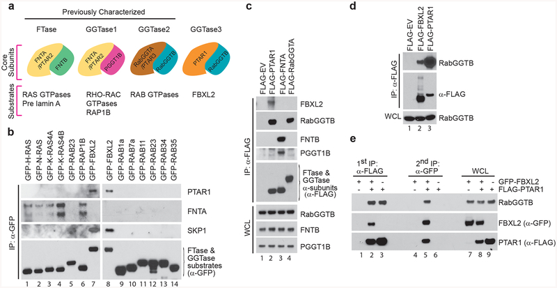
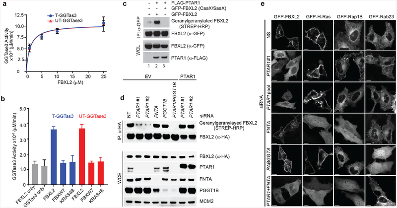
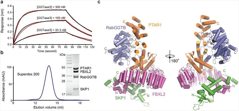
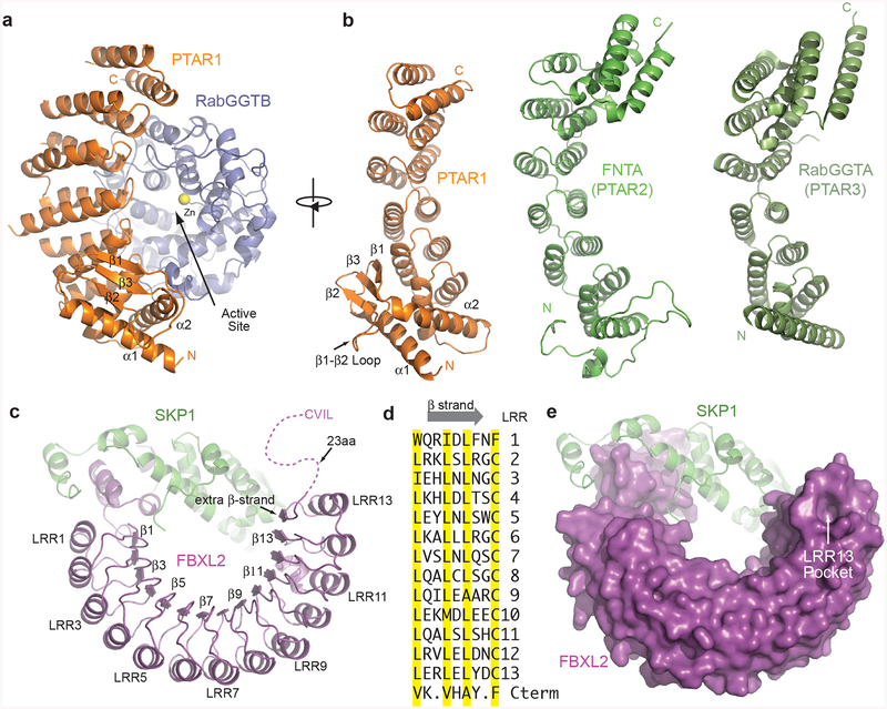
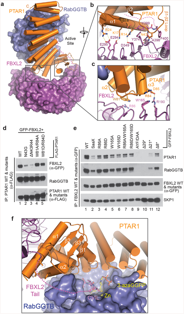
Protein prenylation is believed to be catalyzed by three heterodimeric enzymes: FTase, GGTase1 and GGTase2. Here we report the identification of a previously unknown human prenyltransferase complex consisting of an orphan prenyltransferase α-subunit, PTAR1, and the catalytic β-subunit of GGTase2, RabGGTB. This enzyme, which we named GGTase3, geranylgeranylates FBXL2 to allow its localization at cell membranes, where this ubiquitin ligase mediates the polyubiquitylation of membrane-anchored proteins. In cells, FBXL2 is specifically recognized by GGTase3 despite having a typical carboxy-terminal CaaX prenylation motif that is predicted to be recognized by GGTase1. Our crystal structure analysis of the full-length GGTase3-FBXL2-SKP1 complex reveals an extensive multivalent interface specifically formed between the leucine-rich repeat domain of FBXL2 and PTAR1, which unmasks the structural basis of the substrate-enzyme specificity. By uncovering a missing prenyltransferase and its unique mode of substrate recognition, our findings call for a revision of the 'prenylation code'.


Conflict of interest statement

Competing Interests:

The authors declare no competing financial interests. M.P. is a consultant for BeyondSpring Pharmaceuticals and a member of the scientific advisory boards of CullGen, Inc. and Kymera Therapeutics. N.Z. is a member of the scientific advisory board of Kymera Therapeutics.

Figures

Figure 1.
Figure 1.. PTAR1, an orphan prenyltransferase α subunit, binds FBXL2 and RabGGTB.
(a) Schematic representation of the four human prenyltransferases comprised by combinations of α [PTAR1, FNTA (PTAR2), and RabGGTA (PTAR3)] and β (FNTB, PGGT1B and, RabGGTB) subunits, and their substrates. As shown in this study, PTAR1 and RabGGTB interact to form a prenyltransferase that we named GGTase3. (b) HEK-293T cells were transfected with the indicated GFP-tagged substrates of prenyltransferases for immunoprecipitations and immunoblotting. (c,d) HEK-293T cells were transfected with the indicated plasmids for immunoprecipitations and immunoblotting. WCE: Whole cell extract; EV: empty vector. (e) HEK-293T cells were co-transfected with FLAG-tagged PTAR1 and GFP-tagged FBXL2 as indicated. Immunoprecipitations were carried out sequentially using first an anti-FLAG antibody and then an anti-GFP antibody as described in methods. The first elution was done with a FLAG peptide and the second with 1%SDS. The two eluates were then immunoblotted as indicated.
Figure 2.
Figure 2.. GGTase3 geranylgeranylates FBXL2 and is required for its localization to cellular membranes.
(a) Recombinant GGtase3 geranylgeranylates purified FBXL2. Indicated amounts of purified FBXL2 were incubated with 100 ng of purified GGTase3 (either tagged [T] or untagged [UT] versions) to carry out in vitro geranylgeranylation assay using saturating concentrations of tritiated [H3]-GGPP as described in methods. Each data point represents mean+/− SD of three biological replicates. Michaelis-Menten kinetics was used to generate an apparent Km value of 1.2μM using Prism Graphpad software. (b) In vitro geranylgeranylation assay was carried out and measured as in (a) using 10 μM of purified FBXL2, FBXW7, or K-RAS4B and 100 ng of purified GGTase3. Bar graphs represent mean +/− SD from three biological replicates. Source data for panels a and b are available with the paper online. (c) RPE1-HTERT cells were cotransfected with the indicated plasmids and processed for the detection of geranylgeranylated FBXL2 using a “Click-IT” assay, as described in methods. The experiment was repeated three times. Representative result is shown. Uncropped blot/gel images are shown in Supplementary Data Set 1. (d) HeLa cells were transfected with the indicated siRNA oligos and cDNAs. Twenty-four hours post-transfection cells were incubated with geranylgeranyl-azide for 16 hours, harvested, lysed, and azide selective ligation reaction with sDIBO-Biotin was performed for one hour to label geranylgeranylated proteins via copper-free “Click-IT” reaction. After immunoprecipitation with an anti-HA antibody, immunoblots were carried out. The experiment was repeated four times. Representative result is shown. Uncropped blot/gel images are shown in Supplementary Data Set 1. (e) HeLa cells were transfected first with the indicated siRNA oligos and then with the indicated GFP-tagged proteins. Live cell confocal imaging was carried out as described in methods. Images show representative frames of three independent experiments. Bar size: 10 μm.
Figure 3.
Figure 3.. Overall structure of GGTase3-FBXL2-SKP1 complex.
(a) GGTase3 binding to FBXL2-SKP1 monitored by Octet BioLayer Interferometry analysis in the absence of GGPP or its analog. Sensorgram traces (black lines) of the interactions of His-FBXL2-SKP1 with GGTase3 at three concentrations (300 nM, 100 nM, and 33.3 nM) overlaid with curve fits (red lines). The global fit yields a calculated KD of 69.0 nM ± 1.1 nM. (b) Size exclusion chromatography analysis of the purified GGTase3-FBXL2-SKP1 protein complex with sodium dodecylsulfate-polyacrylamide gel electrophoresis analysis of the central fraction containing the protein complex with all four subunits in equal stoichiometry. (c) Two orthogonal views of the tetrameric complex containing PTAR1 (orange), RabGGTB (slate), FBXL2 (magenta) and SKP1 (green). The zinc ion at the active site of RabGGTB is shown in yellow sphere. The N and C termini of different proteins are labeled N and C in corresponding colors.
Figure 4.
Figure 4.. Structural features of GGTase3 and FBXL2.
(a) Ribbon diagrams of GGTase3 containing PTAR1 (orange) as the α subunit, and RabGGTB (slate) as the β subunit. The catalytic zinc ion is shown in yellow sphere. The unique PTAR1 NTE α/β fold comprising two α helices and three β strands are labeled and numbered. (b) Structural comparison of PTAR1 (orange) to the α subunits of known prenyltransferases, including FNTA (PTAR2) (green) and RabGGTA (PTAR3) (dark green). The N and C termini of different proteins are labeled N and C in corresponding colors. For clarity, the C-terminal IGG and LRR domain of RabGGTA (PTAR3) are not shown. All the α subunits are displayed in a view orthogonal to the PTAR1 shown in (a). (c) Ribbon diagrams of FBXL2 (magenta) in complex with SKP1 (green). Select LRRs are labeled and numbered at their helices and β strands. The disordered C-terminal tail of FBXL2 containing the CVIL motif is shown as dashed magenta line. (d) Sequence alignment of β strands in all the LRRs of FBXL2 and its additional short C-terminal β strands. The characteristic residues in typical LRRs are highlighted in yellow. (e) The unusual LRR13 pocket on the apical ridge of the LRR domain of FBXL2 shown in surface representation.
Figure 5.
Figure 5.. Multivalent FBXL2-GGTase3 interface.
(a) A top view of the overall FBXL2-GGTase3 interface formed by PTAR1 (orange ribbons) and FBXL2 (magenta surface). (b,c) Close-up views of the PTAR1-FBXL2 interface made by the C-terminal and N-terminal FBXL2 LRRs. Select interface residues are shown in sticks. Dashed yellow lines represent a network of hydrogen bonds and polar interactions. The disordered C-terminal tail of FBXL2 is shown as dashed magenta line. (d) HEK-293T cells were co-transfected with GFP-tagged FBXL2 and either wild-type (WT) FLAG-tagged PTAR1 or the indicated mutants. Twenty-four hours post-transfection, cells were harvested for immunoprecipitations and immunoblotting. The experiment was repeated three times. Representative result is shown. Uncropped blot/gel images are shown in Supplementary Data Set 1. (e) HEK-293T cells were transfected with either GFP-tagged WT FBXL2 or the indicated GFP-tagged mutants. Twenty-four hours post-transfection, cells were harvested for immunoprecipitations and immunoblotting. The experiment was repeated three times. Representative result is shown. Uncropped blot/gel images are shown in Supplementary Data Set 1. (f) A model of FBXL2 C-terminal CVIL motif at the active site of GGTase3 in presence of a GGPP analog. The zinc ion is shown in yellow sphere and is buried in the catalytic pocket of RabGGTB (slate surface). The disordered FBXL2 C-tail is shown as magenta dashed line with modeled CVIL motif as magenta sticks. A GGPP analog, 3-azaGGPP, is shown as sticks with the geranylgeranyl group in yellow and the pyrophosphate in orange. The residues in the α1 and α2 helices of PTAR1-NTE forming the hydrophobic patch possibly interacting with the FBXL2 tail are shown in orange sticks.

Similar articles

Cited by

References

    1. Zhang FL & Casey PJ Protein prenylation: molecular mechanisms and functional consequences. Annu Rev Biochem 65, 241–269, doi:10.1146/annurev.bi.65.070196.001325 (1996). - DOI - PubMed
    1. Wang M & Casey PJ Protein prenylation: unique fats make their mark on biology. Nat Rev Mol Cell Biol 17, 110–122, doi:10.1038/nrm.2015.11 (2016). - DOI - PubMed
    1. Wright LP & Philips MR Thematic review series: lipid posttranslational modifications. CAAX modification and membrane targeting of Ras. J Lipid Res 47, 883–891, doi:10.1194/jlr.R600004-JLR200 (2006). - DOI - PubMed
    1. Ahearn IM, Haigis K, Bar-Sagi D & Philips MR Regulating the regulator: post-translational modification of RAS. Nat Rev Mol Cell Biol 13, 39–51, doi:10.1038/nrm3255 (2011). - DOI - PMC - PubMed
    1. Hougland JL & Fierke CA Getting a handle on protein prenylation. Nat Chem Biol 5, 197–198, doi:10.1038/nchembio0409-197 (2009). - DOI - PubMed

Methods-only References

    1. Otwinowski Z & Minor W in Methods in Enzymology Vol. 276 (eds Carter CW & Sweet RM) 307–326 (Academic Press, New York, 1997). - PubMed
    1. CCP4. The CCP4 Suite: programs for protein crystallography. Acta Crystallogr D Biol Crystallogr D50, 760–763 (1994). - PubMed
    1. Adams PD et al. PHENIX: building new software for automated crystallographic structure determination. Acta Crystallogr D Biol Crystallogr 58, 1948–1954, doi:S0907444902016657 [pii] (2002). - PubMed

Publication types