. 2019 Jul;15(7):747-755.
doi: 10.1038/s41589-019-0304-8. Epub 2019 Jun 17.

Harnessing the anti-cancer natural product nimbolide for targeted protein degradation

Affiliations

Harnessing the anti-cancer natural product nimbolide for targeted protein degradation

Jessica N Spradlin et al. Nat Chem Biol. 2019 Jul.

Abstract

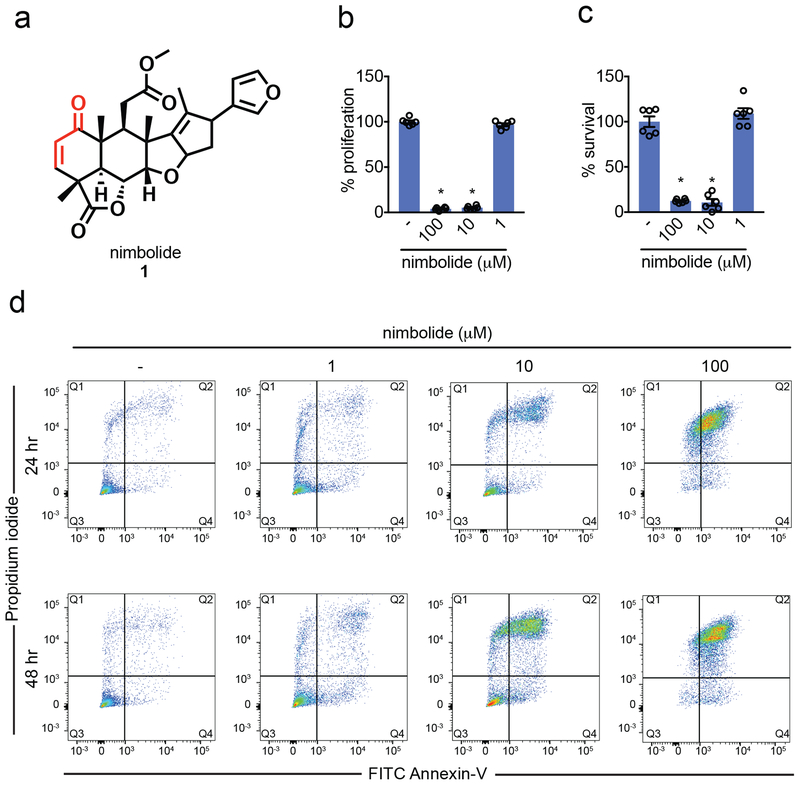
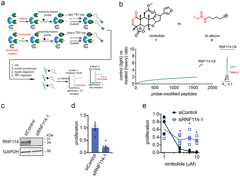
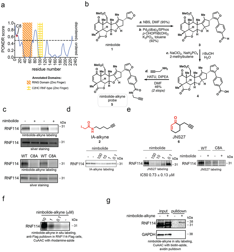
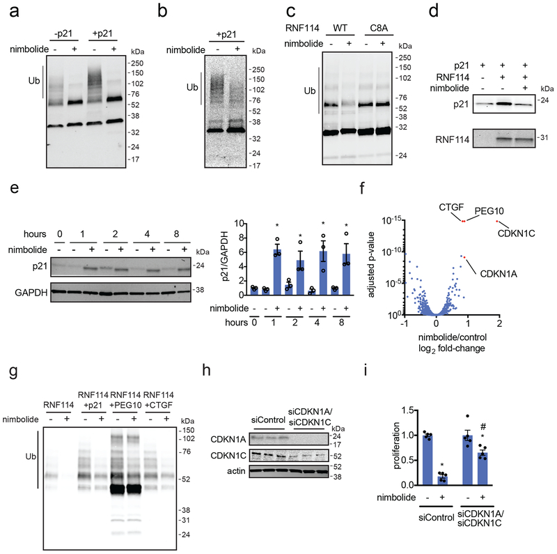
Nimbolide, a terpenoid natural product derived from the Neem tree, impairs cancer pathogenicity; however, the direct targets and mechanisms by which nimbolide exerts its effects are poorly understood. Here, we used activity-based protein profiling (ABPP) chemoproteomic platforms to discover that nimbolide reacts with a novel functional cysteine crucial for substrate recognition in the E3 ubiquitin ligase RNF114. Nimbolide impairs breast cancer cell proliferation in-part by disrupting RNF114-substrate recognition, leading to inhibition of ubiquitination and degradation of tumor suppressors such as p21, resulting in their rapid stabilization. We further demonstrate that nimbolide can be harnessed to recruit RNF114 as an E3 ligase in targeted protein degradation applications and show that synthetically simpler scaffolds are also capable of accessing this unique reactive site. Our study highlights the use of ABPP platforms in uncovering unique druggable modalities accessed by natural products for cancer therapy and targeted protein degradation applications.


Figures

Figure 1.
Figure 1.. Nimbolide impairs breast cancer cell proliferation or survival.
(a) Structure of nimbolide. Nimbolide possesses a cyclic enone that is potentially cysteine-reactive. (b, c) 231MFP breast cancer cell proliferation in serum-containing media (b) and serum-free cell survival (c). Data shown in (b and c) are average ± sem, n=6 biologically independent samples/group. (d) Percent of propidium iodide and Annexin-V-positive (PI+/Annexin-V+) cells assessed by flow cytometry after treating 231MFP cells with DMSO vehicle or nimbolide for 24 or 48 h. Shown are representative FACS data from n=3 biologically independent samples/group. Quantitation of percentage of late-stage apoptotic cells defined as defined as FITC+/PI+ cells are shown in Supplementary Fig. 1d. Statistical significance was calculated with unpaired two-tailed Student’s t-tests. Significance is expressed as *p=7.75×10⁻¹⁴ and 1.14×10⁻¹³ for 100 and 10 μM, respectively in (b) and *p=3.88×10⁻⁸ and 1.53×10⁻⁷ for 100 and 10 μM, respectively in (c) compared to vehicle-treated controls.
Figure 2.
Figure 2.. isoTOP-ABPP analysis of nimbolide in 231MFP breast cancer cell proteomes reveal RNF114 as a target.
(a) Schematic of isoTOP-ABPP in which 231MFP breast cancer cells were treated with DMSO or nimbolide (10 μM, 1.5 h in situ), after which cells were harvested and proteomes were labeled ex situ with IA-alkyne (100 μM, 1 h) followed by the isoTOP-ABPP procedure. (b) isoTOP-ABPP analysis of nimbolide (10 μM) in 231MFP breast cancer cells in situ analyzed as described in (a). Light vs heavy isotopic probe-modified peptide ratios are shown in the left plot where the primary target with the highest ratio was C8 of RNF114. Shown on the right is a representative MS1 light vs heavy peak for the probe-modified peptide bearing C8 of RNF114. (c) RNF114 knockdown by short interfering RNA (siRNA) targeting RNF114 validated by Western blotting of RNF114 compared to siControl 231MFP cells. GAPDH expression is shown as a loading control. Shown gel is a representative gel from n=3 biological replicates/group shown in Supplementary Fig. 7a. (d) 231MFP cell proliferation after 24 h in siControl and siRNF114 cells. (e) Nimbolide effects on 231MFP siControl and siRNF114 231MFP breast cancer cells. Nimbolide effects on 231MFP siControl and siRNF114 231MFP breast cancer cells. Cells were treated with DMSO vehicle or nimbolide for 24 h after which proliferation was assessed. Data for siControl or siRNF114 group was normalized to the respective DMSO vehicle control in each group. Individual biologically independent sample data is shown and the lines indicate mean values. Data shown in (d) is average ± sem. Data shown in (b, c) are from n=3, in (d, e) are from n=5 biologically independent samples/group. Statistical significance in (d, e) was calculated with unpaired two-tailed Student’s t-tests. Significance in (d) is expressed as *p=4.52×10⁻⁵ compared to siControl cells. Significance in (e) is expressed as *p=1.90×10⁻⁵, 2.72×10⁻⁴, and 0.00101 for 10, 6, and 3 μM, respectively compared to the corresponding nimbolide treatment concentration from siControl groups.
Figure 3.
Figure 3.. Nimbolide reacts covalently with C8 of RNF114.
(a) Nimbolide targets an intrinsically disordered region within RNF114 as assessed by PONDR. (b) Route for synthesis of the alkyne-functionalized nimbolide probe. (c) Gel-based ABPP analysis of pure human RNF114 protein labeled with nimbolide probe. In the upper two panels, pure RNF114 protein was pre-incubated with DMSO vehicle or nimbolide (100 μM, 30 min) prior to labeling with the nimbolide probe (10 μM, 1 h) in PBS. In the lower two panels, pure wild-type and C8A mutant RNF114 protein were labeled with the nimbolide probe (10 μM, 1 h) in PBS with 1 mg/ml BSA. Full-length and replicate blots can be found in Supplementary Fig. 7d and 7e. (d) Gel-based ABPP analysis of nimbolide competition against IA-alkyne (10 μM) or JNS27 (50 μM) labeling of pure RNF114 protein. Structures of IA-alkyne and JNS27 probes are shown with reactive moieties highlighted in red. Shown also is gel-based ABPP analysis of nimbolide (50 μM) competition against JNS27 labeling of wild-type and C8A mutant RNF114 protein. In these experiments, DMSO or nimbolide was pre-incubated for 30 min prior to probe labeling for 1 h. Full-length and replicate blots can be found in Supplementary Fig. 7g and 7h. (e) Nimbolide-alkyne labeling of Flag-RNF114 in 231MFP cells. 231MFP cells stably expressing a Flag-tagged RNF114 were treated with DMSO vehicle or nimbolide-alkyne for 2 h. RNF114 was subsequently enriched from harvested cell lysates and then rhodamine-azide was appended onto probe-labeled proteins by CuAAC, after which nimbolide-alkyne labeling was visualized by SDS/PAGE and in-gel fluorescence. Full-length and replicate blots can be found in Supplementary Fig. 7i. (f) Nimbolide-alkyne labeling of endogenous RNF114 in 231MFP cells. 231MFP cells were treated with DMSO vehicle or nimbolide-alkyne (50 μM) for 1.5 h. Biotin-azide was appended to probe-labeled proteins by CuAAC, and these proteins were subsequently avidin-enriched. Resulting pulled down proteins were analyzed by SDS/PAGE and Western blotting for RNF114. Full-length and replicate blots can be found in Supplementary Fig. 7j Gels shown in (c-f) are representative gels from n=3 biologically independent samples/group.
Figure 4.
Figure 4.. Nimbolide inhibits RNF114 activity through disrupting substrate recognition.
(a, b) RNF114 ubiquitination assay with pure GST-Ube1, GST-UBE2D1, and RNF114 protein, Flag-Ubiquitin and ATP with or without addition of p21 and blotting against Flag-ubiquitin (a) or p21 (b). (c) RNF114 autoubiquitination assay with DMSO or nimbolide (100 μM) treatment with wild-type or C8A mutant RNF114. (d) In an in vitro incubation of pure RNF114 and p21 protein, Flag-RNF114 pulldown and p21 enrichment were inhibited by nimbolide (100 μM). Full-length and replicate blots can be found in Supplementary Fig. 8a. (e) 231MFP cells were treated with nimbolide (100 μM). Shown are p21 levels in DMSO control or nimbolide-treated cells. Full-length and replicate blots can be found in Supplementary Fig. 8b. (f) Tandem mass tag (TMT)-based quantitative proteomic profiling of 231MFP breast cancer cells treated with DMSO vehicle or nimbolide (100 nM) for 12 h. Shown in red are proteins that are significantly heightened in levels >2-fold. Data shown in (f) are for 6397 proteins quantified with 2 or more unique peptides in n=3 biologically independent samples/group, see Supplementary Dataset 3 for details. (g) RNF114 ubiquitination assay with pure GST-Ube1, GST-UBE2D1, and RNF114 protein, Flag-Ubiquitin and ATP with the addition of p21 (CDKN1A), PEG10, or CTGF and blotting against Flag-ubiquitin. DMSO or Nimbolide (100 μM) was pre-incubated with RNF114, before the addition of the E1 and E2 enzymes, Flag-ubiquitin and ATP to start the reaction. (h) p21 (CDKN1A) and p57 (CDKN1C) expression in siControl and siCDKN1A/siCDKN1C 231MFP cells, assessed by Western blotting, alongside actin as a loading control. Full-length and replicate blots can be found in Supplementary Fig. 8c. (i) 231MFP cell proliferation in siControl or siCDKN1A/siCDKN1C cells treated with DMSO vehicle or nimbolide (6 μM) for 24 h. Gels shown in (a-e, g) are representative images from n=3 biologically independent samples/group. Quantification for blots shown in (a-d) are in Supplementary Fig. 3a–3d. All three biologically independent samples/group are shown in (h). Data shown in (i) are average ± sem, n=5 biologically independent samples/group. Statistical significance was calculated with unpaired two-tailed Student’s t-tests in (e, i). Significance is expressed as *p=000799, 0.0295, 0.00962, and 0.0135 for 1, 2, 4, and 8h, respectively compared to vehicle-treated control groups for each time point in (e), and p=5.65×10⁻⁸ and 0.0173 compared to vehicle-treated siControl and siCDKN1A/siCDKN1C groups, respectively in (i). Significance expressed as #p=6.70×10⁻⁵ compared to nimbolide-treated siControl cells in (i).
Figure 5.
Figure 5.. Nimbolide can be used to recruit RNF114 for targeted protein degradation of BRD4.
(a) Route for synthesizing XH2, a nimbolide-based degrader consisting of nimbolide as an RNF114 recruiter, a linker, and the BRD4 inhibitor JQ1. (b) Gel-based ABPP analysis of XH2 against RNF114. RNF114 was pre-incubated with DMSO vehicle or XH2 for 30 min prior to JNS27 labeling (50 μM) for 1 h followed by appending rhodamine-azide by CuAAC, SDS/PAGE, and analysis of in-gel fluorescence. Full-length and replicate blots can be found in Supplementary Fig. 9a. (c, d) BRD4 expression in 231MFP breast cancer cells treated with XH2 (c) versus MZ1 (d) treatment for 12 h. Full-length and replicate blots can be found in Supplementary Fig. 9b–c. (e, f) BRD4 expression in 231MFP breast cancer cells. Cells were pre-treated with DMSO vehicle or proteasome inhibitor bortezomib (BTZ) (1 μM) (e) or E1 ubiquitin activating enzyme inhibitor TAK-243 (10 μM) (f) 30 min prior to and also during treatment with MZ1 (1 μM) or XH2 (100 nM) treatment (100 nM) for 12 h. Full-length and replicate blots can be found in Supplementary Fig. 9d–e. (g) RNF114 and loading control GAPDH expression in RNF114 wild-type (WT) and knockout (KO) HAP1 cells. Full-length and replicate blots can be found in Supplementary Fig. 10a. (h) RNF114 and BRD4 expression in RNF114 wild-type (WT) or knockout (KO) HAP1 cells treated with DMSO vehicle, MZ1 (1 μM), or XH2 (100 nM) for 12 h. Full-length and replicate blots can be found in Supplementary Fig. 10b. (i) Tandem mass tag (TMT)-based quantitative proteomic profiling of 231MFP breast cancer cells treated with DMSO vehicle or XH2 (100 nM) for 12 h. Long and short BRD4 isoforms in (c-h) were visualized by SDS/PAGE and Western blotting, quantified by densitometry, and normalized to GAPDH loading controls. Gels shown in (b-h) are representative images from n=3 biologically independent samples/group and quantification for blots in (c, d, e, f, and h) are shown in Supplementary Fig. 4c, d, e, f, and i. Data shown in (i) are for 5797 proteins quantified with 2 or more unique peptides in triplicate treatments, see Supplementary Dataset 3 for details. Statistical significance in (i) are described in the methods section and p-values are reported in Supplementary Dataset 3.

Comment in

  • Stick it to E3s.
    Brand M, Winter GE. Brand M, et al. Nat Chem Biol. 2019 Jul;15(7):655-656. doi: 10.1038/s41589-019-0312-8. Nat Chem Biol. 2019. PMID: 31209352 No abstract available.

References

    1. Nomura DK & Maimone TJ Target Identification of Bioactive Covalently Acting Natural Products. Curr. Top. Microbiol. Immunol (2018). doi:10.1007/82_2018_121 - DOI - PubMed
    1. Drahl C, Cravatt BF & Sorensen EJ Protein-reactive natural products. Angew. Chem. Int. Ed Engl 44, 5788–5809 (2005). - PubMed
    1. Liu J et al. Calcineurin is a common target of cyclophilin-cyclosporin A and FKBP-FK506 complexes. Cell 66, 807–815 (1991). - PubMed
    1. Cohen E, Quistad GB & Casida JE Cytotoxicity of nimbolide, epoxyazadiradione and other limonoids from neem insecticide. Life Sci 58, 1075–1081 (1996). - PubMed
    1. Bodduluru LN, Kasala ER, Thota N, Barua CC & Sistla R Chemopreventive and therapeutic effects of nimbolide in cancer: the underlying mechanisms. Toxicol. Vitro Int. J. Publ. Assoc. BIBRA 28, 1026–1035 (2014). - PubMed

Methods-only References

    1. Jessani N et al. Carcinoma and stromal enzyme activity profiles associated with breast tumor growth in vivo. Proc. Natl. Acad. Sci. U. S. A 101, 13756–13761 (2004). - PMC - PubMed
    1. Anderson KE, To M, Olzmann JA & Nomura DK Chemoproteomics-Enabled Covalent Ligand Screening Reveals a Thioredoxin-Caspase 3 Interaction Disruptor That Impairs Breast Cancer Pathogenicity. ACS Chem. Biol 12, 2522–2528 (2017). - PMC - PubMed
    1. Smith PK et al. Measurement of protein using bicinchoninic acid. Anal. Biochem 150, 76–85 (1985). - PubMed
    1. Xu T et al. ProLuCID: An improved SEQUEST-like algorithm with enhanced sensitivity and specificity. J. Proteomics 129, 16–24 (2015). - PMC - PubMed
    1. Bateman LA et al. Chemoproteomics-enabled covalent ligand screen reveals a cysteine hotspot in reticulon 4 that impairs ER morphology and cancer pathogenicity. Chem. Commun. Camb. Engl 53, 7234–7237 (2017). - PMC - PubMed

Publication types

MeSH terms